Abstract
In 1988, Rudolf Pichlmayr pioneered split liver transplantation (SLT), enabling the transplantation of one donor liver into two recipients - one pediatric and one adult patient. In the same year, Henri Bismuth and colleagues performed the first full right/full left split procedure with two adult recipients. Both splitting techniques were rapidly adopted within the transplant community. However, a SLT is technically demanding, may cause increased perioperative complications, and may potentially transform an excellent deceased donor organ into two marginal quality grafts. Thus, crucial evaluation of donor organs suitable for splitting and careful screening of potential SLT recipients is warranted. Furthermore, the logistic background of the splitting procedure as well as the organ allocation policy must be adapted to further increase the number and the safety of SLT. Under defined circumstances, in selected patients and at experienced transplant centers, SLT outcomes can be similar to those obtained in full organ LT. Thus, SLT is an important tool to reduce the donor organ shortage and waitlist mortality, especially for pediatric patients and small adults. The present review gives an overview of technical aspects, current developments, and clinical outcomes of SLT.
Keywords: Liver transplantation, Organ shortage, In situ split, Extended right lobe, Left lateral lobe, Living donor
Core tip: As of today, split liver transplantation (SLT) is a widely adopted but yet technically demanding approach to enable liver transplantation especially in very young recipients, and to reduce organ shortage and waitlist mortality. In contrast to full organ liver transplantation, many technical evaluations concerning the donor organ, the recipient, as well as the splitting procedure and the organ allocation policy, must be considered before a SLT can safely be performed. The present review gives insight into current controversies, technical challenges, and clinical outcomes of SLT.
INTRODUCTION
The first orthotopic liver transplant (LT), published by Thomas E Starzl, was performed in a 3-year-old patient diagnosed with biliary atresia[1]. During the following years shortage of size-matched grafts for children became an evident dilemma. In the 1980s, Henri Bismuth introduced the method of graft size reduction to enable the use of adult donor grafts for pediatric LT[2]. However, by thus reducing waitlist mortality for pediatric or small adult patients, this technique in parallel increased the organ shortage for adult recipients by sacrificing the remnant liver. In 1988, Rudolf Pichlmayr was the pioneer of split liver transplantation (SLT), enabling the transplantation of one donor liver into two recipients - one pediatric and one adult patient[3]. In the same year Bismuth and colleagues performed the first full right/full left split procedure with two adult recipients[4]. Both split techniques were rapidly adopted within the transplant community and already in 1990, Christoph Broelsch published a report on the outcome of 30 split liver transplants[5].
While in the 1980s the waitlist mortality of pediatric recipients was 40%, SLT and living donor liver transplantation (LDLT) helped reduce it down to 10% in infants and to 5% in older children today[6]. A novel analysis of the development of graft survival rates in LT performed in pediatric patients and reported to the Collaborative Transplant Study (CTS, https://www.ctstransplant.org) is shown in Figure 1. Graft survival in pediatric LT is steadily increasing and current 5-year post-transplant graft survival is nearly 80%. Furthermore, SLT and LDLT enabled LT of very small infants and newborns. Since 1995, the proportion of 0-5-year-old children among recipients of pediatric LT has remained nearly constant at 65%, with a high proportion (almost 30%) in less than 1-year-old infants (Figure 2). As shown in Figure 3A, the ratio of LDLT has been steadily increasing in pediatric LT since 1995. Today, approximately one third of livers transplanted into pediatric recipients are derived from living donors (Figure 3A). On the other hand, the absolute number of livers transplanted into pediatric recipients from deceased donors has not decreased, and thus SLT resulted in an extension of the organ pool and reduction of donor organ scarcity for pediatric as well as adult LT recipients.
Figure 1.
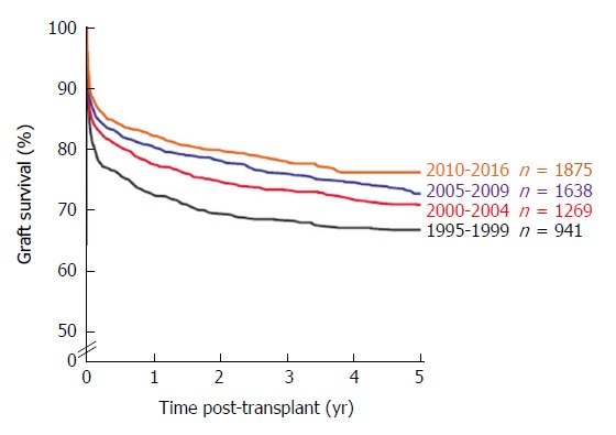
Graft survival of liver transplants in 0-17-year-old pediatric patients by transplant year from 1995-2016. Collaborative Transplant Study data are derived from 95 transplant centers in 22 countries (87% European transplants).
Figure 2.
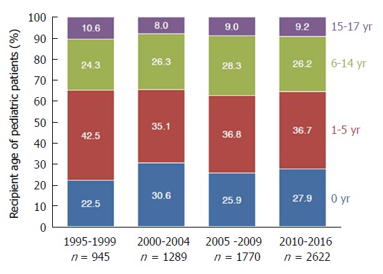
Different pediatric age groups and distribution of liver transplants by transplant year from 1995-2016.
Figure 3.
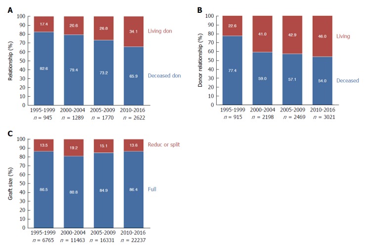
Pediatric (A) and split or reduced liver transplants from living or deceased donors (B) and all liver transplants categorized according to graft size (C) by transplant year from 1995-2016.
Performing a SLT is technically highly demanding in adult patients and may cause increased perioperative complications. The rate of living donors is steadily increasing in patients receiving an SLT and is currently reported to be 46% (Figure 3B). When comparing SLT recipients and full organ recipients, rates of LT using reduced size or split grafts have been steadily increasing until the period of 2000-2004 and since then has leveled off at 13%-15% (Figure 3C). In the first decades of SLT, increased complication rates caused hesitation to further spread the use of SLT. Ethical discussions were raised whether by splitting a liver, an excellent organ is converted into two marginal quality grafts[5,7-9]; whether an adult recipient should be allowed to refuse a split organ remains an unresolved ethical issue. Complication rates of up to 66.7% after SLT versus 45.1% after full organ LT have been described[10,11], especially due to early biliary complications (18.8% in SLT vs 7.5% in full organ LT) and portal vein thrombosis (14.6% in SLT vs 3.6% in full organ LT)[11].
In recent years experienced transplant centers have published favorable SLT outcomes similar to those obtained in full organ LT[12-15], supporting the argument that under defined conditions SLT should continue to be promoted. Therefore, allocation policies focusing on increasing SLT must be developed and transplant centers should be encouraged to perform SLT whenever safe and possible.
SURGICAL TECHNIQUES
Using the “classical” form of SLT, the liver is divided into a left lateral lobe (LLL) graft (segments II + III) for a pediatric or small adult recipient and an extended right lobe (eRL) graft (segments I + IV-VIII) for an adult recipient (Figure 4, yellow line)[16]. During the splitting procedure the hepatoduodenal ligament is first dissected from the left side for adequate identification of the left hepatic artery. Then, the left portal vein is dissected, resulting in a deportalization of segment IV. The following dissection of the liver parenchyma is performed at the right side of the falciform and round ligament, ending between the left and middle hepatic vein. Bile ducts of segments I and IV should be carefully saved while the hilar plate is sharply divided, including segment II and III bile ducts at the longitudinal section of the left portal vein. Finally, the dissected left portal vein, left artery, and left hepatic vein are divided. In the case of very small pediatric recipients, monosegment grafts consisting of segment II or segment III only can be applied.
Figure 4.
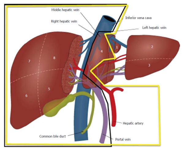
Scheme of a “classical” extended right lobe/left lateral lobe split (yellow line) and a full left/full right split (black line).
In contrast, full size splits consisting of segments I-IV and segments V-VIII (Figure 4, black line) enable transplantation of two adult recipients, although the left lobe (without segment I) is generally only suitable for a small adult. Here, the safe biliary drainage of all segments is especially crucial. In contrast to a “classical split”, the transsection plane of the liver parenchyma is significantly larger. Furthermore, due to the lack of a clear anatomic structure like the falciform ligament to indicate the resection line, in situ splitting (i.e., splitting before cold perfusion) should, whenever possible, be preferred. Potential anatomic variants of blood vessels and bile ducts must be carefully evaluated. Especially in full splits, the principle of avoiding multiple small anastomoses, defined by Henri Bismuth[4], is crucial. A normal left lobe consists of a single portal vein and a single hepatic duct, a venous outflow, which often has a common ostium (left + middle vein), and often multiple branches of small hepatic arteries. A “normal” right lobe consists of a single right artery, but often shows multiple venous branches, hepatic ducts, and sometimes even multiple branches of the portal vein. During the full split procedure it is recommended that the left lobe retains the celiac trunk, whereas the right lobe retains the main hepatic duct, the main portal vein, and the caval vein. For adequate venous drainage the left common ostium of the left and middle vein is anastomosed with the recipient venous cuff created from all three hepatic veins during a piggyback LT technique. For excellent venous outflow of the right lobe, reconstruction of the middle vein must be considered, and for reduction of the risk of a small-for-size syndrome, portal inflow modification of the recipient needs deliberation. Potential modifications are: (1) splenic artery ligation, (2) splenectomy, (3) creating a hemi-portocaval shunt, and (4) (temporary) preservation of preexisting portocaval collaterals. If a relevant small-for-size syndrome still occurs, early retransplantation must be considered.
A crucial aspect to avoid small-for-size (or, in small infants, large-for-size) syndromes is the meticulous evaluation of the donor liver volume. For the Caucasian population, liver volume can be calculated as 1072.8 * body surface area (m2) - 345.7[17]. However, multiple formulae exist to calculate the liver volume of a potential donor, considering weight, body surface area, ethnicity, and thoracoabdominal circumference (see overview in Table 1[17-21]).
Table 1.
Recently published formulae to calculate the liver volume
| Ref. | Formula | Patient group | Technique |
| Urata et al[20], 1995 | LV (mL) = 706.2 × BSA (m2) + 2.4 | 96 patients (65 pediatric) | CT |
| Vauthey et al[21], 2002 | Based on BSA: | 292 adults | CT |
| LV = -794.41 + 1267.28 × BSA (m2) | |||
| r2 = 0.46; P < 0.0001 | |||
| Based on patient weight: | |||
| LV = 191.80 + 18.51 × body weight (kg) | |||
| r2 = 0.49; P < 0.0001 | |||
| Heinemann et al[17], 1999 | LV (mL) = 1072.8 × BSA (m2) - 345.7 | 1332 patients | Autopsy |
| Kokudo et al[19], 2015 | LV = 203.3 - (3.61 × age) + [58.7 × thoracic width (cm)] - [463.7 × race (1 = Asian, 0 = Caucasian)] | 180 Japanese and 160 Swiss patients | CT |
| Herden et al[18], 2013 | Children 0 to ≤ 1 yr: | 388 pediatrics | Autopsy |
| LV (mL) = 143.062973 + 4.274603051 × body length (cm) + 14.78817631 × body weight (kg) | |||
| Children > 1 yr to < 16 yr: | |||
| LV (mL) = 20.2472281 + 3.339056437 × body length (cm) + 13.11312561 × body weight (kg) |
LV: Liver volume; BSA: Body surface area; CT: Computed tomography.
In LDLT, a minimal graft-to-recipient weight ratio of 0.6%-0.8% has been suggested[22-24]. However, since LDLT grafts are derived from very healthy donors under ideal organ procurement conditions that include a minimal cold ischemia time, the minimal graft-to-recipient weight ratio considered safe in deceased donor SLT appears to be 0.8%-1.0%[8,22,25,26]. As a simplified estimation, the liver weight represents 2% of the body weight of a normal weight adult. A LLL split thus consists of approximately 250 mL of liver parenchyma and an eRL of 1100 mL of liver parenchyma. However, due to the often mal- or non-perfused segments IV and I in eRL grafts, their functional liver parenchyma is often smaller. A full left split consists of approximately 400 mL liver parenchyma, thus being sufficient for adult recipients up to a body weight of 40-50 kg. A full right split with 800-1000 mL of liver parenchyma serves recipients up to 80-100 kg body weight, assuming a perfect venous drainage of the whole parenchyma of the right lobe[27,28]. An overview of mostly used grafts in living donor and deceased donor SLT is given in Figure 5. Within the CTS population the majority of SLT performed using living donors are either LLL (38.4%) or extended right splits (41%). In contrast, in 4241 SLTs performed with deceased donor organs from 1995-2016 a significant variation in types of SLTs exists (Figure 5).
Figure 5.
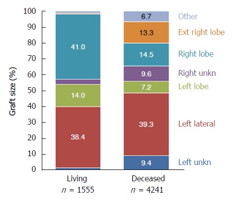
Split or reduced liver transplants from 1995-2016 categorized according to graft size.
The “classical form” of SLT from deceased donors, namely left lateral or eRL SLT, is currently performed in approximately 74% of SLT, whereas full left/right lobe SLT is performed in 17% of SLT (Figure 6A). As illustrated in Figure 6B, left lateral splits are used in 33.9% of the cases for less than 1-year-old recipients and in 50.1% of the cases for 1-5-year-old recipients. A minority of left lateral SLT grafts is used for recipients older than 6 years. Left lobe SLT grafts are used in 12.3% of the cases for less than 1-year-old recipients, in 31.6% of the cases for 1-5-year-old recipients, and in 37.7% of the cases for 6-14-year-old recipients. In contrast, almost 90% of right lobe or eRL grafts are used for adult recipients (Figure 6B), and 16.8% of left lobe splits are used for (small) adult recipients.
Figure 6.
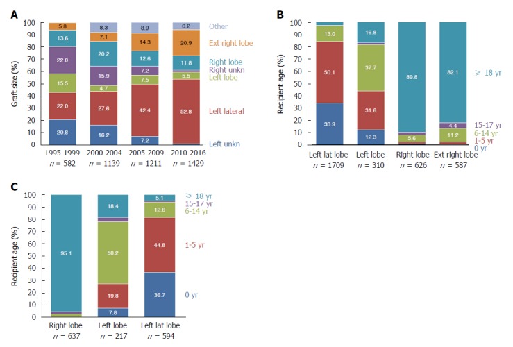
Split or reduced liver transplants from 1995-2016 from deceased donors categorized according to (A) graft size, (B) recipient age, and (C) from living donors categorized according to recipient age.
When SLT from living donor organs were analyzed within the CTS population, 637 right lobe, 217 left lobe, and 594 LLL donations were performed from 1995-2016 (Figure 6C). In adult recipients, 95.1% of right lobes, 18.4% of left lobes, and 5.1% of LLL were used, whereas 27.6% of left lobes and more than 80% of LLL were used for 0-5-year-old infants (Figure 6C).
The decision making for deceased donor SLT must factor in careful donor selection, logistic aspects concerning the split procedure, and the condition of the recipient. Concerning donor selection, Liu et al[28] recommended the following criteria for “classical” SLT: hemodynamically stable patients younger than 55 years, duration of ICU treatment less than 5 d, fatty degeneration of the liver of less than 30%, GGT < 50 U/L, GPT < 60 U/L, and serum sodium < 160 mmol/L. For full right/left SLT, donors should weigh more than 70 kg, be younger than 40 years, duration of ICU treatment less than 3 d, and fatty degeneration of the liver should be less than 10%[28]. These criteria slightly differ between transplant programs world-wide (see Table 2). Analyzing the CTS data, the median age of deceased donor livers used for SLT is 28-30 years and has remained constant since 1995 (Figure 7A). However, the median age of the deceased donor livers slightly increases in relation to the age of the recipient (Figure 7B). This reflects ongoing discussions of the influence of donor age on long-term outcomes after LT and SLT[29]. At the same time these data show that the maximum donor age tolerated for SLT in most transplant programs (see Table 2) is not exhausted and decision making for SLT may be stricter than needed[29].
Table 2.
Criteria and rates of split liver transplantation in different transplant programs according to the transplant programs homepages
| Program | Rate of SLT | Donor age | Weight/BMI | Transaminases | Other criteria |
| UNOS | 1%-4% (but according to UNOS criteria > 10% eligible) | < 40 | < 28 kg/m2 | < 3 × ULN | Single vasopressor |
| ET | 6% | < 50 | > 50 kg | ||
| United Kingdom | 10.6% | < 40 | > 50 kg | < 5 d ICU | |
| Argentina/Brazil | 10% | < 47 | umbilical perimeter < 92 cm | AST < 42 U/L | |
| ALT < 29 U/L | |||||
| Oceania | 6% | ||||
| Scandia-transplant | ? | < 51 | < 26 kg/m2 | ALT/AST < 3 × normal | < 4 d ICU |
| Saudi-Arabia | 5.6% | ||||
| South Africa | 3% | ||||
| Japan | 1.8% | ||||
| Italy | 8% (Northern Italy: 20%) | < 60 | Near-normal liver function tests | < 5 d ICU Low inotropic support |
ALT: Alanine transaminase; AST: Aspartate aminotransferase; ET: Eurotransplant; ICU: Intensive care unit; SLT: Split liver transplantation.
Figure 7.

Distribution of donor age according to transplant year (A) and recipient age (B) in deceased donor split or reduced liver transplantations performed from 1995-2016.
ETHICAL ISSUES
Because splitting a liver is a technically demanding procedure, outstanding surgical expertise is warranted. If the split procedure is performed in situ, up to 3 h more operating time during organ procurement is needed and might result in quality impairment regarding other organs procured from the same donor. If the split is performed after donor hepatectomy, i.e., ex situ in the recipient center, a respective longer cold ischemia time of the donor liver will result.
A current analysis of 37333 deceased donor LT in the United States revealed that 2352 (6.3%) of these livers met strict criteria for potential split, however only 1418 livers (3.8%) were indeed split. During the study period, less children died on the waitlist than livers were procured that could potentially have been split and used for SLT. Thus, an infant waitlist mortality of 10% and pediatric waitlist mortality of 5% in the United States could theoretically be eliminated by promoting SLT[6]. An overview of rates of SLT and criteria to perform a SLT in different transplant programs is given in Table 2. While most programs show SLT of up to 6%, programs in the United Kingdom, Brazil, and Argentina reach 10%. The highest rate of SLT is in the Northern Italian region at a rate of 20%. According to ideal evaluation regimes of donor organs, in situ splitting and a modified allocation policy promoting SLT in Northern Italy, this region is a pioneer in promoting SLT with a very high outcome quality in selected patients (see Outcome section). When comparing the rates of SLT reported to the International Registry in Organ Donation and Transplantation (IRODaT) for 2016, variations of SLT rates between 0% and 20% have been published (Table 3). A weakness of this database is the voluntary reporting of SLT rates to the IRODaT, resulting in underrated SLT rates and a lack of deceased donor organs due to religious and cultural reasons in Asian countries (where in contrast a huge expertise in LDLT exists). Nevertheless, based on published criteria to perform an SLT in different transplant programs and reported SLT numbers, a significant discrepancy can be stated in most transplant programs, and SLT should be further promoted.
Table 3.
Numbers of liver transplantations, living-donor liver transplantations, deceased donor liver transplantations, and split liver transplantations and the rate of split liver transplantation in deceased donor liver transplantation performed in different transplant programs in 2016 according to the International Registry in Organ Donation and Transplantation
| Total number | Living donors | Deceased donors | Split-liver | Split-liver (%) | |
| UNOS | 8082 | 367 | 7715 | 0 | 0 |
| Eurotransplant | |||||
| Germany | 821 | 61 | 760 | 74 | 9.01 |
| Austria | 154 | 2 | 152 | 6 | 3.90 |
| Croatia | 121 | 0 | 121 | 0 | 0 |
| Netherlands | 147 | 0 | 147 | 0 | 0 |
| Belgium | 302 | 46 | 256 | 1 | 0.33 |
| Luxemburg | 0 | 0 | 0 | 0 | 0 |
| Hungary | 74 | 0 | 74 | 0 | 0 |
| Slovenia | 27 | 0 | 27 | 0 | 0 |
| Scandiatransplant | 0 | ||||
| Sweden | 199 | 2 | 197 | 0 | 0 |
| Norway | 100 | 0 | 100 | 0 | 0 |
| Finland | 61 | 0 | 61 | 0 | 0 |
| Iceland | 0 | 0 | 0 | 0 | 0 |
| Denmark | 57 | 0 | 57 | 0 | 0 |
| China | - | - | - | - | |
| India | - | - | - | - | |
| Japan | 438 | 381 | 57 | 8 | 1.83 |
| South Korea | 1473 | 965 | 508 | 36 | 2.44 |
| Australia | 314 | 2 | 312 | 65 | 20.70 |
| Brazil | 2037 | 157 | 1880 | 0 | 0 |
| Argentina | 349 | 37 | 312 | 0 | 0 |
| Mexico | 178 | 3 | 175 | 0 | 0 |
| Canada | 582 | 73 | 509 | 0 | 0 |
| South Africa | 69 | 15 | 54 | 2 | 2.90 |
| United Kingdom | 953 | 32 | 921 | 101 | 10.60 |
| France | 1317 | 5 | 1312 | 0 | 0 |
| Spain | 1159 | 28 | 1131 | 0 | 0 |
| Italy | 1220 | 7 | 1213 | 98 | 8.03 |
| Poland | 345 | 28 | 317 | 0 | 0 |
| Czech Republic | 179 | 1 | 178 | 0 | 0 |
| Balttransplant | 33 | 0 | 33 | 0 | 0 |
OUTCOMES
In a recent Korean analysis, 86 eRL deceased donor split LT in adult recipients were compared to 303 deceased donor full organ LT. Of note, less than 25% of LT in South Korea are performed using deceased donors, i.e., a great surgical expertise in in situ splitting for LDLT exists. Groups were matched for recipient age, MELD score, duration of ischemia, and graft-to-recipient weight ratio. Donors of eRL splits were significantly younger. There was no significant difference in complication rates and 5-year graft survival rates between deceased donor SLT and full organ LT (89% vs 93%, P > 0.05). However, the 5-year overall survival rate and graft failure-free survival rate (both 63%) of eRL splits were significantly worse than in the full organ LT group (79%, P = 0.05). Factors resulting in a reduced overall survival in eRL recipients were a MELD-score greater than 30 and donor-recipient weight ratio less than 1.0. In subgroup analyses the outcome of both groups was equivalent if a donor-recipient weight ratio greater than 1.0 was observed in the eRL recipients[30].
A recent European Liver Transplant Registry analysis of 1500 pediatric recipients of left lateral split LT between 2006 and 2014 described SLT as safe when identified risk factors were avoided. Risk factors for graft failure in multivariate analyses were urgency of SLT, recipient body weight less than 6 kg, donor age greater than 50 years, and a prolonged cold ischemia time (HR = 1.07/h). The authors concluded that recipients less than 6 kg and recipients needing urgent SLT needed cold ischemia times less than 6 h and careful graft/recipient size matching[31]. Another recent analysis of the European Liver Transplant Registry showed the impact of the Eurotransplant (ET) allocation policy on the outcome of eRL SLT. Current SLT allocation by ET allocates a splittable organ primarily to the pediatric LT center. After splitting, eRL splits are mostly reallocated to a second transplant center. Of the 5351 LTs analyzed, 269 were eRL SLTs. Patient survival rates showed no significant differences in eRL SLT vs whole organ LT (5-year overall survival of 73.6% after whole organ as well as after eRL LT). However, cold ischemia times were significantly longer in eRL recipients (12.1 ± 3.3 h vs 8.3 ± 2.8 h, P < 0.001) and eRL recipients had a significantly higher risk for retransplantation (14.4% after eRL SLT; 10.2% after whole organ LT, P = 0.02). Furthermore, overall survival correlated with a MELD-score greater than 14 in eRL recipients, whereas this correlation was only seen for MELD-scores greater than 20 in whole organ LT recipients[32].
A critical point of this analysis was that splitting was mainly performed ex situ in the transplant center accepting the LLL, resulting in reallocation and prolonged cold ischemia time of the eRL. In contrast, Northern Italy where the splitting procedures are mostly performed as in situ splits, reported excellent outcomes of 382 eRL SLTs in a multicenter analysis[33]. Since this publication, all deceased donor livers from donors less than 50 years of age and not allocated to high urgency patients in Italy are now evaluated for SLT. If SLT is done, allocation of the eRL is performed center-specific outside the MELD allocation system[34]. Recently, the first long-term analysis of 119 matched-pair recipients of whole LT recipients vs eRL SLT recipients in Italy showed contradictive results; full organ recipients had significantly longer 1-year, 5-year and 10-year overall survival rates compared to SLT recipients (1 year: 93% vs 73%, 5 years: 87% vs 65%; 10 years: 83% vs 60%, P < 0.001)[35]. Also graft survival rates were substantially better in full organ recipients (1 year: 90% vs 76%; 5 years: 84% vs 57%; 10 years; 81% vs 52%, P < 0.001). When analyzing the subgroup of patients who survived the first year after LT, 5- and 10-year patient and graft survival did not show more significant differences in this publication. The authors identified the following risk factors for SLT recipients: (1) donor age exceeding 50 years, (2) graft-to-recipient weight ratio less than 1.0, (3) retransplantation and (4) recipients with UNOS status I-IIa. The authors concluded that SLT using eRL grafts, if used after careful evaluation and in selected patients, can achieve long-term outcomes similar to full organ LT, but should not be performed in patients with risk factors[35].
THE FUTURE OF SLT
Recent clinical research has investigated the influence of normothermic machine perfusion on procured livers. A first report on feasibility and safety of normothermic recovery of an initially rejected liver graft was published by the Birmingham group in 2016[36], and they subsequently reported on five additional similar cases[37]. In 2018, the same group published the first splitting procedure (eRL/LLL) during normothermic machine perfusion[38]. Although this marginal organ was not used for LT after the split procedure, further research and development of this technique may positively impact future numbers of SLT in a monitored and safe manner.
Another ongoing discussion is the need to modify organ allocation to promote SLT. The sickest first policy, represented by the Model of End Stage Liver Disease (MELD) was implemented in UNOS in 2002 and in ET at the end of 2006[39,40]. SLT allocation according to the MELD system allocates a splittable organ primarily to the center of the recipient with highest priority (mostly the pediatric patient). After splitting is confirmed, the unused split is reallocated to a second transplant center. This allocation practice results in reduced rates of in situ splitting, prolonged cold ischemia times, and potentially higher complication rates with SLT. Center-based allocation is performed in countries with few transplant centers, e.g., Australia and the United Kingdom. In these programs reported SLT rates significantly exceed SLT rates in MELD-based allocation regions. As the Northern Italian experience has shown, all deceased donor livers meeting criteria to perform SLT, and not allocated to high urgency patients, should be offered for SLT. If SLT is performed, then the allocation of both splits should be performed center-specific outside the MELD allocation system to create another incentive for the transplant centers willing and able to perform an SLT. In situ splitting should be performed whenever possible, but we realize that such a new policy may result in further centralization of transplant surgery or even discrimination of centers not able to perform SLT. Also, a center-based allocation system is more prone to subjective decision making and should be critically monitored. Strasberg and colleagues have suggested a nation- or transplant program-wide focus of organ allocation based on the number of lives saved, rather than the MELD-based sickest first policy[41]. Due to the increasing shortage of donor organs and still relevant waitlist mortality, such a policy would be an essential step towards improved quantities without reduction of quality in LT.
CONCLUSION
SLT, although technically demanding, is a routine and safe procedure resulting in increased numbers of LT, increased feasibility of LT in very young recipients, and in reduced waitlist mortality. Short and long-term outcomes and survival can be similar to whole organ LT if meticulous evaluations of donor organs and SLT recipients are performed, and logistics of organ allocation and splitting procedures are adapted. Because donor organ scarcity remains a primary problem in LT today, SLT is a valid solution and should be further promoted by both transplant program regulations and participating transplant centers/surgeons. Organ allocation policies should be adapted to create further incentives for LT centers willing and able to perform SLT. Safety must continue to have highest priority in LT and SLT.
ACKNOWLEDGEMENTS
The authors thank Professor Edward Geissler for language editing.
Footnotes
Manuscript source: Invited manuscript
Specialty type: Gastroenterology and hepatology
Country of origin: Germany
Peer-review report classification
Grade A (Excellent): 0
Grade B (Very good): 0
Grade C (Good): C, C, C
Grade D (Fair): 0
Grade E (Poor): 0
Conflict-of-interest statement: The authors declare no conflicts of interest.
Peer-review started: July 10, 2018
First decision: July 18, 2018
Article in press: October 21, 2018
P- Reviewer: Bramhall S, Hilmi I, Tao R S- Editor: Gong ZM L- Editor: Filipodia E- Editor: Huang Y
Contributor Information
Christina Hackl, Department of Surgery, University Hospital Regensburg, Regensburg 93053, Germany. christina.hackl@ukr.de.
Katharina M Schmidt, Department of Surgery, University Hospital Regensburg, Regensburg 93053, Germany.
Caner Süsal, Collaborative Transplant Study (CTS), Institute of Immunology, Heidelberg University, Heidelberg 69120, Germany.
Bernd Döhler, Collaborative Transplant Study (CTS), Institute of Immunology, Heidelberg University, Heidelberg 69120, Germany.
Martin Zidek, Department of Surgery, University Hospital Regensburg, Regensburg 93053, Germany.
Hans J Schlitt, Department of Surgery, University Hospital Regensburg, Regensburg 93053, Germany.
References
- 1.Starzl TE, Marchioro TL, Vonkaulla KN, Hermann G, Brittain RS, Waddell WR. Homotransplantation of the liver in humans. Surg Gynecol Obstet. 1963;117:659–676. [PMC free article] [PubMed] [Google Scholar]
- 2.Bismuth H, Houssin D. Reduced-sized orthotopic liver graft in hepatic transplantation in children. Surgery. 1984;95:367–370. [PubMed] [Google Scholar]
- 3.Pichlmayr R, Ringe B, Gubernatis G, Hauss J, Bunzendahl H. [Transplantation of a donor liver to 2 recipients (splitting transplantation)--a new method in the further development of segmental liver transplantation] Langenbecks Arch Chir. 1988;373:127–130. [PubMed] [Google Scholar]
- 4.Azoulay D, Castaing D, Adam R, Savier E, Delvart V, Karam V, Ming BY, Dannaoui M, Krissat J, Bismuth H. Split-liver transplantation for two adult recipients: feasibility and long-term outcomes. Ann Surg. 2001;233:565–574. doi: 10.1097/00000658-200104000-00013. [DOI] [PMC free article] [PubMed] [Google Scholar]
- 5.Broelsch CE, Emond JC, Whitington PF, Thistlethwaite JR, Baker AL, Lichtor JL. Application of reduced-size liver transplants as split grafts, auxiliary orthotopic grafts, and living related segmental transplants. Ann Surg. 1990;212:368–375; discussion 375-377. doi: 10.1097/00000658-199009000-00015. [DOI] [PMC free article] [PubMed] [Google Scholar]
- 6.Perito ER, Roll G, Dodge JL, Rhee S, Roberts JP. Split liver transplantation and pediatric waitlist mortality in the United States: potential for improvement. Transplantation. 2018 doi: 10.1097/TP.0000000000002249. Epub ahead of print. [DOI] [PMC free article] [PubMed] [Google Scholar]
- 7.Vagefi PA, Parekh J, Ascher NL, Roberts JP, Freise CE. Outcomes with split liver transplantation in 106 recipients: the University of California, San Francisco, experience from 1993 to 2010. Arch Surg. 2011;146:1052–1059. doi: 10.1001/archsurg.2011.218. [DOI] [PubMed] [Google Scholar]
- 8.Hashimoto K, Quintini C, Aucejo FN, Fujiki M, Diago T, Watson MJ, Kelly DM, Winans CG, Eghtesad B, Fung JJ, et al. Split liver transplantation using Hemiliver graft in the MELD era: a single center experience in the United States. Am J Transplant. 2014;14:2072–2080. doi: 10.1111/ajt.12791. [DOI] [PubMed] [Google Scholar]
- 9.Emond JC, Whitington PF, Thistlethwaite JR, Cherqui D, Alonso EA, Woodle IS, Vogelbach P, Busse-Henry SM, Zucker AR, Broelsch CE. Transplantation of two patients with one liver. Analysis of a preliminary experience with ‘split-liver’ grafting. Ann Surg. 1990;212:14–22. doi: 10.1097/00000658-199007000-00003. [DOI] [PMC free article] [PubMed] [Google Scholar]
- 10.Diamond IR, Fecteau A, Millis JM, Losanoff JE, Ng V, Anand R, Song C; SPLIT Research Group. Impact of graft type on outcome in pediatric liver transplantation: a report From Studies of Pediatric Liver Transplantation (SPLIT) Ann Surg. 2007;246:301–310. doi: 10.1097/SLA.0b013e3180caa415. [DOI] [PMC free article] [PubMed] [Google Scholar]
- 11.Hackl C, Schlitt HJ, Melter M, Knoppke B, Loss M. Current developments in pediatric liver transplantation. World J Hepatol. 2015;7:1509–1520. doi: 10.4254/wjh.v7.i11.1509. [DOI] [PMC free article] [PubMed] [Google Scholar]
- 12.Moussaoui D, Toso C, Nowacka A, McLin VA, Bednarkiewicz M, Andres A, Berney T, Majno P, Wildhaber BE. Early complications after liver transplantation in children and adults: Are split grafts equal to each other and equal to whole livers? Pediatr Transplant. 2017:21. doi: 10.1111/petr.12908. [DOI] [PubMed] [Google Scholar]
- 13.Doyle MB, Maynard E, Lin Y, Vachharajani N, Shenoy S, Anderson C, Earl M, Lowell JA, Chapman WC. Outcomes with split liver transplantation are equivalent to those with whole organ transplantation. J Am Coll Surg. 2013;217:102–112; discussion 113-114. doi: 10.1016/j.jamcollsurg.2013.03.003. [DOI] [PubMed] [Google Scholar]
- 14.Cauley RP, Vakili K, Fullington N, Potanos K, Graham DA, Finkelstein JA, Kim HB. Deceased-donor split-liver transplantation in adult recipients: is the learning curve over? J Am Coll Surg. 2013;217:672–684.e1. doi: 10.1016/j.jamcollsurg.2013.06.005. [DOI] [PMC free article] [PubMed] [Google Scholar]
- 15.Battula NR, Platto M, Anbarasan R, Perera MT, Ong E, Roll GR, Ferraz Neto BH, Mergental H, Isaac J, Muiesan P, et al. Intention to Split Policy: A Successful Strategy in a Combined Pediatric and Adult Liver Transplant Center. Ann Surg. 2017;265:1009–1015. doi: 10.1097/SLA.0000000000001816. [DOI] [PubMed] [Google Scholar]
- 16.Busuttil RW, Goss JA. Split liver transplantation. Ann Surg. 1999;229:313–321. doi: 10.1097/00000658-199903000-00003. [DOI] [PMC free article] [PubMed] [Google Scholar]
- 17.Heinemann A, Wischhusen F, Püschel K, Rogiers X. Standard liver volume in the Caucasian population. Liver Transpl Surg. 1999;5:366–368. doi: 10.1002/lt.500050516. [DOI] [PubMed] [Google Scholar]
- 18.Herden U, Wischhusen F, Heinemann A, Ganschow R, Grabhorn E, Vettorazzi E, Nashan B, Fischer L. A formula to calculate the standard liver volume in children and its application in pediatric liver transplantation. Transpl Int. 2013;26:1217–1224. doi: 10.1111/tri.12198. [DOI] [PubMed] [Google Scholar]
- 19.Kokudo T, Hasegawa K, Uldry E, Matsuyama Y, Kaneko J, Akamatsu N, Aoki T, Sakamoto Y, Demartines N, Sugawara Y, et al. A new formula for calculating standard liver volume for living donor liver transplantation without using body weight. J Hepatol. 2015;63:848–854. doi: 10.1016/j.jhep.2015.05.026. [DOI] [PubMed] [Google Scholar]
- 20.Urata K, Kawasaki S, Matsunami H, Hashikura Y, Ikegami T, Ishizone S, Momose Y, Komiyama A, Makuuchi M. Calculation of child and adult standard liver volume for liver transplantation. Hepatology. 1995;21:1317–1321. [PubMed] [Google Scholar]
- 21.Vauthey JN, Abdalla EK, Doherty DA, Gertsch P, Fenstermacher MJ, Loyer EM, Lerut J, Materne R, Wang X, Encarnacion A, et al. Body surface area and body weight predict total liver volume in Western adults. Liver Transpl. 2002;8:233–240. doi: 10.1053/jlts.2002.31654. [DOI] [PubMed] [Google Scholar]
- 22.Hashimoto K, Fujiki M, Quintini C, Aucejo FN, Uso TD, Kelly DM, Eghtesad B, Fung JJ, Miller CM. Split liver transplantation in adults. World J Gastroenterol. 2016;22:7500–7506. doi: 10.3748/wjg.v22.i33.7500. [DOI] [PMC free article] [PubMed] [Google Scholar]
- 23.Kaido T, Mori A, Ogura Y, Hata K, Yoshizawa A, Iida T, Yagi S, Uemoto S. Lower limit of the graft-to-recipient weight ratio can be safely reduced to 0.6% in adult-to-adult living donor liver transplantation in combination with portal pressure control. Transplant Proc. 2011;43:2391–2393. doi: 10.1016/j.transproceed.2011.05.037. [DOI] [PubMed] [Google Scholar]
- 24.Selzner M, Kashfi A, Cattral MS, Selzner N, Greig PD, Lilly L, McGilvray ID, Therapondos G, Adcock LE, Ghanekar A, et al. A graft to body weight ratio less than 0.8 does not exclude adult-to-adult right-lobe living donor liver transplantation. Liver Transpl. 2009;15:1776–1782. doi: 10.1002/lt.21955. [DOI] [PubMed] [Google Scholar]
- 25.Lee WC, Chan KM, Chou HS, Wu TJ, Lee CF, Soong RS, Wu TH, Lee CS. Feasibility of split liver transplantation for 2 adults in the model of end-stage liver disease era. Ann Surg. 2013;258:306–311. doi: 10.1097/SLA.0b013e3182754b8e. [DOI] [PubMed] [Google Scholar]
- 26.Hong JC, Yersiz H, Busuttil RW. Where are we today in split liver transplantation? Curr Opin Organ Transplant. 2011;16:269–273. doi: 10.1097/MOT.0b013e328346572e. [DOI] [PubMed] [Google Scholar]
- 27.Lee SG. Living-donor liver transplantation in adults. Br Med Bull. 2010;94:33–48. doi: 10.1093/bmb/ldq003. [DOI] [PubMed] [Google Scholar]
- 28.Liu H, Li R, Fu J, He Q, Li J. Technical Skills Required in Split Liver Transplantation. Ann Transplant. 2016;21:408–415. doi: 10.12659/aot.896351. [DOI] [PubMed] [Google Scholar]
- 29.Lué A, Solanas E, Baptista P, Lorente S, Araiz JJ, Garcia-Gil A, Serrano MT. How important is donor age in liver transplantation? World J Gastroenterol. 2016;22:4966–4976. doi: 10.3748/wjg.v22.i21.4966. [DOI] [PMC free article] [PubMed] [Google Scholar]
- 30.Yoon KC, Song S, Jwa EK, Lee S, Kim JM, Kim OK, Hong SK, Yi NJ, Lee KW, Kim MS, et al. Survival Outcomes in Split Compared to Whole Liver Transplantation. Liver Transpl. 2018 doi: 10.1002/lt.25196. Epub ahead of print. [DOI] [PubMed] [Google Scholar]
- 31.Angelico R, Nardi A, Adam R, Nadalin S, Polak WG, Karam V, Troisi RI, Muiesan P; European Liver and Intestine Transplant Association (ELITA) Outcomes of left split graft transplantation in Europe: report from the European Liver Transplant Registry. Transpl Int. 2018;31:739–750. doi: 10.1111/tri.13147. [DOI] [PubMed] [Google Scholar]
- 32.Andrassy J, Wolf S, Lauseker M, Angele M, van Rosmalen MD, Samuel U, Rogiers X, Werner J, Guba M; Eurotransplant Liver Advisory Committee. Higher retransplantation rate following extended right split-liver transplantation: An analysis from the eurotransplant liver follow-up registry. Liver Transpl. 2018;24:26–34. doi: 10.1002/lt.24980. [DOI] [PubMed] [Google Scholar]
- 33.Maggi U, De Feo TM, Andorno E, Cillo U, De Carlis L, Colledan M, Burra P, De Fazio N, Rossi G; Liver Transplantation and Intestine North Italy Transplant Study Group. Fifteen years and 382 extended right grafts from in situ split livers in a multicenter study: Are these still extended criteria liver grafts? Liver Transpl. 2015;21:500–511. doi: 10.1002/lt.24070. [DOI] [PubMed] [Google Scholar]
- 34.Cillo U, Burra P, Mazzaferro V, Belli L, Pinna AD, Spada M, Nanni Costa A, Toniutto P; I-BELT (Italian Board of Experts in the Field of Liver Transplantation) A Multistep, Consensus-Based Approach to Organ Allocation in Liver Transplantation: Toward a “Blended Principle Model”. Am J Transplant. 2015;15:2552–2561. doi: 10.1111/ajt.13408. [DOI] [PubMed] [Google Scholar]
- 35.Ross MW, Cescon M, Angelico R, Andorno E, Rossi G, Pinna A, De Carlis L, Baccarani U, Cillo U, Colledan M, et al. A matched pair analysis of multicenter longterm follow-up after split-liver transplantation with extended right grafts. Liver Transpl. 2017;23:1384–1395. doi: 10.1002/lt.24808. [DOI] [PubMed] [Google Scholar]
- 36.Perera T, Mergental H, Stephenson B, Roll GR, Cilliers H, Liang R, Angelico R, Hubscher S, Neil DA, Reynolds G, et al. First human liver transplantation using a marginal allograft resuscitated by normothermic machine perfusion. Liver Transpl. 2016;22:120–124. doi: 10.1002/lt.24369. [DOI] [PubMed] [Google Scholar]
- 37.Mergental H, Perera MT, Laing RW, Muiesan P, Isaac JR, Smith A, Stephenson BT, Cilliers H, Neil DA, Hübscher SG, et al. Transplantation of Declined Liver Allografts Following Normothermic Ex-Situ Evaluation. Am J Transplant. 2016;16:3235–3245. doi: 10.1111/ajt.13875. [DOI] [PubMed] [Google Scholar]
- 38.Stephenson BTF, Bonney GK, Laing RW, Bhogal RH, Marcon F, Neil DAH, Perera MTPR, Afford SC, Mergental H, Mirza DF. Proof of concept: liver splitting during normothermic machine perfusion. J Surg Case Rep. 2018;2018:rjx218. doi: 10.1093/jscr/rjx218. [DOI] [PMC free article] [PubMed] [Google Scholar]
- 39.Malinchoc M, Kamath PS, Gordon FD, Peine CJ, Rank J, ter Borg PC. A model to predict poor survival in patients undergoing transjugular intrahepatic portosystemic shunts. Hepatology. 2000;31:864–871. doi: 10.1053/he.2000.5852. [DOI] [PubMed] [Google Scholar]
- 40.Said A, Williams J, Holden J, Remington P, Gangnon R, Musat A, Lucey MR. Model for end stage liver disease score predicts mortality across a broad spectrum of liver disease. J Hepatol. 2004;40:897–903. doi: 10.1016/j.jhep.2004.02.010. [DOI] [PubMed] [Google Scholar]
- 41.Strasberg SM, Lowell JA, Howard TK. Reducing the shortage of donor livers: what would It take to reliably split livers for transplantation into two adult recipients? Liver Transpl Surg. 1999;5:437–450. doi: 10.1002/lt.500050508. [DOI] [PubMed] [Google Scholar]


