Key Points
The combination of ruxolitinib and decitabine was generally well tolerated in patients with accelerated and blast-phase MPN.
The composite overall response rate by protocol-defined criteria was 53% (42.9% by intention-to-treat analysis) in advanced phase MPN.
Abstract
Myeloproliferative neoplasms (MPN), including polycythemia vera, essential thrombocythemia, and primary myelofibrosis, have a propensity to evolve into accelerated and blast-phase disease (MPN-AP/BP), carrying a dismal prognosis. Conventional antileukemia therapy has limited efficacy in this setting. Thus, MPN-AP/BP is an urgent unmet clinical need. Modest responses to hypomethylating agents and single-agent ruxolitinib have been reported. More recently, combination of ruxolitinib and decitabine has demonstrated synergistic in vitro activity in human and murine systems. These observations led us to conduct a phase 1 study to explore the safety of combined decitabine and dose-escalated ruxolitinib in patients with MPN-AP/BP. A total of 21 patients were accrued to this multicenter study. Ruxolitinib was administered at doses of 10, 15, 25, or 50 mg twice daily in combination with decitabine (20 mg/m2 per day for 5 days) in 28-day cycles. The maximum tolerated dose was not reached. The most common reasons for study discontinuation were toxicity/adverse events (37%) and disease progression (21%). Fourteen patients died during study treatment period or follow-up. The median overall survival for patients on study was 7.9 months (95% confidence interval, 4.1-not reached). Among evaluable patients, the overall response rate by protocol-defined criteria (complete remission with incomplete count recovery + partial remission) was 9/17 (53%) and by intention-to-treat analysis was 9/21 (42.9%). The combination of decitabine and ruxolitinib was generally well tolerated by patients with MPN-AP/BP and demonstrates potentially promising clinical activity. A phase 2 trial evaluating the efficacy of this combination regimen is ongoing within the Myeloproliferative Disorder Research Consortium.
Visual Abstract
Introduction
Myeloproliferative neoplasm (MPN) carries an inherent risk of progression to an accelerated-phase disease (AP; 10%-19% blasts in the peripheral blood or bone marrow), as well as to blast-phase disease (BP; ≥20% blasts in the peripheral blood or bone marrow).1 The risk of disease progression is affected by various clinical, biological, and genomic factors. The estimated risk of transformation of essential thrombocythemia (ET) and polycythemia vera (PV), respectively, at 10 years from diagnosis is 1% and 4%. The risk of transformation of myelofibrosis at 10 years from diagnosis is ∼20%.2
The prognosis of patients with MPN-AP/BP remains very poor, with a median survival of 2.6 months.3,4 Importantly, chemotherapy regimens used to treat acute myeloid leukemia (AML) appear to have limited efficacy in this setting, with 1 series reporting a median survival of 3.9 months for those treated with standard induction chemotherapy; the study also reported treatment-related mortality of 35%.3 To date, approaches using the hypomethylating agents azacitidine (Vidaza) and decitabine (Dacogen) have been evaluated in small series and have demonstrated activity in MPN-BP. Azacitidine was evaluated in a group of MPN patients who had progressed to AML or myelodysplastic syndrome and demonstrated a 24% complete remission (CR) rate as well as an overall response rate (ORR) of 52%.5 The use of decitabine in patients with MPN-BP has demonstrated efficacy with a median survival beyond 9 months at last reported follow-up.6 Data supporting the use of the JAK1/2 inhibitor ruxolitinib (Jakafi) in MPN-BP demonstrated a response to therapy in 3 of 18 AML patients (2 CR and 1 CR with insufficient recovery of blood counts).7 In addition, case reports have been published regarding the use of ruxolitinib in combination with azacitidine or low-dose cytarabine in MPN-BP and have demonstrated activity of these combinations.8
Recently, we described the first murine model of JAK2 V617F–driven AML. Using this model, we tested several therapeutic concepts in vitro, including the combination of ruxolitinib and decitabine. These experiments demonstrated synergistic efficacy of this combination regimen when compared with either agent alone.9 This observation, together with prior clinical observations of the utility of hypomethylating agents and ruxolitinib as single-agent therapy in MPN-BP, led us to conduct a phase 1 study to test the safety and efficacy of combination ruxolitinib and decitabine (using escalating doses of ruxolitinib and a fixed dose of decitabine) in patients with MPN-AP/BP, as well as to identify a recommended phase 2 dose of ruxolitinib to be used in combination with decitabine (NCT02076191). Ruxolitinib is not approved alone or in combination with any other drug for treatment of patients with MPN-AP/BP. Here we describe the safety and clinical outcome data for this study.
Methods
This study was conducted in accordance with the principles of the Declaration of Helsinki and Good Clinical Practice guidelines. The trial was designed and monitored by the Myeloproliferative Disease Research Consortium. The protocol was approved by institutional review boards at participating institutions. Written informed consent was obtained from all patients before screening.
Patients aged 18 years or older with MPN-AP as defined by 10% to 19% blasts in the peripheral blood or bone marrow or a diagnosis of MPN-BP as defined by ≥20% blasts in the blood or bone marrow, following a previous diagnosis of ET, PV, or primary myelofibrosis (MF), were recruited to the study. Eastern Cooperative Oncology Group (ECOG) performance status scores of 0 through 3 were eligible.
The primary aim of the study was to establish the MTD and recommended phase 2 dose of ruxolitinib in combination with decitabine by identifying the optimal dose of ruxolitinib that can be administered concurrently with decitabine with an incidence <33% nonhematologic grade 3 or higher toxicities as assessed using the National Cancer Institute Common Terminology Criteria for Adverse Events, version 4.0. For the purposes of assessing safety, dose-limiting toxicities (DLT) were defined as those adverse events (AE) occurring in the first 5 weeks (35 days) after initiation of therapy that are not clearly related to disease or intercurrent illness. DLT were defined as grade 3 or higher nonhematologic toxicity events not clearly related to disease and grade 4 hematologic events with a bone marrow cellularity of ≤5% and no evidence of leukemia.
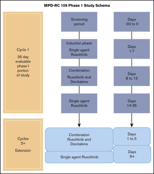
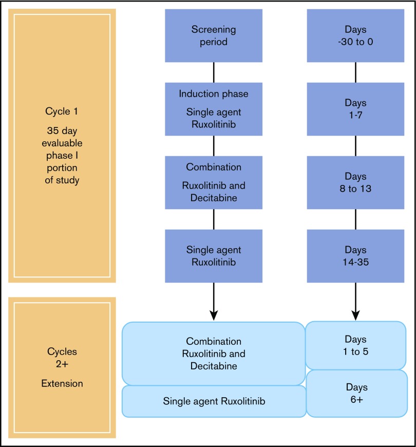
Patients were enrolled in a standard 3 + 3 phase 1 design with an MTD defined as a dose with <33% DLT rate. Ruxolitinib was administered in dose cohorts of 10, 15, 25, or 50 mg every 12 hours in combination with decitabine at a dose of 20 mg/m2 intravenously daily for 5 days and repeated every 28 days. During cycle 1, ruxolitinib was administered as a single agent for 7 days; decitabine was added on day 8 (Figure 1). Response assessment was carried out every cycle using modified Cheson criteria10: CR required 0% peripheral blood blasts, white blood cell ≥4 × 10⁹/L, hemoglobin ≥10 g/L, and platelets ≥100 × 109/L; CRi required 0% peripheral blood blasts with incomplete count recovery; and partial remission (PR) required ≥50% decrease in peripheral blood blasts regardless of blood counts (supplemental Table 1). ORR included CR, complete remission with incomplete count recovery (CRi), and PR. Overall survival (OS) was defined as the time from first dose of ruxolitinib to death by any cause. Patients were considered censored at the last known date alive, if death was not documented. OS was estimated using the Kaplan-Meier method.
Figure 1.
Myeloproliferative Disease Research Consortium phase 1 study schema.
The bone marrow aspirate is often unobtainable in patients with MF and MPN-AP/BP because of fibrosis, thus preventing application of classic World Health Organization therapeutic response assessment. Further, discordance between bone marrow blast abundance and peripheral blood blast count is often observed in MPN-AP/BP, which may be due to extramedullary hematopoiesis.11 To date, no validated uniform response criteria for MPN-AP/BP have been established.12 We therefore adapted current World Health Organization AML response criteria to determine responses based on peripheral blood counts; thus, response assessments are based on clearance or decrease of peripheral blood blasts only. Best responses were assessed at any point during treatment on or after cycle 2 day 1 in 17 patients that were evaluable for response assessment. Because of the use of peripheral blood blasts for response assessment, 2 patients who had peripheral blasts of 0% at baseline were unevaluable for therapeutic response. These 2 patients had 10% blasts in the bone marrow, thus meeting eligibility criteria, and were evaluable for toxicity assessment.
Genomic and cytogenetic analysis
Assessment of genomic alterations was carried out in all patients at baseline using the HemePACT assay, as previously described.13 Bone marrow metaphase cells were obtained using standard technology and interphase fluorescence in situ hybridization scoring and analysis was previously reported.14
Pharmacokinetic analysis
Plasma levels of ruxolitinib were assessed at predose and 0.5, 2.0, and 4.0 hours after ruxolitinib administration during both the ruxolitinib run-in phase (day 1) and the combination phase of ruxolitinib and decitabine (day 8). Ruxolitinib plasma concentrations were analyzed by validated liquid chromatography tandem mass spectometry method as previously described. The quantification limit of ruxolitinib was 0.3 ng/mL. The pharmacokinetic parameters (time to reach maximum concentration, maximum serum concentration, and area under the curve from time 0 to last quantifiable concentration [AUClast]) were calculated using noncompartmental analysis by Phoenix WinNonlin (version 6.3, Certara USA, Princeton, NJ). The drug–drug interactions were analyzed using a linear mixed effects model. The model included treatment (combination of ruxolitinib + decitabine or ruxolitinib alone) as fixed effects and subject as a random effect. Estimates and confidence intervals (CI) were first constructed in the logarithmic scale. By taking antilogarithms, estimates and CI for the geometric means and ratios of geometric means were derived.
Results
Patient characteristics
A total of 21 patients were accrued to study (Table 1; supplemental Table 2). The median age was 63 years (range, 48-88). At the time of study enrollment, 8 (38.1%) carried a diagnosis of MPN-AP, and 13 (61.9%) carried a diagnosis of MPN-BP. Six (29%) and 5 (24%) patients had prior exposure to ruxolitinib and decitabine, respectively. One patient had failed prior allogeneic hematopoietic stem cell transplantation (HSCT). At MPN presentation, 4 (19.1%) patients were diagnosed with MPN-AP/BP, 8 (38%) with MF (primary or secondary), 8 (38%) with PV, and 1 (4.8%) with ET. The median duration of an MPN diagnosis before enrollment on study was 85.3 months (range, 0.8-408). The median palpable spleen size below the left costal margin at time of enrollment was 6 cm (range, 0-19). The majority of patients had a performance status of ECOG 0-1 (15; 71.4%). JAK-STAT activating mutations were observed in 18/21 (85.7%) of patients. The most frequent non–JAK-STAT mutations occurred in splicing factors (7/21, 33.3%), RUNX1 (5/21, 23.8%), TP53 (4/21, 19%), and TET2/IDH1/2 (4/21, 19%). The median number of variants observed per patient was 3 (range, 1-7). Normal karyotype was observed in 10/21 patients (47.6%); abnormal karyotype was detected in 9/21 patients (43%). Cytogenetics was not available or inadequate in the remaining 2 patients. Complex karyotype with or without monosomal karyotype was detected in 6 of 9 patients with an abnormal karyotype (67% among abnormal, 28.5% of total), consistent with the frequency of chromosomal abnormalities observed in advance stages of MF. Four of these patients had monosomy or deletion of chromosome 7, whereas 3 patients exhibited monosomy or del (5q). Sole monosomy 7 was seen in 1 patient. One case of monosomy 17 resulting in loss of TP53 in 64% of interphase cells detected by fluorescence in situ hybridization was observed in a patient with a concomitant TP53 truncating mutation (supplemental Table 3).
Table 1.
Baseline patient demographics
| 10 mg bid (N = 6) | 15 mg bid (N = 3) | 25 mg bid (N = 6) | 50 mg bid (N = 6) | Total (N = 21) | |
|---|---|---|---|---|---|
| Age, median (range), y | 62.0 (58.0-74.0) | 63.0 (58.0-79.0) | 66.5 (48.0-81.0) | 72.0 (56.0-88.0) | 63.0 (48.0-88.0) |
| Female/male, n (%) | 3 (50.0)/3 (50.0) | 1 (33.3)/2 (66.7) | 2 (33.3)/4 (66.7) | 2 (33.3)/4 (66.7) | 8 (38.1)/13 (61.9) |
| ECOG 0-1, n (%) | 3 (50.0) | 3 (100.0) | 5 (83.3) | 4 (66.7) | 15 (71.4) |
| ECOG 2-3, n (%) | 3 (50.0) | — | 1 (16.7) | 2 (33.3) | 6 (28.6) |
| Disease duration before therapy, median (range), mo | 137.8 (14.3-408.0) | 114.6 (24.7-116.6) | 168.1 (0.8-360.2) | 27.0 (1.5-148.6) | 85.3 (0.8-408.0) |
| Accelerated phase, n (%) | 2 (33.3) | — | 3 (50.0) | 3 (50.0) | 8 (38.1) |
| Blast phase, n (%) | 4 (66.7) | 3 (100.0) | 3 (50.0) | 3 (50.0) | 13 (61.9) |
| Spleen by palpation, median (range) | 13 (0-19) | 6 (6-6) | 8.5 (0-15) | 0 (0-13) | 6 (0-19) |
| Prior ruxolitinib, n (%) | 2 (33.3) | — | 1 (16.7) | 3 (50.0) | 6 (28.6) |
| Prior decitabine, n (%) | 1 (16.7) | 2 (66.7) | 2 (33.3) | — | 5 (23.8) |
| Prior HSCT, n (%) | — | — | 1 (16.7) | — | 1 (4.8) |
bid, twice daily.
Pharmacokinetic and drug–drug interaction analysis
Following oral administration of ruxolitinib alone or combination administration of ruxolitinib and decitabine, time to reach maximum concentration occurred from 0 to 4 hours postdose. Mean (± standard deviation) AUClast values for ruxolitinib administered alone or as combination with decitabine were 1310 (± 1970) and 998 (± 864) hours × ng/mL, respectively. Geometric mean percentage ratios (90% CI) of AUClast and maximum serum concentration (ruxolitinib + decitabine/ruxolitinib) were 97.47% (58.76%-161.67%) and 96.32% (59.31%-56.43%), respectively, indicating that the pharmacokinetics of ruxolitinib and decitabine combination treatment is similar to ruxolitinib alone and not affected by the combination treatment (supplemental Figure 1).
Safety
All 21 enrolled patients were evaluable for AE assessment. No DLT were observed in the 15- and 50-mg dose cohorts. One DLT event (grade 3 laryngeal mucositis) occurred at the 10-mg dose level in the first cohort of 3 patients. Thus, an additional 3 patients were enrolled without the occurrence of a second DLT. One DLT event (grade 3 AST increased) occurred at the 25-mg dose level in the first cohort of 3 patients. Thus, an additional 3 patients were enrolled at this dose level without the occurrence of second DLT. The MTD was not reached. The most frequent treatment emergent AE experienced by patients (TEAEs) regardless of attribution (all grades) included; neutropenia (10 patients, 47.6%), thrombocytopenia (10 patients, 47.6%), and febrile neutropenia (7 patients, 33.3%). Hematologic toxicity constituted the majority of grades 3 and TEAE. Grade 3/4 hematologic AE occurring in >5% of patients included neutropenia (7 patients, 33.3%), lymphopenia (4 patients, 19%), thrombocytopenia (4 patients, 19%), and anemia (3 patients, 14%). Grades 3 and 4 nonhematologic TEAE regardless of attribution occurring in more than 5% of patients included febrile neutropenia (7 patients, 33.3%), pneumonia (6 patients, 29%), sepsis (3 patients, 14%), respiratory failure (2 patients, 9.5%), hypertension (2 patients, 9.5%), cellulitis (2 patients, 9.5%), gastrointestinal bleeding (2 patients, 9.5%), and squamous cell carcinoma of the skin (2 patients, 9.5%) (Table 2). Grade 3 and higher TEAE were comparable between patients with MPN-AP vs MPN-BP (supplemental Table 4).
Table 2.
Adverse events by ruxolitinib dose level
| 10 mg bid (N = 6) | 15 mg bid (N = 3) | 25 mg bid (N = 6) | 50 mg bid (N = 6) | Total (N = 21) | |
|---|---|---|---|---|---|
| Hematologic, n (%) | |||||
| Neutropenia | 2 (33.4) | 1 (33.3) | 2 (33.3) | 2 (33.3) | 7 (33.3) |
| Grade 3 | 1 | 1 | 2 | 2 | 6 |
| Grade 4 | 1 | 1 | |||
| Febrile neutropenia | 2 (33.3) | 1 (33.3) | 2 (33.3) | 2 (33.3) | 7 (33.3) |
| Grade 3 | 2 | 1 | 2 | 2 | 7 |
| Lymphocytopenia | 2 (33.3) | 1 (16.7) | 1 (16.7) | 4 (19.0) | |
| Grade 3 | 2 | 1 | 1 | 4 | |
| Thrombocytopenia | 2 (33.3) | 1 (33.3) | 1 (16.7) | 4 (19.0) | |
| Grade 3 | 1 | 1 | 1 | 2 | |
| Grade 4 | 1 | 2 | |||
| Anemia | 1 (16.7) | 1 (33.3) | 1 (16.7) | 3 (14.3) | |
| Grade 3 | 1 | 1 | 1 | 3 | |
| Nonhematologic, n (%) | |||||
| Pneumonia | 3 (50) | 1 (33.3) | 1 (16.7) | 1 (16.7) | 6 (28.6) |
| Grade 3 | 3 | 1 | 1 | 1 | 6 |
| Respiratory failure | 2 (33.3) | 2 (9.5) | |||
| Grade 3 | 1 | 1 | |||
| Grade 4 | 1 | 1 | |||
| Sepsis | 1 (33.3) | 1 (16.7) | 1 (16.7) | 3 (14.3) | |
| Grade 4 | 1 | 1 | 1 | ||
| Squamous cell carcinoma | 2 (33.3) | 2 (9.5) | |||
| Grade 3 | 2 | 2 | |||
| Cellulitis | 1 (16.7) | 1 (16.7) | 2 (9.5) | ||
| Grade 3 | 1 | 1 | |||
| Gastrointestinal bleeding | 1 (33.3) | 1 (16.7) | 2 (9.5) | ||
| Grade 3 | 1 | 1 | |||
| Grade 4 | |||||
| Hypertension | 1 (16.7) | 1 (16.7) | 2 (9.5) | ||
| Grade 3 | 1 | 1 | |||
| Grade 4 | |||||
Response
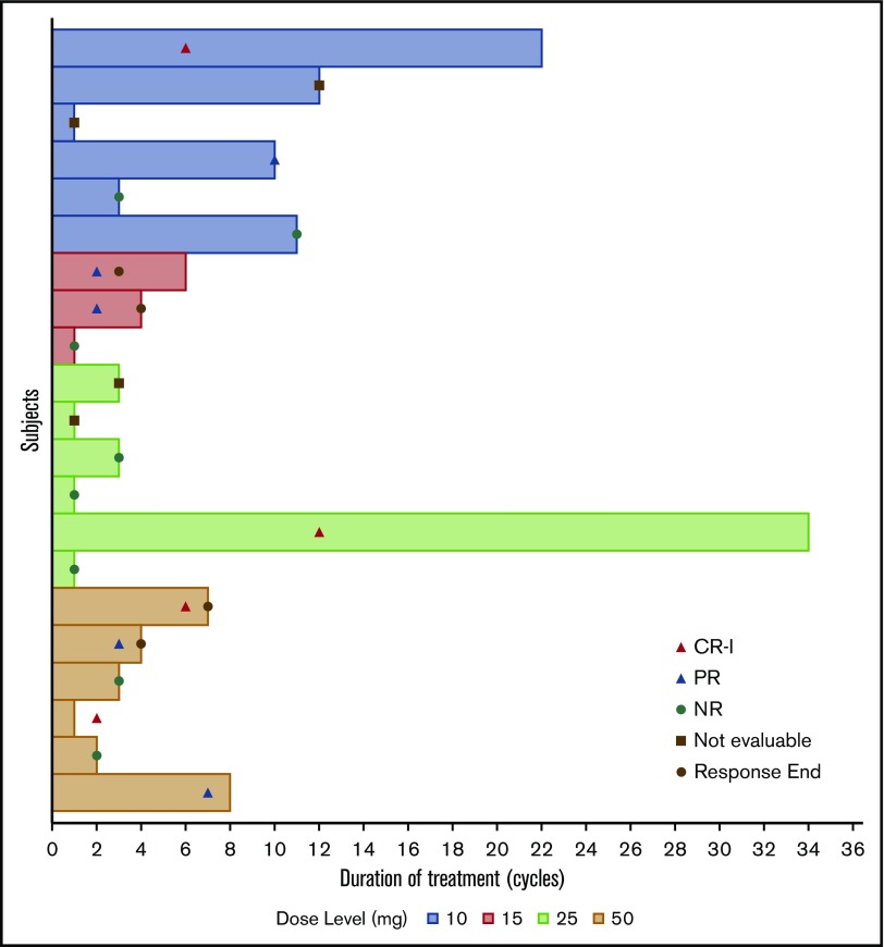
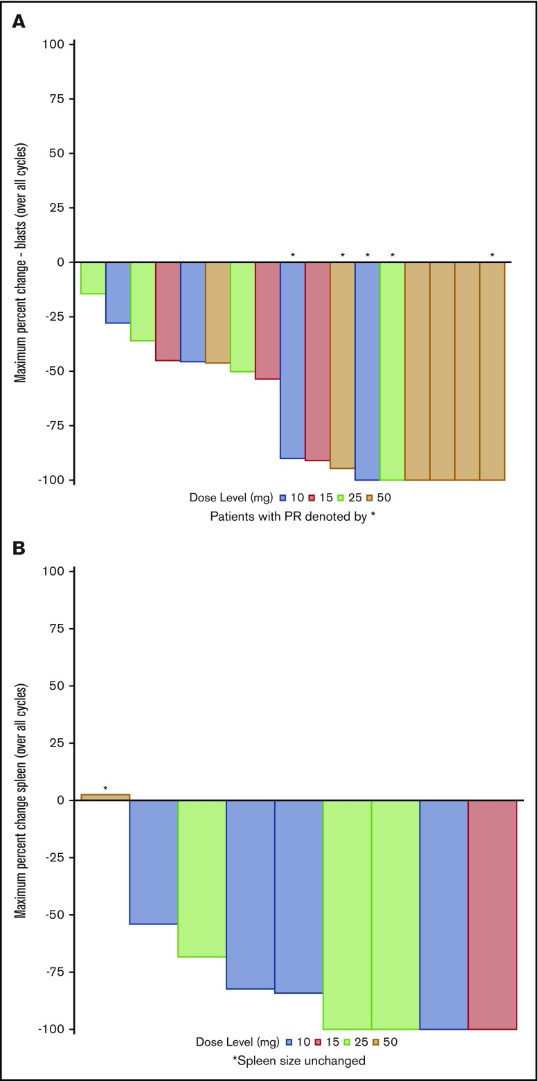
Seventeen patients were evaluable for response. Four patients were unevaluable for response assessment because of an absence of baseline blasts (2 patients) and not completing cycle 1 day 35 (2 patients). CRi was observed in 4 (23.5%; 95% CI, 6.8-49.9) patients (occurring at dose level 10 mg bid cycle 6, dose level 25 mg bid cycle 12, and dose level 50 mg bid cycles 2 and 6), PR was observed in 5 (29.4%; 95% CI, 10.3-56.0) patients, and no response was observed in 8 (47.1%; 95% CI, 23.0-72.2) of treated patients. Response (CR + CRi + PR) rate was 9/17 (53%; 95% C, 27.8-77.0) overall and was 4/6 (66.7%; 95% CI, 22.3-95.7) in MPN-AP and 5/11 (45.5%; 95% CI, 16.8-76.6) in MPN-BP patients. Using intention-to-treat analysis, the ORR was 9/21 (42.9%; 95% CI, 21.8-66.0); 4/8 (50.0%; 95% CI, 15.7%-84.3) with MPN-AP and 5/13 (38.5%; 95% CI, 13.9-68.4) with MPN-BP. The median number of cycles of therapy administered across all dose cohorts was 3 (range, 1-34). Median peripheral blood blasts at baseline was 10.5% (range, 0-76). Patients in the 10-mg ruxolitinib dose cohort received the greatest number of cycles (median, 10.5; range, 1-22) (Figure 2). The median blast percentage for these patients at baseline was 10.5% and at time of response was 2.0%, consistent with reversion to chronic phase disease (<10% blasts). Reduction in peripheral blood blast percentage (Figure 3A) was observed in all dosing cohorts. A median blast count reduction of 57% (range, 8.4-100), in terms of maximum blast reduction across all patients, was observed. The largest reduction in blast percentage was observed in the 50-mg ruxolitinib dose cohort (median, 91%; range, −100 to 50) because 5 of 6 patients had blast reductions at this dose followed by the 10-mg dosing cohort (median, 60%; range, −100 to 33). Importantly, among patients attaining PR, all demonstrated a blast count ≤5% in the peripheral blood at the time of response assessment. The median reduction in spleen size across all dosing cohorts was 84.2% (range, 0-100), with greatest reductions observed in the 15- and 25-mg ruxolitinib doses (Figure 3B). Clinical responses (CRi or PR) were observed in 1 of 2 evaluable patients (50%) with TP53 mutations, 2 of 4 evaluable patients (50%) with RUNX1 mutations, and 3 of 5 evaluable patients (60%) with splicing factor mutations. Clinical responses were observed in 1 of 3 evaluable patients (33.3%) with chromosome 7 abnormalities, 0 of 2 evaluable patients (0%) with chromosome 17 abnormalities, and 5 of 9 patients (55.6%) with a normal karyotype.
Figure 2.
Duration of treatment by ruxolitinib dose level. Corresponding clinical responses are indicated by symbols. End of response is defined as peripheral blood blast count exceeding baseline value. NR, no response.
Figure 3.
Maximum change from baseline. Peripheral blood blast count per patient (A) and spleen length by palpation in the midclavicular line per patient (B) in patients with palpable splenomegaly at baseline. Dosing cohort designated by color. *Unchanged spleen size.
To date, 19 patients have ended treatment, 2 patients still currently remain on therapy (1 patient in the 25-mg and 1 patient in the 50-mg cohorts). Reasons for ending treatment included: AE, 7 (36.8%); disease progression as assessed by the treating physician, 4 (21.1%); patient refused further treatment, 3 (15.8%); death, 2 (10.5%); other treatment, 2 (10.6%, including 1 patient who underwent HSCT and 1 patient who received induction therapy for AML);and investigator decision, 1 (5.3%). There have been 14 deaths to date. Causes of death were noted as sepsis/pneumonia, 5 (35.7%); relapse/progression, 4 (28.6%); unknown,: 2 (14.2%); gastrointestinal hemorrhage, 1 (7.1%); circulatory failure, 1 (7.1%); and cardiac arrest, 1 (7.1%). No deaths occurred within 30 days of treatment. There were 2 deaths occurring <40 days after treatment started. One death occurred at 36 days secondary to sepsis and a second patient death occurred at 38 days because of circulatory failure.
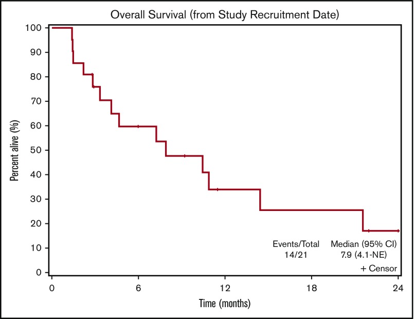
The median OS for patients on study was 7.9 months (95% CI, 4.1 months-not reached) (Figure 4). Survival did not differ by dosing cohort. Median OS for MPN-AP patients was 16.0 months (95% CI, 4.7 -not reached) and was 7.2 months (95% CI, 2.2-not reached) in MPN-BP patients. Median OS in responding patients was 10.9 months (95% CI, 7.9-not reached) vs 7.2 months in nonresponders (95% CI, 3.3-14.4). No clear pattern of response based on molecular genetic characteristics, spleen size, or baseline peripheral blood blast count was observed (supplemental Tables 5 and 6).
Figure 4.
Overall survival from the time of study enrollment for the cohort as a whole.
Discussion
Therapy for patients with MPN-AP/BP remains a major unmet need. In this multicenter phase 1 dose escalation trial, we demonstrated that ruxolitinib administered up to a dose of 50 mg bid can be combined with a fixed dose of decitabine without establishing an MTD. Additionally, the ORR rate of 53% (42.9% by intention-to-treat analysis) is encouraging considering the historical data of standard AML induction chemotherapy, which has minimal benefit outside of consolidation HSCT. Indeed, data from Mesa et al demonstrate that patients with MPN-BP had a median survival of 3.9 months when treated with induction therapy vs 2.1 months for those not receiving induction therapy.3 More recent retrospective data from Kennedy et al15 demonstrate that MPN-BP patients treated with curative intent (induction therapy followed by HSCT if donor identified) had a median survival of 9.4 months vs 2.3 months for those treated with noncurative intent (low-intensity therapy). However, no significant difference in OS was detected between patients treated with intensive chemotherapy who achieved a response but did not go on to receive HSCT vs those patients treated with nonintensive regimens (median survival, 9.4 vs 6.6 months, respectively). Importantly, induction therapy–related mortality was ∼15%. These data suggest that in the absence of a viable HSCT donor, a noninduction-based treatment regimen may offer a less toxic and less clinically burdensome treatment approach. Our data, demonstrating a median survival of 7.9 months compare favorably with these data. Limitations of the study include the heterogeneity of the population studied (comprising both MPN-AP and MPN-BP), and the reliance on peripheral blood blast count for response assessment owing to a lack of standard response criteria as well as technical challenges involved in bone marrow assessments in this patient population. Prospective data in a phase 2 setting is required to better assess the clinical benefit of this combination therapy regimen. In addition, the clinical effect of peripheral blast count clearance on long-term outcome remains to be determined. Furthermore, data regarding the use of this regimen before HSCT are needed to fully understand the applicability of this approach to all patients with MPN-AP/BP. Finally, validated criteria for clinical response assessment in this patient population is needed for future clinical studies.
Based on the results of this study, we are conducting a phase 2 trial of decitabine in combination with ruxolitinib at 25 mg bid for an initial cycle followed by 10 mg bid for subsequent cycles. The rationale for this approach is based on the observation that although patients treated with the 25 mg and 50 mg of ruxolitinib attained the greatest blast count and spleen size reductions, patients treated with 10 mg bid of ruxolitinib were able to continue on therapy for a longer duration (10.5 vs 3.5 median number of cycles received in 10-mg cohort vs 50-mg cohort respectively), suggesting improved tolerability. Through this approach, we hypothesize that we may be able to limit potential toxicity of therapy while still administering clinically efficacious dosing of the treatment regimen. Finally, given that these data have demonstrated that the combination of ruxolitinib and decitabine was safe and tolerable in this patient population, this regimen may serve as a basis to which other novel/targeted therapeutic agents may be added to further improve efficacy.
Supplementary Material
The full-text version of this article contains a data supplement.
Acknowledgments
The authors thank Martin S. Tallman for insightful commentary.
This study was supported by grants from the National Institutes of Health, National Cancer Institute (1 P01 CA108671-01A2 [principal investigator: R.H.] and 1K08CA188529-01 [R.K.R.]); Cancer Center Support Grant/Core Grant to Memorial Sloan Kettering Cancer Center (P30 CA008748); and Incyte Corporation.
Authorship
Contribution: R.K.R., J.O.M., J.D.G., L.P., A.C.D., M.L.H., R.L.L., and R.H. designed the study; R.K.R., J.O.M., D.B., E.H., C.N.A., M.K., M.L.H., and R.A.M. carried out the clinical investigations; A.C.D. and L.S. provided regulatory support and guidance for the study; H.E.K. and A.C.D. performed statistical analysis; M.E.S. performed central pathology review; R.K.R. and K.M. performed molecular genetic analysis; V.N. performed cytogenetic analysis; R.S.W. performed sample procurement and curation; R.K.R., J.O.M., H.E.K., and A.C.D. analyzed data; and R.K.R. and J.O.M. wrote the manuscript.
Conflict-of-interest disclosure: R.K.R. has received consulting fees from Incyte, Celgene, Agios Pharmaceuticals, Apexx Oncology, and Jazz Pharmaceuticals, and has received research funding from Constellation Pharmaceuticals, Incyte, and Stemline Therapeutics. J.O.M. has received research funding from Incyte, Novartis, Merck, CTI Biopharma, Roche, Celgene, Janssen, and Promedior, and consulting fees from Novartis, Celgene, CTI Biopharma, and Incyte. E.H. has served as an advisor and received research support from Blueprint Medicines and Novartis Oncology. M.K. has received research funding from Incyte, Celgene, Constellation, and Blueprint Medicines, and consulting fees from La Jolla Pharmaceutical. R.A.M. has received consulting fees from Novartis, Sierra Oncology, and La Jolla Pharmaceutical, and research support from Celgene, Incyte, AbbVie, Samus, and Genentech. R.L.L. is on the supervisory board of Qiagen and is a scientific advisor to Loxo, Imago, C4 Therapeutics, and Isoplexis, which each include an equity interest. He receives research support from and consulted for Celgene and Roche, has received research support from Prelude Therapeutics, and has consulted for Novartis and Janssen. He has received honoraria from Lilly and Amgen for invited lectures and from Gilead for grant reviews. The remaining authors declare no competing financial interests.
Correspondence: John Mascarenhas, Adult Leukemia Program, Myeloproliferative Disorders Clinical Research Program, Tisch Cancer Institute, Division of Hematology/Oncology, Associate Professor of Medicine, Icahn School of Medicine at Mount Sinai, 1 Gustave L. Levy Pl, Box 1079, New York, NY 10029; e-mail: john.mascarenhas@mssm.edu.
References
- 1.Mascarenhas J. A concise update on risk factors, therapy, and outcome of leukemic transformation of myeloproliferative neoplasms. Clin Lymphoma Myeloma Leuk. 2016;16(Suppl):S124-S129. [DOI] [PubMed] [Google Scholar]
- 2.Cervantes F, Tassies D, Salgado C, Rovira M, Pereira A, Rozman C. Acute transformation in nonleukemic chronic myeloproliferative disorders: actuarial probability and main characteristics in a series of 218 patients. Acta Haematol. 1991;85(3):124-127. [DOI] [PubMed] [Google Scholar]
- 3.Mesa RA, Li CY, Ketterling RP, Schroeder GS, Knudson RA, Tefferi A. Leukemic transformation in myelofibrosis with myeloid metaplasia: a single-institution experience with 91 cases. Blood. 2005;105(3):973-977. [DOI] [PubMed] [Google Scholar]
- 4.Tefferi A, Mudireddy M, Mannelli F, et al. . Blast phase myeloproliferative neoplasm: Mayo-AGIMM study of 410 patients from two separate cohorts. Leukemia. 2018;32(5):1200-1210. [DOI] [PMC free article] [PubMed] [Google Scholar]
- 5.Thepot S, Itzykson R, Seegers V, et al. ; Groupe Francophone des Myelodysplasies (GFM). Treatment of progression of Philadelphia-negative myeloproliferative neoplasms to myelodysplastic syndrome or acute myeloid leukemia by azacitidine: a report on 54 cases on the behalf of the Groupe Francophone des Myelodysplasies (GFM). Blood. 2010;116(19):3735-3742. [DOI] [PubMed] [Google Scholar]
- 6.Mascarenhas J, Navada S, Malone A, Rodriguez A, Najfeld V, Hoffman R. Therapeutic options for patients with myelofibrosis in blast phase. Leuk Res. 2010;34(9):1246-1249. [DOI] [PubMed] [Google Scholar]
- 7.Eghtedar A, Verstovsek S, Estrov Z, et al. . Phase 2 study of the JAK kinase inhibitor ruxolitinib in patients with refractory leukemias, including postmyeloproliferative neoplasm acute myeloid leukemia. Blood. 2012;119(20):4614-4618. [DOI] [PMC free article] [PubMed] [Google Scholar]
- 8.Mwirigi A, Galli S, Keohane C, et al. . Combination therapy with ruxolitinib plus 5-azacytidine or continuous infusion of low dose cytarabine is feasible in patients with blast-phase myeloproliferative neoplasms. Br J Haematol. 2014;167(5):714-716. [DOI] [PubMed] [Google Scholar]
- 9.Rampal R, Ahn J, Abdel-Wahab O, et al. . Genomic and functional analysis of leukemic transformation of myeloproliferative neoplasms. Proc Natl Acad Sci USA. 2014;111(50):E5401-E5410. [DOI] [PMC free article] [PubMed] [Google Scholar]
- 10.Cheson BD, Bennett JM, Kopecky KJ, et al. ; International Working Group for Diagnosis, Standardization of Response Criteria, Treatment Outcomes, and Reporting Standards for Therapeutic Trials in Acute Myeloid Leukemia. Revised recommendations of the International Working Group for Diagnosis, Standardization of Response Criteria, Treatment Outcomes, and Reporting Standards for Therapeutic Trials in Acute Myeloid Leukemia [published correction appears in J Clin Oncol. 2004;22(3):576]. J Clin Oncol. 2003;21(24):4642-4649. [DOI] [PubMed] [Google Scholar]
- 11.Rampal R, Mascarenhas J. Pathogenesis and management of acute myeloid leukemia that has evolved from a myeloproliferative neoplasm. Curr Opin Hematol. 2014;21(2):65-71. [DOI] [PubMed] [Google Scholar]
- 12.Mascarenhas J, Heaney ML, Najfeld V, et al. ; Post-myeloproliferative neoplasm acute myeloid leukemia consortium. Proposed criteria for response assessment in patients treated in clinical trials for myeloproliferative neoplasms in blast phase (MPN-BP): formal recommendations from the post-myeloproliferative neoplasm acute myeloid leukemia consortium. Leuk Res. 2012;36(12):1500-1504. [DOI] [PMC free article] [PubMed] [Google Scholar]
- 13.Kucine N, Viny AD, Rampal R, et al. . Genetic analysis of five children with essential thrombocytosis identified mutations in cancer-associated genes with roles in transcriptional regulation. Haematologica. 2016;101(6):e237-e239. [DOI] [PMC free article] [PubMed] [Google Scholar]
- 14.Najfeld V, Montella L, Scalise A, Fruchtman S. Exploring polycythaemia vera with fluorescence in situ hybridization: additional cryptic 9p is the most frequent abnormality detected. Br J Haematol. 2002;119(2):558-566. [DOI] [PubMed] [Google Scholar]
- 15.Kennedy JA, Atenafu EG, Messner HA, et al. . Treatment outcomes following leukemic transformation in Philadelphia-negative myeloproliferative neoplasms. Blood. 2013;121(14):2725-2733. [DOI] [PubMed] [Google Scholar]
Associated Data
This section collects any data citations, data availability statements, or supplementary materials included in this article.


