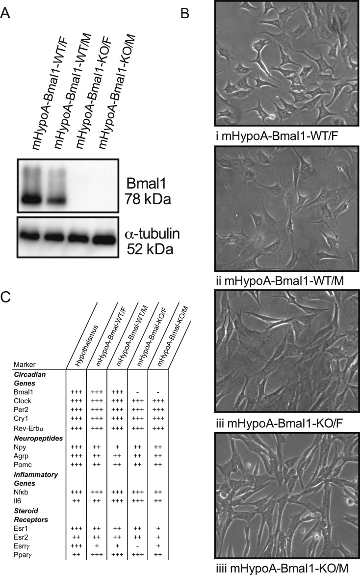
Figure 3.
Characterization of the mHypoA-Bmal1-WT and -KO cell models. mHypoA-Bmal1-KO/F and mHypoA-Bmal1-KO/M cell lines do not express BMAL1. (A) Expression or absence of BMAL1 protein in the WT vs KO cell lines was verified with Western blotting with a BMAL1 antibody and alpha-tubulin used as a loading control. (B) Cell lines were imaged with an Olympus CKX41 microscope (10× lens objective) with the Tucsen 10.0 MP IS1000 USB camera and exhibit neuronal morphology. (C) Summary of circadian, neuropeptide, inflammatory marker and steroid receptor mRNA expression in hypothalamus tissue, mHypoA-Bmal1-WT/F, mHypoA-Bmal1-WT/M, mHypoA-Bmal1-KO/F, and mHypoA-Bmal1-KO/M cell lines. RNA was isolated from untreated hypothalamic tissue or cells before cDNA synthesis and analysis by qRT-PCR. Relative expression is denoted by + or −, where − (not expressed) cycle at threshold (CT) ≤ 35, + CT = 30 to 34.9, ++ CT = 25 to 29.9, +++ (highly expressed) CT = 20 to 24.9. Esr1, estrogen receptor α;Esr2, estrogen receptor β;Esrrγ, estrogen-related receptor γ;Nfkb, nuclear factor κb.

