Abstract Abstract
An account is given of the karyotypes of Hydramagnipapillata Itô, 1947, H.oxycnida Schulze, 1914, and Pelmatohydraoligactis (Pallas, 1766) (Cnidaria, Hydrozoa, Hydridae). A number of different techniques were used: conventional karyotype characterization by standard staining, DAPI-banding and C-banding was complemented by the physical mapping of the ribosomal RNA (18S rDNA probe) and H3 histone genes, and the telomeric (TTAGGG)n sequence by fluorescence in situ hybridization (FISH). We found that the species studied had 2n = 30; constitutive heterochromatin was present in the centromeric regions of the chromosomes; the “vertebrate” telomeric (TTAGGG)n motif was located on both ends of each chromosome and no interstitial sites were detected; 18S rDNA was mapped on the largest chromosome pair in H.magnipapillata and on one of the largest chromosome pairs in H.oxycnida and P.oligactis; in H.magnipapillata, the major rRNA and H3 histone multigene families were located on the largest pair of chromosomes, on their long arms and in the centromeric areas respectively. This is the first chromosomal mapping of H3 in hydras.
Keywords: Hydra , Pelmatohydra , Hydridae , karyotype, chromosomes, FISH, (TTAGGG)n, 18S rDNA, histone H3
Introduction
Hydras are simple freshwater invertebrates belonging to one of the most ancient members of the animal kingdom, the phylum Cnidaria (class Hydrozoa, order Hydrida, family Hydridae). Hydras are of general interest since they display fundamental principles that underlie development, differentiation, regeneration and symbiosis (e.g. Bosch 2007, 2008, Khalturin et al. 2009, Augustin et al. 2010, Bosch et al. 2010). Some species of hydras are relatively easy animals to culture and maintain in the laboratory, then, they have been used as model organisms in many different areas of biological research, primarily in developmental biology often referred to as “evo-devo”, i.e. evolutionary developmental biology research (Slobodkin and Bossert 2001, Galliot 2012).
Without detailed knowledge of these basal metazoans, it is impossible to provide an effective comparative framework for animal evolution (Zacharias et al. 2004). Nevertheless, the species level diversity, taxonomy and phylogenetic relationships of the hydra species are far from well understood. Jankowski et al. (2008) suggested 12–15 really different hydra species, whereas Bouillon et al. (2006) reported approximately 30 valid species, and the World Register of Marine Species lists 40 species (Schuchert 2018). All hydras were originally included in the single genus Hydra Linnaeus, 1758. However Schulze (1914, 1917) divided hydras into three genera, Hydra, Chlorohydra Schulze, 1914, and Pelmatohydra Schulze, 1914, and their validity was substantiated elsewhere (e.g. Collins 2000, Stepanjants et al. 2000, Anokhin 2002).
During the past decade or so, several molecular phylogenetic studies using mitochondrial and nuclear genes shed light on the diversity within Hydra sensu Linnaeus, 1758 (Hemmrich et al. 2007, Kawaida et al. 2010, Martínez et al. 2010, Schwentner and Bosch 2015). The genome of one species, Hydramagnipapillata Itô, 1947, has been recently assembled (Chapman et al. 2010).
Chromosomes are known to be the carriers of genetic material, and chromosome changes provide the basis of speciation (White 1973). As many as 8 species from all three above-mentioned hydra genera have been karyotyped so far (Xinbai et al. 1987, Ovanesyan and Kuznetsova 1995, Anokhin et al. 1998, 2010, Anokhin and Kuznetsova 1999, Anokhin 2002, 2004, Anokhin and Nokkala 2004, Zacharias et al. 2004, Stepanjants et al. 2006, Traut et al. 2007). These species were mainly studied using conventional chromosome staining techniques, including C-banding. They were shown to have 2n = 30, almost exclusively meta/submetacentric (m/sm) chromosomes of similar size, and C-heterochromatin blocks localized in the centromeric regions of the chromosomes. Sex chromosomes were not distinguished in any species. Thus, hydras can now been considered as the group with the greatest stability in their karyotype, at least regarding the number of chromosomes. In two studies only (Traut et al. 2007, Anokhin et al. 2010), the fluorescence in situ hybridization (FISH) was used to characterize hydras in terms of telomeric sequences and the chromosomal distribution of the rRNA and some other genes.
Our study was aimed to add new data on hydra chromosomes studied using C-banding and FISH with probes for the “vertebrate” telomere motif (TTAGGG)n, 18S rDNA, and histone H3. We adopt here the generic hydra classification of Schulze (1914, 1917).
Material and methods
Experiments were carried out with three species, Hydramagnipapillata, H.oxycnida Schulze, 1914, and Pelmatohydraoligactis (Pallas, 1766). H.magnipapillata (strain 105) was obtained from the Institute of Zoology, University of Kiel (Germany); H.oxycnida and P.oligactis were collected from nature (58°48'46.9"N, 29°59'02.7"E, the Oredezh river, Leningrad Province, Russia). Polyps were cultured at 18 ± 0.5 °C for a long period of time in the case of H.magnipapillata or for one-two weeks in the cases of H.oxycnida and P.oligactis. They were fed regularly with freshly hatched nauplii of Artemiasalina (Linnaeus, 1758) (Crustacea, Branchiopoda).
Different methods were tried to characterize the chromosomes of the above-mentioned species: C-banding for H.magnipapillata and P.oligactis; FISH mapping of 18S rRNA and histone H3 genes for H.magnipapillata and of the “vertebrate” telomere motif (TTAGGG)n for H.oxycnida and P.oligactis.
Spread chromosome preparations were made from asexual polyps. Hydras were subjected to a hypoosmotic shock with 0.4% trisodium citrate for 30 min followed by fixation in ethanol and acetic acid (3:1) for 15 min. Specimens were transferred to a drop of 70% ethanol on the glass slides and dissected with needles. The cell suspension was spread by the warm air stream (37–70 °C).
In DNA isolation, 18S rDNA and (TTAGGG)n probes generation and FISH experiments we followed the protocol described in Anokhin et al. (2010). The probe for the histone H3 was PCR amplified and labeled by Rhodamine-5-dUTP (GeneCraft, Germany) using primers H3F: 5’-ATG GCT CGT ACC AAG CAG ACV GC-3’ and H3R: 5’-ATA TCC TTR GGC ATR ATR GTG AC-3’ (Huang et al. 2011).
Microscopic images were taken using a Leica DM 6000B microscope with a 100× objective, Leica DFC 345 FX camera and Leica Application Suite 3.7 software with an Image Overlay module (Leica Microsystems Wetzlar GmbH, Germany). The filter sets applied were A, L5, N21 (Leica Microsystems, Wetzlar, Germany).
Results
Cytogenetic analyses were carried out on 10 specimens of every species (asexual forms), Hydramagnipapillata, H.oxycnida, and P.oligactis. Representative mitotic images of the species subjected to routine chromosome staining, C-banding, and FISH with the 18S rDNA, histone H3 and telomere (TTAGGG)n probes are shown in Figures 1–3.
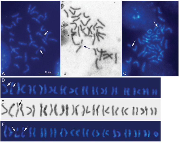
Figure 1.
Mitotic chromosomes of Hydramagnipapillata after C- banding (A), Hydraoxycnida after routine staining (B), and Pelmatohydraoligactis after C- banding (C). C-bands are visible in the centromeric areas of the chromosomes. Karyograms of H.magnipapillata (D), H.oxycnida (E) and P.oligactis (F). Arrows indicate achromatic gaps.
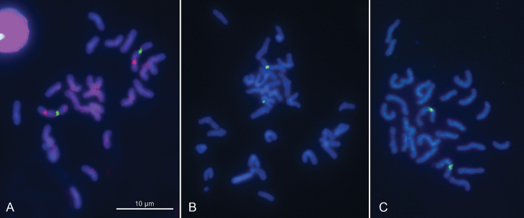
Figure 3.
FISH with the 18S rDNA (green signals) and H3 histone (red signals) probes on mitotic chromosomes of Hydramagnipapillata (A), and with the 18S rDNA probe only on mitotic chromosomes of Hydraoxycnida (B) and Pelmatohydraoligactis (C). In H.magnipapillata, the FISH signals derived from the 18S and H3 probes are visible on the largest pair of chromosomes, on their long arms and in the centromeric areas respectively. Chromosomes are counterstained with DAPI.
Hydra magnipapillata
The karyotype was found to consist of 30 m/sm chromosomes (2n = 30), it is symmetrical in structure, with chromosomes showing a regular gradation in size. No heteromorphic chromosome pair (putative sex chromosomes) is identified. The homologues of the largest pair carry achromatic gaps on their long arms. C-banding procedure revealed blocks of constitutive heterochromatin (C-blocks) localized in the centromere areas of the chromosomes (Fig. 1 A, D). FISH mapping of the 18S rDNA and histone H3 probes revealed hybridization signals on the largest pair of autosomes, on their long arms and around the centromeres respectively (Fig. 3A). The rDNA signals position corresponds to that of achromatic gaps, that’s to be expected (Fig. 1 A, D).
Hydra oxycnida
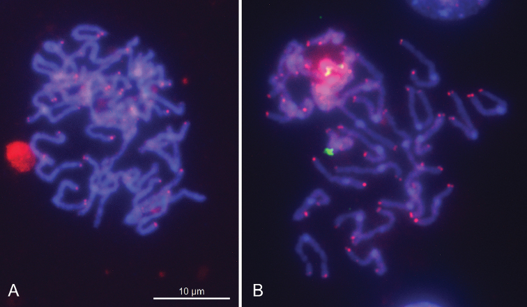
As with H.magnipapillata, this species has 2n = 30; its karyotype is symmetrical in structure, with chromosomes showing a regular gradation in size, and no heteromorphic chromosome pair is observed. One of the largest chromosome pairs (the largest or the second largest) carries secondary constrictions on the long arm of every homologue (Fig. 1 B, E). Furthermore, the 18S rDNA signals were detected on the long arms of one of largest chromosome pairs (Fig. 3 B). Again, as in the routinely stained preparations, more precise identification of this pair, whether it is the largest or the second largest one, appeared to be difficult. The (TTAGGG)n probe hybridized to the termini of every chromosome suggesting this sequence to be characteristic of the species (Fig. 2 A).
Figure 2.
FISH with the “vertebrate” (TTAGGG)n telomeric probe (red signals) on mitotic chromosomes of H.oxycnida (A) and P.oligactis (B). The chromosomes are counterstained with DAPI.
Pelmatohydra oligactis
As with both above-mentioned species, this species has 2n = 30; its karyotype is symmetrical in structure, with chromosomes showing a regular gradation in size, and no heteromorphic chromosome pair is observed. C-banding procedure followed by DAPI staining revealed C-blocks in the centromere regions of the chromosomes. All but one chromosome pairs were found to be m/sm. The exception was the smallest pair of chromosomes with very short arms which can be preliminarily identified as a subtelocentric/acrocentric pair (st/a). One of the largest chromosome pairs (the largest but maybe the second largest one) carries secondary constrictions on the long arm of every homologue (Fig. 1 C, F). Furthermore, the 18S rDNA signals were detected on the long arms of one of largest chromosome pairs (Fig. 3 C). Again, as in the routinely stained preparations, more precise identification of this pair, whether it is the largest or the second largest one, appeared to be difficult. The (TTAGGG)n probe hybridized to the termini of every chromosome suggesting this sequence to be characteristic of the species (Fig. 2 B).
Discussion
Characterization of karyotypes using standard staining and C-banding technique
Basic features of karyotypes revealed here in Hydramagnipapillata, H.oxycnida, and Pelmatohydraoligactis agree with those reported for these species previously (Anokhin and Kuznetsova 1999, Anokhin and Nokkala 2004, Anokhin et al. 2010). All hydra species studied so far have 2n = 30 with chromosomes showing a regular gradation in size, suggesting thus these features are under stabilizing natural selection. Among chromosomes, there is no pair to be taken as that of sex chromosomes. The centromere position is generally difficult to distinguish after conventional staining, and only C-banding is able to solve this question since C-heterochromatin in the hydra chromosomes is invariably located in the centromere regions (Anokhin and Nokkala 2004, Zacharias et al. 2004, present paper). The karyotypes of H.magnipapillata and P.oligactis as well as karyotypes of previously studied H.circumcincta Schulze, 2014 and H.vulgaris Pallas, 1766 (Anokhin and Nokkala 2004) are symmetrical and consist of mainly m/sm chromosomes. At the same time, a comparison between C-banded karyotypes of P.oligactis and H.magnipapillata showed that the former species had two subtelo/acrocentric (st/a) chromosomes, whereas the last-mentioned species had m/sm chromosomes only. This observation makes it apparent that some chromosome rearrangements have occurred during hydra species evolution, and thus, the species with the same chromosome number can differ one from another in chromosome morphology. The resolving of the issue needs to study in depth.
Characterization of karyotypes using FISH with the “vertebrate” (TTAGGG)n telomeric probe
Previous studies on Hydravulgaris (Traut et al. 2007) and H.magnipapillata (Anokhin et al. 2010) have shown that these species possess the “vertebrate” (TTAGGG)n motif of telomeres. Our FISH analyses also showed the presence of this motif at the ends of chromosomes of H.oxycnida and Pelmatohydraoligactis. Furthermore, the “vertebrate” telomeric sequence is present in representatives of all basal metazoan groups (Traut et al. 2007) and, with some notable exceptions (nematodes and arthropods), is conserved in most Metazoa. Bearing in mind that the “vertebrate” TTAGGG telomeric repeat is widely distributed and is present in most major eukaryotic groups, it is assumed to be the ancestral motif of telomeres in eukaryotes as a whole (Traut et al. 2007, Gomes et al. 2010, Fulnečková et al. 2013).
Characterization of karyotypes using FISH with 18S rDNA and H3 probes
The chromosomal location of the 18S rRNA genes was studied here in all three species. Hydramagnipapillata was shown to have 18S rDNA sites on the large arms of the largest chromosome pair. In H.oxycnida and Pelmatohydraoligactis, these sites were revealed on one of the largest pairs, the largest or maybe on the second largest one. In every case, the location of these sites coincides with the achromatic gaps, which are generally referred to as secondary constrictions, the nucleolus organizer region (NOR) involved in the formation of nucleolus (McStay 2016). The chromosomal location of the histone H3 gene family was studied in H.magnipapillata only. Noteworthy that mapping of H3 has been achieved for the first time in hydras. H.magnipapillata showed the H3 sites in the centromeric areas of the largest pair of chromosomes. It is the species that has received the most study by FISH to investigate the chromosomal distribution of different genes and sequences including genes coding for 18S rRNA and 28S rRNA, a head-specific gene ks1, a gene family DMRT suggested to be involved in sex determination and Tol2- like transposable element (Anokhin et al. 2010). The rRNA genes were shown to be co-localized on the homologues of the largest pair of chromosomes, on their long arms. A sex-related gene DMRT was revealed on a pair of chromosomes suggesting thus that it is a dose-regulated sex-determining gene in hydras. Probes specific for the ks1 hybridized to three distinct chromosome pairs, and multiple copies of a Tol2 transposable element gene were found on every chromosome. We have shown here that the major rDNA and the H3 genes are positioned on the same pair of chromosomes of H.magnipapillata, on their long arms and in the centromeres respectively, and should be thus inherited together. Furthermore, our results suggest that, in H.magnipapillata, the canonical histone H3 appears in the form of its centromere-specific variant CENH3, which is known to be the key histone component of the centromere in eukaryotes (Malik et al. 2002, Black and Bassett 2008).
In conclusion, this study delivers insight into the organization of genomes of hydras by reporting first data on (1) the chromosomal location of the H3 histone genes by the example of Hydramagnipapillata; (2) the telomere motif and the distribution of the 18S rRNA genes on chromosomes of Hydraoxycnida and Pelmatohydraoligactis. Our results provide a foundation for further studying the mechanisms involved in the chromosome evolution of this phylogenetically important group having an ancient origin within Metazoa.
Acknowledgements
The study was performed within the framework of the state research projects No. AAAAA17-117030310018-5 and AAAA-A17-117030310207-3, and was mainly financially supported by the program of fundamental research of the Presidium of the RAS “Biodiversity of Natural Systems”, the subprogram “Genofunds of living nature and their conservation”. Developing appropriate methodology for the H3 histone study was supported by the grant No. 14-14-00541 from the Russian Science Foundation. We thank Dr. S. Grozeva and Dr. N. Golub for their valuable remarks and suggestions to improve the paper.
Citation
Anokhin BA, Kuznetsova VG (2018) FISH-based karyotyping of Pelmatohydra oligactis (Pallas, 1766), Hydra oxycnida Schulze, 1914, and H. magnipapillata Itô, 1947 (Cnidaria, Hydrozoa). Comparative Cytogenetics 12(4): 539–548. https://doi.org/10.3897/CompCytogen.v12i2.32120
References
- Anokhin BA. (2002) Redescription of the endemic Baikalian species Pelmatohydrabaikalensis (Cnidaria: Hydrozoa, Hydrida, Hydridae) and assessment of the hydra fauna of Lake Baikal. Annales Zoologici (Warszawa) 52(4): 195–201. [Google Scholar]
- Anokhin BA. (2004) Revision of Hydrida (Cnidaria, Hydrozoa): comparative morphological, karyological and taxonomical aspects. PhD Thesis, Zoological Institute, Russian Academy of Sciences. St. Petersburg, 190 pp. [in Russian] [Google Scholar]
- Anokhin BA, Kuznetsova VG. (1999) Chromosome morphology and banding patterns in Hydraoligactis Pallas and H.circumcincta Schulze (Hydroidea, Hydrida). Folia Biologica (Kraków) 47(3–4): 91–96. 10.1080/00087114.2004.10589387 [DOI] [Google Scholar]
- Anokhin B, Nokkala S. (2004) Characterization of C-heterochromatin in four species of Hydrozoa (Cnidaria) by sequence specific fluorochromes Chromomycin A3 and DAPI. Caryologia 57(2): 167–170. 10.1080/00087114.2004.10589387 [DOI] [Google Scholar]
- Anokhin B, Hemmrich-Stanisak G, Bosch TCG. (2010) Karyotyping and single-gene detection using fluorescence in situ hybridization on chromosomes of Hydramagnipapillata. Comparative Cytogenetics 4(2): 97–110. 10.3897/compcytogen.v4i2.41 [DOI] [Google Scholar]
- Anokhin BA, Stepanjants SD, Kuznetsova VG. (1998) Hydra fauna of Leningrad region and adjacent territory: taxonomy with the karyological analysis. Proceedings of the Zoological Institute RAS 276: 19–26. [Google Scholar]
- Augustin R, Fraune S, Bosch TCG. (2010) How Hydra senses and destroys microbes. Seminars in Immunology 22: 54–58. 10.1016/j.smim.2009.11.002 [DOI] [PubMed] [Google Scholar]
- Black BE, Bassett EA. (2008) The histone variant CENP-A and centromere specification. Current Opinion in Cell Biology 20(1): 91–100. 10.1016/j.ceb.2007.11.007 [DOI] [PubMed] [Google Scholar]
- Bosch TCG. (2007) Why polyps regenerate and we don´t: towards a cellular and molecular framework for Hydra regeneration. Development Biology 303: 421–433. 10.1016/j.ydbio.2006.12.012 [DOI] [PubMed] [Google Scholar]
- Bosch TCG. (2008) Stem cells: from Hydra to man. Springer Netherlands, 192 pp. 10.1007/978-1-4020-8274-0 [DOI]
- Bosch TCG, Anton-Erxleben F, Hemmrich G, Khalturin K. (2010) The Hydra polyp: nothing but an active stem cell community. Development, Growth & Differentiation 52(1): 15–25. 10.1111/j.1440-169X.2009.01143.x [DOI] [PubMed] [Google Scholar]
- Bouillon J, Gravili C, Pagès F, Gili JM, Boero F. (2006) An introduction to Hydrozoa. Paris: Publications Scientifiques du Muséum, Paris, 591 pp. [Google Scholar]
- Chapman JA, Kirkness EF, Simakov O, Hampson SE, Mitros T, Weinmaier T, Rattei T, Balasubramanian PG, Borman J, Busam D, Disbennett K, Pfannkoch C, Sumin N, Sutton GG, Viswanathan LD, Walenz B, Goodstein DM, Hellsten U, Kawashima T, Prochnik SE, Putnam NH, Shu S, Blumberg B, Dana CE, Gee L, Kibler DF, Law L, Lindgens D, Martinez DE, Peng J, Wigge PA, Bertulat B, Guder C, Nakamura Y, Ozbek S, Watanabe H, Khalturin K, Hemmrich G, Franke A, Augustin R, Fraune S, Hayakawa E, Hayakawa S, Hirose M, Hwang JS, Ikeo K, Nishimiya-Fujisawa C, Ogura A, Takahashi T, Steinmetz PRH, Zhang X, Aufschnaite R, Eder M-K, Gorny A-K, Salvenmoser W, Heimberg AM, Wheeler BM, Peterson KJ, Böttger A, Tischler P, Wolf A, Gojobori T, Remington KA, Strausberg RL, Venter JC, Technau U, Hobmayer B, Bosch TCG, Holstein TW, Fujisawa T, Bode HR, David CN, Rokhsar DS, Steele RE. (2010) The dynamic genome of Hydra. Nature 464: 592–596. 10.1038/nature08830 [DOI] [PMC free article] [PubMed] [Google Scholar]
- Collins AG. (2000) Towards understanding the phylogenetic history of Hydrozoa: hypothesis testing with 18S gene sequence data. Scienta Marina 64 (Supl. 1): 5–22. 10.3989/scimar.2000.64s15 [DOI]
- Fulnečková J, Ševčíková T, Fajkus J, Lukešová A, Lukeš M, Vlček Č, Lang BF, Kim E, Eliáš M, Sýkorová E. (2013) A broad phylogenetic survey unveils the diversity and evolution of telomeres in eukaryotes. Genome Biology and Evolution 5(3): 468–483. 10.1093/gbe/evt019 [DOI] [PMC free article] [PubMed] [Google Scholar]
- Galliot B. (2012) Hydra, a fruitful model system for 270 years. The International Journal of Developmental Biology 56: 411–423. 10.1387/ijdb.120086bg [DOI] [PubMed] [Google Scholar]
- Gomes NM, Shay JW, Wright WE. (2010) Telomere biology in Metazoa. FEBS Letters 584: 3741–3751. 10.1016/j.febslet.2010.07.031 [DOI] [PMC free article] [PubMed] [Google Scholar]
- Hemmrich G, Anokhin B, Zacharias H, Bosch TCG. (2007) Molecular phylogenetics in Hydra, a classical model in evolutionary developmental biology. Molecular Phylogenetics and Evolution 44: 281–290. 10.1016/j.ympev.2006.10.031 [DOI] [PubMed] [Google Scholar]
- Huang D, Licuanan WY, Baird AH, Fukami H. (2011) Cleaning up the ‘Bigmessidae’: molecular phylogeny of scleractinian corals from Faviidae, Merulinidae, Pectiniidae and Trachyphylliidae BMC Evolutionary Biology 11: 37. 10.1186/1471-2148-11-37 [DOI] [PMC free article] [PubMed]
- Jankowski T, Collins AG, Campbell R. (2008) Global diversity of inland water cnidarians. Hydrobiologia 595(1): 35–40. 10.1007/s10750-007-9001-9 [DOI] [Google Scholar]
- Kawaida H, Shimizu H, Fujisawa T, Tachida H, Kobayakawa Y. (2010) Molecular phylogenetic study in genus Hydra. Gene 468: 30–40. 10.1016/j.gene.2010.08.002 [DOI] [PubMed] [Google Scholar]
- Khalturin K, Hemmrich G, Fraune S, Augustin R, Bosch TCG. (2009) More than just orphans: are taxonomically restricted genes important in evolution? Trends in Genetics 25(9): 404–413. 10.1016/j.tig.2009.07.006 [DOI] [PubMed]
- McStay B. (2016) Nucleolar organizer regions: genomic ‘dark matter’ requiring illumination. Genes & Development 30: 1598–1610. 10.1101/gad.283838.116 [DOI] [PMC free article] [PubMed] [Google Scholar]
- Malik HS, Vermaak D, Henikoff S. (2002) Recurrent evolution of DNA-binding motifs in the Drosophila centromeric histone. Proceedings of the National Academy of Sciences of the United States of America 99: 1449–1454. 10.1073/pnas.032664299 [DOI] [PMC free article] [PubMed] [Google Scholar]
- Martínez DE, Iñiguez AR, Percell KM, Willner JB, Signorovitch J, Campbell RD. (2010) Phylogeny and biogeography of Hydra (Cnidaria: Hydridae) using mitochondrial and nuclear DNA sequences. Molecular Phylogenetics and Evolution 57: 403–410. 10.1016/j.ympev.2010.06.016 [DOI] [PubMed] [Google Scholar]
- Ovanesyan I, Kuznetsova VG. (1995) The karyotype of Hydravulgaris Pall. and the survey of the karyotype data on other Hydridae species (Cnidaria, Hydrozoa, Hydroidea, Hydrida). Proceedings of the Zoological Institute RAS 2: 95–101. [In Russian].
- Schwentner M, Bosch TCG. (2015) Revisiting the age, evolutionary history and species level diversity of the genus Hydra (Cnidaria: Hydrozoa). Molecular Phylogenetics and Evolution 91: 41–55. 10.1016/j.ympev.2015.05.013 [DOI] [PubMed] [Google Scholar]
- Schuchert P. (2018) World Hydrozoa Database. Hydra Linnaeus, 1758. World Register of Marine Species. http://www.marinespecies.org/aphia.php?p=taxdetails&id=267491 [accessed 09. December 2018]
- Schulze P. (1914) Bestimmungstabelle der deutschen Hydraarten. Sitzungsberichte der Gesellschaft naturforschender Freunde zu Berlin 9: 395–398. [Google Scholar]
- Schulze P. (1917) Neue Beiträge zu einer Monographie der Gattung Hydra. Archives für Biontologie 4(2): 29–119. [Google Scholar]
- Slobodkin LB, Bossert PE. (2001) Cnidaria. In: Thorp JH, Covich AP. (Eds) Ecology and Classification of North American Freshwater Invertebrates.Academic Press, 135–154. 10.1016/B978-012690647-9/50006-5 [DOI]
- Stepanjants SD, Anokhin BA, Kuznetsova VG. (2000) Hydrida composition and place in the system of Hydroidea (Cnidaria: Hydrozoa). Proceedings of the Zoological Institute RAS 286: 155–162. [Google Scholar]
- Stepanjants SD, Anokhin BA, Kuznetsova VG. (2006) Cnidarian fauna of relict Lakes Baikal, Biwa and Khubsugul. Hydrobiologia 568(Suppl.): 225–232. 10.1007/s10750-006-0310-1 [DOI]
- Traut W, Szczepanowski M, Vítková M, Opitz C, Marec F, Zrzavý J. (2007) The telomere repeat motif of basal Metazoa. Chromosome Research 15(3): 371–382. 10.1007/s10577-007-1132-3 [DOI] [PubMed] [Google Scholar]
- White MJD. (1973) Animal Cytology and Evolution. Cambridge University Press, London 961 pp.
- Xinbai S, Shuwei D, Xueming F, Hongling Z, Jiaying L. (1987) The characteristics of Hydrarobusta and its difference from H.oligactis. Acta Zoologica Sinica 33(2): 174–179. [Google Scholar]
- Zacharias H, Anokhin B, Khalturin K, Bosch TCG. (2004) Genome sizes and chromosomes in the basal metazoan Hydra. Zoology 107: 219–227. 10.1016/j.zool.2004.04.005 [DOI] [PubMed]


