FIGURE 6.

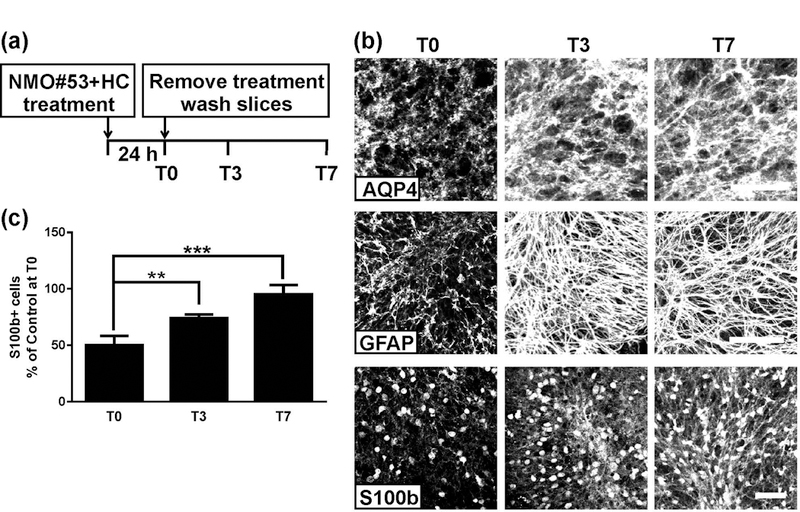
Astrocyte repair after treatment with NMO#53+HC. (a) Schematic depicting the treatment and recovery periods. Cerebellar slice cultures prepared from PLP-eGFP mice were treated with 20 μg/ml NMO#53 rAb plus 10% HC for 24 h. Following treatment, rAb and HC were removed by media exchange, and slices were allowed recover in culture media (in the absence of rAb+HC). Slices were fixed, stained, and imaged at T0, T3, and T7. (b) Confocal images of slices stained with AQP4, GFAP or S100b at the indicated time points. (c) Quantification of S100b+ cell number in slices. Cell count was normalized to control (ISO+HC treated) slice at T0. Statistical analyses were performed by unpaired Student’s t test. **: p<0.01, ***: p<0.001, n=3–4. Scale bars: 50µm.
