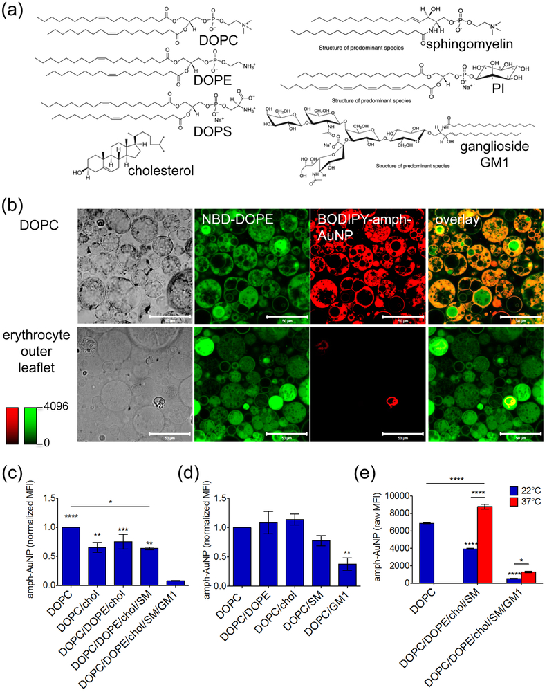
Figure 5.
(a) Synthetic lipids used to make model erythrocyte membranes. (b) Confocal micrographs of all-DOPC GMVs and erythrocyte outer leaflet GMVs treated with BODIPY-MUS:OT 2:1 amph-AuNPs (batch B, 0.07 mg/mL) for 1 hr at 22°C, scale bars 50 µm. (c, d) Amph-AuNPs (batch B, 0.07 mg/mL) were incubated with incremental GMV formulations (c) or GMVs incorporating individual membrane components (d) for 1 hr at 22°C, and analyzed for NP/membrane association by flow cytometry. Median fluorescence intensities of particles associated with GMVs (error bars for SEM between individual sample replicates). In (c), significance noted directly above columns is compared to the DOPC/DOPE/chol/SM/GM1 case. In (d), significance noted directly above columns is compared to the all-DOPC case. (e) Amph-AuNPs (batch B, 0.07 mg/mL) incubated with GMVs with or without GM1 present at 22°C and 37°C and assessed after 1 hr by flow cytometry. Significance noted directly above columns is compared to the all-DOPC case (22°C). Statistics were performed with a one-way ANOVA test, followed by Tukey’s multiple comparison test (P < 0.05 is significant, *** indicates P < 0.0001). All experiments were performed with at least 3 biological replicates per condition.

