Abstract
Background:
Femoral head osteonecrosis is a progressive disease with disabling outcomes in hip joint if not treated. This study was designed to compare the effects of zoledronic acid plus vitamin E versus zoledronic acid alone in surgical induced femoral head osteonecrosis in rabbits.
Methods:
26 Japanese white adult normal male rabbits at 28-32 weeks old were undertaken surgical femoral dislocation to devastate the femoral neck vessels; the femoral neck vessels were ligated and the hip was relocated. Next, the first 10 rabbits received zoledronic acid injections at 1st and the 4th weeks; the second group (10 rabbits) received zoledronic acid injections at 1st and the 4th week along with daily oral vitamin E for 12 weeks; and the third group was considered as non-treated control group. Radiographic and postmortem pathological assessments including the Ficat classification, epiphyseal quotient (EQ), new bone formation, and residual necrotic bone (RNB) were performed and compared after week 12.
Results:
A significant difference was found between the combination therapy group and the control group in Ficat classification at 12th weeks (P=0.048), but, the difference between monotherapy and combination therapy groups at 12th weeks was nonsignificant (P=0.37). Also, both treated groups had significant difference with the control group for RNB (P=0.015). There were no significant differences between the three groups for Ficat classification at the 6th week (P=0.65); EQ at 6th (P=0.59) and 12th week (P=0.64); and NBF (P=0.55).
Conclusion:
Although zoledronic acid therapy along with vitamin E could improve some radiologic and pathological indices related to femoral head osteonecrosis, vitamin E showed a relative impact.
Level of evidence:
I
Key Words: ONFH, Osteonecrosis of femoral head, Vitamin E, Zoledronic acid
Introduction
It is estimated that 20,000-30,000 new cases are diagnosed with osteonecrosis annually in the United States (1). Osteonecrosis of femoral head (ONFH) is known as a progressive disease and leads to the collapse of femoral head when untreated (2, 3). Trauma or slipped capital femoral epiphysis (SCFE) at adolescence both can be associated with an increased risk of ONFH (2, 4, 5). Besides trauma, there is a long list of conditions that may reason or are related to ONFH including steroids utilization, alcohol abuse and etc.; however, some cases are considered idiopathic (2, 5-7). ONFH may be painless at early stage; while, it may be followed by an unexpected beginning of severe pain at groin with progression of the disease. The eventual presentation is painful limitation of hip motion and collapse and fracture of the femoral head, leading to end-stage degenerative changes may be expected (2, 3, 8). Accordingly, preventing the progression of the disease, especially the collapse is one of the treatment goals.
Complete evaluation of the patient’s history and physical exam are required to diagnose the ONFH. For years plain radiographs have been the basic imaging assessment tools for recognition and staging of ONFH though bone scan demonstrates high sensitivity for early detection and MRI is considered the method of choice for detection and staging of the disease (5).
Despite the vast knowledge, little agreement has been made on factors that trigger the avascular necrosis of bone. Intra- and extra-luminal obliterations are the possible involved mechanisms; however, cytotoxicity and genetic factors may be implicated (7). Intraluminal obliteration of blood vessels can occur by microscopic fat emboli, sickle cells, nitrogen bubbles (caisson disease), and focal clotting due to procoagulant abnormalities. Extraluminal obliteration may result from increased marrow pressure or fat (7). The process of avascular necrosis of bone seems multifactorial even though the final mechanism is always critical ischemia (7).
There is no gold standard treatment for ONFH and multidisciplinary approaches are usually necessary (1). The therapeutic approaches consist of pharmacologic agents as well as biophysical and surgical treatments (1). Despite various non operative treatment options, the most favorable results have been seen by surgical interventions. Surgery has been the most common decision by orthopedic surgeons in current practice. Majority of the surgical approaches consist of a core decompression part. In this regard, many investigations are going on animal models to evaluate the efficacy of various pharmaceutical or surgical therapeutic methods. Several studies have been designed to evaluate the use of medications such as enoxaparin, lovastatin, nitrate patch, rifampicin, vitamin E, bisphosphonates, sodium ferulate, and ACTH (9-17). Bisphosphonates including alendronate and zoledronic acid seem effective in treatment of femoral head osteonecrosis and Legg-Calve´-Perthes disease-like in rat models, respectively (13-15, 18). Furthermore, animal intervention studies have revealed that bisphosphonate therapy could improve bone volume and mineral density and preserve a better femoral head shape (13). In addition, oral vitamin E (α-tocopherol) supplementation at a safe dose can suppress corticoste-roid-induced osteonecrosis in rabbits (12).
We intended to evaluate the effect of zoledronic acid alone and in combination with vitamin E on surgicalinduced ONFH in rabbit.
Materials and Methods
Experimental animals
The clearance to conduct this study was provided by the animal ethics committee of Iran University of Medical Sciences (Tehran, Iran) (IR.IUMS.REC.1394.9211242016). 26 healthy Japanese normal white adult male rabbits at 28-32 weeks old (weight, 2 to 4.250 kg) were purchased from Pasteur Institute, Iran. The animals were raised in separate cages on a 12/12-hour light/dark cycle and 23±2oC temperature, fed with a standard laboratory diet and water (ORC4: Oriental Yeast Co. Ltd, Tokyo, Japan). The Epiphysis and normal pelvis closure were confirmed by radiography. Healthy animals were considered to have closed epiphysis with no density changes, sclerosis, or other osteonecrosis symptoms in femoral head in radiographs. Also, 3 rabbits in each group underwent MRI for further confirmation. All the processes were performed at Shaheed Rajaei Cardiovascular Medical and Research Center, Tehran, Iran.
Induction of ONFH: Femoral head Osteonecrosis was inducted by surgical dislocation and ligation of the left hip vessels according to the method used by a previous study (19). In brief, general anesthesia was induced by IV administration of ketamine (1 mg/kg) and midazolam (0.5 mg/kg) under veterinary supervision with an anesthetic technician. The animals received IV cephazoline (25mg/kg pre-op and 25mg/kg bd for 3 day). After prep and drape, the animal was laid on lateral position; a 4 cm curve incision was made on anterolateral area of the left hip. The fasciae between tensor fasciae latae muscle and gluteus maximus was opened; the gluteus maximus muscle was pulled over and hip capsulotomy was performed. The hip teres ligament was cut and the femur head was dislocated to anterior position. The superior vessels of the femur neck were destroyed by cutting the periosteum all around the femoral neck and the femur neck vessels were ligated. Finally, the hip was relocated; the capsule was closed; and the fasciae and skin were sutured in separate layers. Hip joint reduction was confirmed by radiography at the end of surgical procedure.
Treatment procedure
The rabbits were divided into three groups. Animals in the first group (n=10) were treated with zoledronic acid; the second group (n=10) received a combination of zoledronic acid and vitamin E supplementation; and the 6 rabbits were considered as control. Zoledronic acid (1mg/kg-subcutaneous injection) was injected for both test groups at the 1st and the 4th week. In addition, vitamin E (600 mg/kg, per oral) was added to the food of one of the treated groups similar to a previous study (12). The rabbits were assessed for 12 weeks after the surgery. The radiographic evaluation was performed at the end of the 6th and 12th weeks. The extent of femoral head involvement and severity of femoral head osteonecrosis was determined according to the Ficat classification (20, 21). Considering the proportionally small sample size in this study, a modification was made on the radiographic classification based on the femoral head collapse. All cases were divided into two groups: group one consisted of pre-collapse cases including stage I and IIa in Ficat classification, while, post-collapse cases included IIb, III, and IV stages in Ficat classification. The data were grouped into good (Ficat stage: 0, I and IIa) and poor (Ficat stage: IIb, III and IV).
Also, the femoral head collapse was evaluated by radiographic assessment of epiphyseal quotient (EQ). The EQ was calculated by dividing the maximum height of the osseous epiphysis of the femoral head by the maximum diameter (22).
All rabbits were sacrificed at the end of the 12th week. New Bone Formation (NBF) and Residual Necrotic Bone (RNB) in femur head were determined and scored by a pathologist. According to NBF, the results were divided into good (stage or grade 2 and 3) and poor (stage or grade 0 and 1) groups. Also, based on RNB, the results were grouped into good (stage or grade 0 and 1) and poor (stage or grade 2 and 3) groups.
Statistical analysis
The difference in Ficat classification at the end of the 6th and 12th week and the changes according to RNB and NBF at the end of 12th week was evaluated by chi-square test. Alterations in mean EQ at the end of 6th week and 12th week were evaluated by Paired t-tests. Furthermore, independent t-test and Fisher’s test were used for other comparisons. A P<0.05 was considered as statistically significant. Statistical analyses were performed using SPSS 22.0 software.
Results
This study examines the therapeutic effects of zoledronic acid ± vitamin E in treatment of femoral head osteonecrosis in rabbits. The radiologic (Ficat and EQ) and pathologic (NBF and RNB) indices show the effect of the therapeutic plan during a 12-week course.
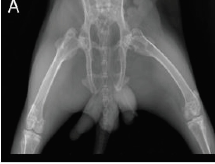
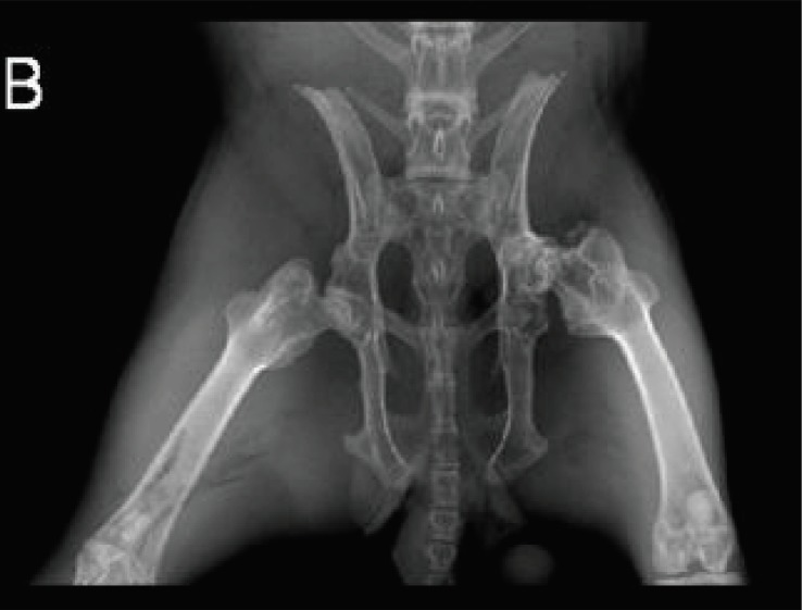
The radiographs of femoral head before surgical intervention demonstrate the closure of epiphysis and normal pelvis also at the end of the 6th and 12th weeks indicate the radiologic indices in all three groups [Figures 1; 2].
Figure 1.
The radiograph of femoral head before surgical intervention.
Figure 2.
The radiograph of femoral head after 12th week in control group.
The results of zoledronic acid monotherapy and combination therapy with vitamin E on femoral head osteonecrosis based on Ficat classification are presented in table 1.
Table 1.
The results of zoledronic acid monotherapy and combination therapy with vitamin E based on Ficat classification
|
Ficat at the end of 6
th
week
|
Ficat at the end of 12
th
week
|
|||
|---|---|---|---|---|
| good | poor | good | poor | |
| Zoledronic acid | 50% | 50% | 30% | 70% |
| Zoledronic acid and vitamin E | 70% | 30% | 55.6%* | 44.4% |
| Control | 33% | 67% | 16% | 84% |
P<0.05 in comparison with control group according to Ficat (good) at the end of 12th week.
There was no significant difference between the two treatment groups (P=0.67) as well as between the treatment groups and controls based on Ficat evaluation at the end of the 6th week (P=0.65). Also, no significant difference was found neither between the monotherapy and combination therapy groups (P=0.37) nor between monotherapy and control based on Ficat evaluation at the end of the 12th week (P=0.54). However, the results of Ficat (good) was statistically significant between the zoledronic acid plus vitamin E combination therapy group and control at the end of the 12th week (P=0.048) [Table 1].
No difference was found in the mean EQ between the end of the 6th and 12th week in zoledronic acid monotherapy group (P=1) [Table 2]. Although the mean EQ in combination therapy group increased at the end of 6th and 12th week, this difference was not statistically significant (P=0.59) [Table 2]. Also, the mean EQ decreased at the end of 6th and 12th week in control group, this difference was not statistically significant (P=0.36) [Table 2].
Table 2.
The results of zoledronic acid monotherapy and combination therapy with vitamin E is expressed as EQ mean ± SD
| EQ Mean at the end of 6 th week | EQ Mean at the end of 12 th week | |
|---|---|---|
| Zoledronic acid | 2.811 ± 0.242 | 2.811 ± 0.278 |
| Zoledronic acid and vitamin E | 2.990 ± 0.280 | 3.030 ± 0.297 |
| Control | 2.635 ± 0.227 | 2.440 ± 0.282 |
The results in table 3 reveals that the change of mean EQ in rabbits with poor Ficat in the monotherapy, combination therapy and control groups is not so much at the end of the 12th week [Table 3]. Independent t-test revealed no significant difference between the three groups (P=0.63).
Table 3.
The results of zoledronic acid monotherapy and combination therapy with vitamin E based on EQ mean and Fiact (poor) at the end of 12th week
| Ficat at the end of 12 th week | EQ Mean at the end of 12 th week | |
|---|---|---|
| Zoledronic acid | poor | 2.850 ± 0.200 |
| Zoledronic acid and vitamin E | poor | 3.085 ± 0.258 |
| Control | poor | 2.440 ± 0.272 |
Regarding the NBF, 70% of the monotherapy, 77.8% of the combination therapy and 33% of the control groups had good NBF [Table 4]. The rabbits with good NBF in each group were compared with the other groups. Yet, there was no significant difference neither between the treated groups and control (P=0.287), nor between the two treated groups (P=0.342).
Table 4.
The results of zoledronic acid monotherapy and combination therapy with vitamin E based on good NBF and good RNB.
P< 0.05 in comparison with control base on RNB (good).
According to RNB, 90% of the monotherapy group, 90% of combination therapy group and 33% of the control groups had good RNB [Table 4]. Also, the rabbits with good RNB in each group were compared with the other groups. Accordingly, the difference between the treated groups and control was significant (P=0.015).
Seventy percent of the monotherapy group had poor Ficat scores at the end of the 12th week; in addition, 70% and 90% of the monotherapy group had good NBF and good RNB, respectively [Table 5]. Also, 44.4% of combination therapy group was poor based on Ficat at the end of the 12th week; 77.8% and 90% of the combination therapy group had good NBF and good RNB, respectively [Table 5]. However, 84% of the control group were poor based on Ficat at the end of the 12th week; good NBF contained 33% of the rabbits in control group; also, good RNB included the same percent [Table 5]. Fisher’s test was used to compare the results presented in table 5. The comparison of the results of good NBF and good RNB with the result of poor Ficat at the end of the 12th week in monotherapy group revealed that the difference was not significant (P=0.117). Likewise, similar comparison of the mentioned indices presented no significant difference neither in combination therapy group (P=0.154) nor in control (P=0.172). The results of zoledronic acid monotherapy and combination therapy with vitamin E based on EQ mean for NBF (good) and RNB (good) at the end of 12th week presented in table 6. Nonetheless, the independent t-test revealed that there was no significant difference in comparing of the three groups (P=0.14).
Table 5.
The results of zoledronic acid monotherapy and combination therapy with vitamin E based on Ficat (poor), NBF (good) and RNB (good) at the end of 12th week
|
Ficat at the end of 12
th
week
Poor |
NBF
Good |
RNB
Good |
|
|---|---|---|---|
| Zoledronic acid | 70% | 70% | 90% |
| Zoledronic acid and vitamin E | 44.4% | 77.8% | 90% |
| Control | 84% | 33% | 33% |
Table 6.
The results of zoledronic acid monotherapy and combination therapy with vitamin E based on EQ mean for NBF (good) and RNB (good) at the end of 12th week
|
EQ Mean at 12
th
week for
good NBF and good RNB |
|
|---|---|
| Zoledronic acid | 2.751 ± 0.287 |
| Zoledronic acid and vitamin E | 3.091 ± 0.296 |
| Control | 2.380 ± 0.273 |
Discussion
a vascular femoral capital osteonecrosis
According to the increasing frequency of avascular femoral capital osteonecrosis, we evaluated a pharmacologic approach for treatment of ONFH (23). In this study, the therapeutic regimes consisting of zoledronic acid monotherapy and combination therapy of zoledronic acid and vitamin E were used for treatment of surgical induced ONFH in rabbits.
Major findings of the study
The results of this study revealed that Ficat (stage: 0, I and IIa) was the only radiologic index presented a statistical significant difference between the zoledronic acid plus vitamin E combination therapy group and control at the end of the 12th week (P=0.048). Both treatment groups showed less progression to the higher stages of Ficat classification (stage: IIb, III and IV) in comparison with the control group at the end of the 12th week. This finding suggests that less femoral head destruction might have been occurred. Also, the combination therapy group had less progression to the higher stages of Ficat classification in comparison with the zoledronic acid monotherapy group in 12 weeks. Other, notable outcome of this study was resulted from evaluation of RNB, the pathologic index. 90% of the group received monotherapy with the zoledronic acid and also 90% the group received combination therapy group with the zoledronic acid and vitamin E had good RNB; furthermore, comparing of RNB of the groups received the mentioned therapeutic agents with the control revealed a statistical significant difference (P=0.015). This confirms the effect of treatment on decreasing femoral head necrosis. However, according to RNB index, there was not any numerical difference between the results of monotherapy with combination therapy groups. It seems that treatment with zoledronic acid with/without vitamin E can be effective for ONFH.
Comparing with previous articles
In accord with our results, a ten-year prospective observational study on human demonstrated that 3-year-long oral alendronate therapy was a useful choice to delay the requirement for arthroplasty in the young and active patients (14). In this regard, another study on human by Agarwala et al. showed an improvement in the clinical function, a decline in the rate of collapse and reducing in demand for total hip replacement (15). Also, they indicated that improvement is principally marked if the treatment is initiated before collapse (15). Moreover, because the result with significant statistical difference according to Ficat classification was obtained at the end of a 12-week period (P=0.048), it seems that the time course of treatment may be another factor, which can influence the therapeutic effect of zoledronic acid plus vitamin E for ONFH. Furthermore, the finding of this study is similar to the other experimental studies performed on rat and rabbit by using alendronate, which is another nitrogen-containing bisphosphonate. Likewise, surgical osteonecrosis of the rat femoral head was induced by interruption of blood circulation and the rats received alendronate injections of 200 µg/kg/day for 42 days; the evaluation of femoral head by computed-assisted morphometry revealed that treatment with alendronate prevented the distortion and destruction of the femoral head (24). Also, the effect of alendronate was studied by the experimental surgical-induced osteonecrosis of the hip in adult rabbits; the animals received alendronate 150 μg/kg/day via subcutaneous injections, three times a week; then, the rabbits were euthanized at 6 and 12 months postoperatively (25). The result revealed that bone mass in the trabecular region of the femoral head was significantly increased due to inhibition of bone resorption by alendronate treatment during repair of the osteonecrotic femoral head; also, subchondral resorption was inhibited and cartilage degeneration in the acetabulum was reduced (25).
However, evaluating the results of EQ, another radiologic index, and NBF, the pathological index, were not statistically significant different in this study. In addition, according to the findings of this study, the comparison of the results of radiologic assessments with the pathologic findings in three groups was not statistically significant. Although the mean EQ differences were not statistically significant, more decrease of mean EQ in control group in contrast to the treated groups at the end of 12th week may indicate that the treatment might impede the femoral head collapse. If we consider the results of similar study used alendronate and lasted for 6 to 12 months, it seems that increasing the treatment duration with bisphosphonate may help to confirm its effect on experimental ONFH models (25). By considering the mean EQ index in this study, the combination therapy containing vitamin E in comparison with zoledronic acid monotherapy was not statistically significant, but the numerical rise of mean EQ of combination therapy at the end of 12th week may suggest that increasing the experimental sample size might improve the statistical significance.
According to NBF index, the high percentage of the rabbits received treatments was put in good NBF stage and the less percentage of control group was in good NBF stage. These results can point to the effects of treatment; by the way, increasing the experimental sample size may confirm not only the effects of zoledronic acid therapy but also the usefulness of vitamin E in combination with bisphosphonates.
Even though no significant statistical difference was found in comparing the results of monotherapy with combination therapy groups, it seems that accompanying of vitamin E may relatively improve the zoledronic acid therapy. In this regard, Kuribayashi et al. suggested another approach for the prevention of corticosteroid-induced osteonecrosis by giving vitamin E to rabbits and their findings was in agreement with the results of this study, which we used vitamin E in combination with zoledronic acid (12). However, the method of the present study was surgical induced osteonecrosis instead of corticosteroid-induced osteonecrosis. By the way, it can be suggested using vitamin E separately in surgical induced osteonecrosis model in another study to clarify its effect.
Limitation of this study
Small sample size which was chosen based on previous studies. Limitation on performing MRI every two weeks. Special conditions for keeping rabbits. The diet which was based on standard diet laboratory veterinary protocol.
Advantage of this study
The main advantage of this study included simultaneous and accurate study of radiographic and MRI findings with pathological findings, their comparison as well.
Suggest direction for future research
Conducting study with large sample size as well as investigating therapeutic effects of zoledronic acid ± vitamin E in corticosteroid-induced osteonecrosis instead of surgical induced osteonecrosis.
This study demonstrated that there was a significant statistical difference between zoledronic acid plus vitamin E combination therapy group with the control according to Ficat at the end of 12th week. Furthermore, RNB presented a significant statistical difference between each treated group with the control. Thus, it can be concluded that both therapeutic regimes including zoledronic acid plus vitamin E as well as zoledronic acid monotherapy were effective in treatment of ONFH based on RNB pathological index. However, the Ficat radiologic index was in favor of zoledronic acid plus vitamin E combination therapy. Besides, comparison of the combination therapy with the monotherapy revealed that vitamin E had beneficial effect in treatment although the statistical difference was not significant. Certainly, the animal studies with increased sample size will augment the results and lead to plan the related clinical trials.
Disclosure: The authors report no conflict of interest concerning the materials or methods used in this study or the findings specified in this paper.
Funding: This research received no specific grant from any funding agency in the public, commercial, or not-for-profit sectors.
References
- 1.Scaglione M, Fabbri L, Celli F, Casella F, Guido G. Hip replacement in femoral head osteonecrosis: current concepts. Clin Cases Miner Bone Metab. 2015;12(Suppl 1):51–4. doi: 10.11138/ccmbm/2015.12.3s.051. [DOI] [PMC free article] [PubMed] [Google Scholar]
- 2.Houdek MT, Wyles CC, Martin JR, Sierra RJ. Stem cell treatment for avascular necrosis of the femoral head: current perspectives. Stem Cells. 2014;7(1):65–70. doi: 10.2147/SCCAA.S36584. [DOI] [PMC free article] [PubMed] [Google Scholar]
- 3.Sen RK. Management of avascular necrosis of femoral head at pre-collapse stage. Indian J Orthop. 2009;43(1):6–16. doi: 10.4103/0019-5413.45318. [DOI] [PMC free article] [PubMed] [Google Scholar]
- 4.Rached E, Akkari M, Braga SR, Minutti MF, Santili C. Slipped capital femoral epiphysis: reduction as a risk factor for avascular necrosis. J Pediatr Orthop B. 2012;21(4):331–4. doi: 10.1097/BPB.0b013e32835368a7. [DOI] [PubMed] [Google Scholar]
- 5.Malizos KN, Karantanas AH, Varitimidis SE, Dailiana ZH, Bargiotas K, Maris T. Osteonecrosis of the femoral head: etiology, imaging and treatment. Eur J Radiol. 2007;63(1):16–28. doi: 10.1016/j.ejrad.2007.03.019. [DOI] [PubMed] [Google Scholar]
- 6.Karimova EJ, Rai SN, Howard SC, Neel M, Britton L, Pui CH, et al. Femoral head osteonecrosis in pediatric and young adult patients with leukemia or lymphoma. J Clin Oncol. 2007;25(12):1525–31. doi: 10.1200/JCO.2006.07.9947. [DOI] [PubMed] [Google Scholar]
- 7.Lafforgue P. Pathophysiology and natural history of avascular necrosis of bone. Joint Bone Spine. 2006;73(5):500–7. doi: 10.1016/j.jbspin.2006.01.025. [DOI] [PubMed] [Google Scholar]
- 8.Babhulkar S. Osteonecrosis of femoral head: treatment by core decompression and vascular pedicle grafting. Indian J Orthop. 2009;43(1):27–35. doi: 10.4103/0019-5413.45320. [DOI] [PMC free article] [PubMed] [Google Scholar]
- 9.Kang P, Gao H, Pei F, Shen B, Yang J, Zhou Z. Effects of an anticoagulant and a lipid-lowering agent on the prevention of steroid-induced osteonecrosis in rabbits. Int J Exp Pathol. 2010;91(3):235–43. doi: 10.1111/j.1365-2613.2010.00705.x. [DOI] [PMC free article] [PubMed] [Google Scholar]
- 10.Drescher W, Beckmann R, Kasch R, Pufe M, Knobe M, Kweider N, et al. Nitrate patch prevents steroid-related bone necrosis. J Orthop Res. 2011;29(10):1517–20. doi: 10.1002/jor.21420. [DOI] [PubMed] [Google Scholar]
- 11.Han N, Yan ZQ, Guo CA, Shen F, Liu J, Shi YX, et al. Effect of rifampicin on the risk of steroid-induced osteonecrosis of the femoral head. Orthop Surg. 2010;2(2):124–33. doi: 10.1111/j.1757-7861.2010.00075.x. [DOI] [PMC free article] [PubMed] [Google Scholar]
- 12.Kuribayashi M, Fujioka M, Takahashi KA, Arai Y, Ishida M, Goto T, et al. Vitamin E prevents steroid-induced osteonecrosis in rabbits. Acta Orthop. 2010;81(1):154–60. doi: 10.3109/17453671003628772. [DOI] [PMC free article] [PubMed] [Google Scholar]
- 13.Young ML, Little DG, Kim HK. Evidence for using bisphosphonate to treat Legg-Calve-Perthes disease. Clin Orthop Relat Res. 2012;470(9):2462–75. doi: 10.1007/s11999-011-2240-0. [DOI] [PMC free article] [PubMed] [Google Scholar]
- 14.Agarwala S, Shah SB. Ten-year follow-up of avascular necrosis of femoral head treated with alendronate for 3 years. J Arthroplasty. 2011;26(7):1128–34. doi: 10.1016/j.arth.2010.11.010. [DOI] [PubMed] [Google Scholar]
- 15.Agarwala S, Shah S, Joshi VR. The use of alendronate in the treatment of avascular necrosis of the femoral head. J Bone Joint Surg Br. 2009;91(8):1013–8. doi: 10.1302/0301-620X.91B8.21518. [DOI] [PubMed] [Google Scholar]
- 16.Tian L, Dang XQ, Wang CS, Yang P, Zhang C, Wang KZ. Effects of sodium ferulate on preventing steroid-induced femoral head osteonecrosis in rabbits. J Zhejiang Univ Sci B. 2013;14(5):426–37. doi: 10.1631/jzus.B1200311. [DOI] [PMC free article] [PubMed] [Google Scholar]
- 17.Zaidi M, Sun L, Robinson LJ, Tourkova IL, Liu L, Wang Y, et al. ACTH protects against glucocorticoid-induced osteonecrosis of bone. Proc Natl Acad Sci U S A. 2010;107(19):8782–7. doi: 10.1073/pnas.0912176107. [DOI] [PMC free article] [PubMed] [Google Scholar]
- 18.Little DG, McDonald M, Sharpe IT, Peat R, Williams P, McEvoy T. Zoledronic acid improves femoral head sphericity in a rat model of perthes disease. J Orthop Res. 2005;23(4):862–8. doi: 10.1016/j.orthres.2004.11.015. [DOI] [PubMed] [Google Scholar]
- 19.Kim HK, Su PH, Qiu YS. Histopathologic changes in growth-plate cartilage following ischemic necrosis of the capital femoral epiphysis An experimental investigation in immature pigs. J Bone Joint Surg Am. 2001;83-A(5):688–97. doi: 10.2106/00004623-200105000-00007. [DOI] [PubMed] [Google Scholar]
- 20.Chan KL, Mok CC. Glucocorticoid-induced avascular bone necrosis: diagnosis and management. Open Orthop J. 2012;6(1):449–57. doi: 10.2174/1874325001206010449. [DOI] [PMC free article] [PubMed] [Google Scholar]
- 21.Smith SW, Meyer RA, Connor PM, Smith SE, Hanley EN Jr. Interobserver reliability and intraobserver reproducibility of the modified Ficat classification system of osteonecrosis of the femoral head. J Bone Joint Surg Am. 1996;78(11):1702–6. doi: 10.2106/00004623-199611000-00010. [DOI] [PubMed] [Google Scholar]
- 22.Kim HK, Randall TS, Bian H, Jenkins J, Garces A, Bauss F. Ibandronate for prevention of femoral head deformity after ischemic necrosis of the capital femoral epiphysis in immature pigs. J Bone Joint Surg Am. 2005;87(3):550–7. doi: 10.2106/JBJS.D.02192. [DOI] [PubMed] [Google Scholar]
- 23.Boss JH, Misselevich I, Bejar J, Norman D, Zinman C, Reis DN. Experimentally gained insight–based proposal apropos the treatment of osteonecrosis of the femoral head. Med Hypotheses. 2004;62(6):958–65. doi: 10.1016/j.mehy.2003.12.036. [DOI] [PubMed] [Google Scholar]
- 24.Peled E, Bejar J, Zinman C, Reis DN, Boss JH, Ben-Noon H, et al. Alendronate preserves femoral head shape and height/length ratios in an experimental rat model: a computer-assisted analysis. Indian J Orthop. 2009;43(1):22–6. doi: 10.4103/0019-5413.44630. [DOI] [PMC free article] [PubMed] [Google Scholar]
- 25.Hofstaetter JG, Wang J, Yan J, Glimcher MJ. The effects of alendronate in the treatment of experimental osteonecrosis of the hip in adult rabbits. Osteoarthritis Cartilage. 2009;17(3):362–70. doi: 10.1016/j.joca.2008.07.013. [DOI] [PubMed] [Google Scholar]


