. 2018 Dec 12;7:e42150. doi: 10.7554/eLife.42150

Figure 2. Architecture of the DEP-DHEX module.

(a) Superposition of the DEP domains of RGS7 (pink), RGS9 (green; PDB entry 2pbi), EPAC (yellow; PDB entry 2byv), pleckstrin (brown; PDB entry 1w4m), and Dvl (grey; PDB entry 1fsh) reveals diversity in the conformational organization of the β-hairpin arm. (b) Comparison of the DEP-DHEX domains of RGS7 and RGS9 shows differences in several loops and in the orientation of the DHEX domain. In RGS7, the DHEX domain is lifted upward thus generating a wide space between the domains. (c) Evaluation of the electrostatic surface potentials of the DEP-DHEX domains of RGS7 (left) and RGS9 (right). A strong basic patch (blue) is formed by three loops in the RGS7 structure but differences in the organization of these loops result in a more scattered distribution of basic charges in the RGS9 structure.

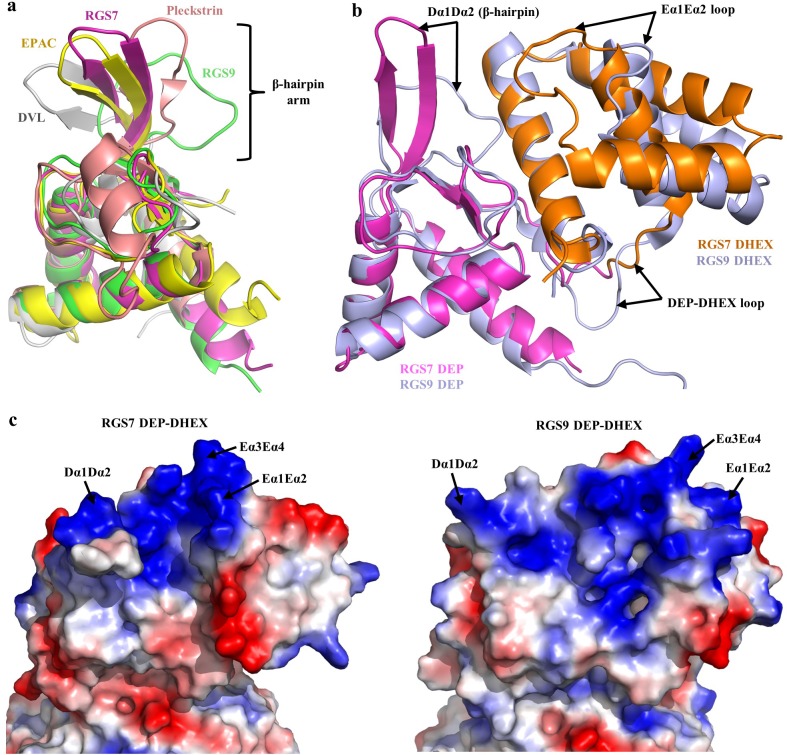
Figure 2.

Figure 2—figure supplement 1. Homology of the DEP domains and loop flexibility in the DEP-DHEX domain of RGS7.

Figure 2—figure supplement 1.

(a) Multiple sequence alignments of the DEP domain containing proteins. The β-hairpin shows no sequence conservation. The DEP domain sequences are taken from the structures of RGS7, RGS9 (PDB entry 2pbi), EPAC (PDB entry 2byv), pleckstrin (PDB entry 1w4m), and Dvl (PDB entry 1fsh). This Figure was generated with ESPript 3.0. (b) Comparison of the loop flexibilities of the DEP-DHEX domains of RGS7. Superposition of the two molecules in the asymmetric unit of the RGS7-Gβ5 dimer shows the loop variability in the β-hairpin and the Eα1Eα2 loop is indicated by black arrows. The DEP and DHEX domains are colored in pink and orange, respectively.
Figure 2—figure supplement 2. Comparison of the Dvl2 DEP domain with the Dvl2 DEP domain-µ2 complex.

Figure 2—figure supplement 2.

Cartoon representation of the superposed Dvl2 DEP domain (cyan, PDB entry 1fsh) with the Dvl2 DEP domain (green)-µ2 subunit of the AP-2 clathrin adaptor (pink) complex (PDB entry 3ml6) is shown. The involvement in the complex formation and conformational changes of the β-hairpin arm are indicated by a black arrow.