Abstract
Thrombosis is the main cause of morbidity and mortality in patients with JAK2V617F myeloproliferative neoplasms. Recent studies have reported the presence of JAK2V617F in endothelial cells of some patients with myeloproliferative neoplasms. We investigated the role of endothelial cells that express JAK2V617F in thrombus formation using an in vitro model of human endothelial cells overexpressing JAK2V617F and an in vivo model of mice with endothelial-specific JAK2V617F expression. Interestingly, these mice displayed a higher propensity for thrombus. When deciphering the mechanisms by which JAK2V617F-expressing endothelial cells promote thrombosis, we observed that they have a pro-adhesive phenotype associated with increased endothelial P-selectin exposure, secondary to degranulation of Weibel-Palade bodies. We demonstrated that P-selectin blockade was sufficient to reduce the increased propensity of thrombosis. Moreover, treatment with hydroxyurea also reduced thrombosis and decreased the pathological interaction between leukocytes and JAK2V617F-expressing endothelial cells through direct reduction of endothelial P-selectin expression. Taken together, our data provide evidence that JAK2V617F-expressing endothelial cells promote thrombosis through induction of endothelial P-selectin expression, which can be reversed by hydroxyurea. Our findings increase our understanding of thrombosis in patients with myeloproliferative neoplasms, at least those with JAK2V617F-positive endothelial cells, and highlight a new role for hydroxyurea. This novel finding provides the proof of concept that an acquired genetic mutation can affect the pro-thrombotic nature of endothelial cells, suggesting that other mutations in endothelial cells could be causal in thrombotic disorders of unknown cause, which account for 50% of recurrent venous thromboses.
Introduction
Myeloproliferative neoplasms (MPN) are acquired clonal hematopoietic stem cell disorders, characterized by an increase in one or more myeloid lineages. The Philadelphia chromosome-negative MPN include polycythemia vera with an excess of red blood cells, essential thrombocythemia with an increase of platelets, and primary myelofibrosis.1 More than 90% of patients with polycythemia vera and half of those with essential thrombocythemia and primary myelofibrosis carry a mutation in the Janus kinase 2 (JAK2) gene, i.e. JAK2V617F.2–5 JAK2 is a tyrosine kinase that initiates intracellular signaling of various type 1 cytokine receptors, such as erythropoietin and thrombopoietin receptors.6 The JAK2V617F mutation is responsible for constitutive activation of JAK2 kinase, resulting in subsequent activation of its downstream signaling pathways, ultimately leading to overproduction of myeloid cells.
Arterial and venous thromboses are the main causes of morbidity in Philadelphia chromosome-negative MPN with reported incidences ranging from 12–39% in polycythemia vera and 11–25% in essential thrombocythemia.7 The pathogenesis of thrombosis in patients with MPN is complex and still largely elusive.8 A variety of blood cells have been reported to participate in the pathophysiology of thrombosis in these neoplasms: (i) platelets isolated from MPN patients show signs of enhanced in vivo activation;9 (ii) leukocytes are activated and hyperleukocytosis is an independent risk factor for thrombosis;9–11 and (iii) red blood cells from patients with polycythemia vera display increased adhesion to the endothelium.12 However, there is evidence that JAK2V617F can be present not only in blood cells but also in endothelial cells (EC) from JAK2V617F-positive MPN patients.13–15
Under physiological conditions, the endothelium maintains a hemostatic balance between pro-thrombotic and anti-thrombotic factors. When stimulated by extrinsic factors such as inflammatory cytokines, hypoxia or antiphospholipid antibodies, EC become activated and promote thrombosis.16 Whether EC can become pro-thrombotic due to intrinsic modifications such as genetic mutations, has not yet been demonstrated.
In the current study, we tested whether vascular EC expression of JAK2V617F is sufficient to promote a pro-thrombotic state. Specifically, we investigated the hemostatic properties of JAK2V617F-expressing EC (hereafter, simply JAK2V617F EC) and these cells’ role in thrombus formation using human EC overexpressing human JAK2V617F, and mice with EC-specific JAK2V617F expression.
Methods
In vitro static adhesion of normal blood cells on endothelial cells
Blood from healthy volunteers was collected into test-tubes containing EDTA after informed consent had been obtained. Mononuclear cells and neutrophils were isolated by Pancoll density gradient or neutrophil separation medium (Polymorphprep, Fresenius Kabi) and marked with CellTracker Orange. Cells (105) were plated over confluent human umbilical vein endothelial cells (HUVEC) for 1 h at 37°C. After removal of non-adherent cells, the adherent cells were visualized using a fluorescence microscope (AxioObserver, Zeiss), and images were analyzed by ZEN imaging software (Zeiss). Experiments were performed using three to six wells per condition with 12 images taken in each well. The method of quantification has been described elsewhere15,17 Where noted, we added 10 mg/mL P-selectin blocking antibody (AK4 clone, BioLegend) 30 min before plating blood cells on HUVEC.
In vitro neutrophil adhesion on human umbilical vein endothelial cells under flow conditions
Channels (Vena8 Endothelial + Cellix) were coated with human fibronectin (100 ng/mL) before infusing 3×10⁶/mL HUVEC.18 After 2 h at 37°C, cells were exposed for 48 h to two-period flow (Kima Pump, Cellix) with alternating perfusion for 3 min (at 600 μL/min) and resting for 20 min (no flow). After 48 h, a confluent monolayer had developed. Neutrophils (3×10⁶/mL) were infused with a 10 μL/min venous flux and imaged using a Zeiss microscope, 20X in phase contrast, for 5 min. Channels were washed with medium and images of every channel were recorded. Neutrophil quantification was performed blindly and the neutrophil velocity was studied with FIJI (Image J).
In vivo leukocyte adhesion to mesenteric venules
Pdgfb-iCreERT2;JAK2V617F/WT mice and Pdgfb-iCreERT2;JAK2V617F/WT mice were used 15–20 days after tamoxifen injection. Mice were injected, intraperitoneally, with 250 ng tumor necrosis factor-alpha (TNF-α) and, 4h later, anesthetized with ketamine/xylazine. Leukocytes were stained with 3.6 mg/kg Rhodamine 6G (Sigma-Aldrich). For each mouse, five mesenteric venules of 150 to 250 μm diameter were observed for 90 sec within 30 min after the surgical procedure, using a fluorescent microscope (AXIO Zoom.V16, Zeiss). Where noted, 25 μg of P-selectin blocking antibody (RB40 clone, BD Biosciences) or isotype control (A110-1 clone, BD Biosciences) were injected 5 min before starting the analysis.
Mouse model of thrombosis
To induce platelet activation, the mice were given an intraperitoneal injection of 75 μg/kg collagen (Nycomed Pharma) and 30 μg/kg epinephrine (Helena Laboratories) 3 min before euthanasia.19,20 To increase inflammation, 250 ng TNF-α (RD Systems) were injected 4 h before the animals were sacrificed. After euthanasia, intracardiac puncture was performed and phosphate-buffered saline was perfused for 3 min, followed by 10% formalin for another 3 min. Where noted, 25 mg P-selectin blocking antibody were injected 4 h prior to euthanasia.
Statistics
Results are expressed as the mean ± standard error of mean (SEM). Statistical significance was calculated using the Student t test or Mann-Whitney statistical test to compare differences between two groups. For multiple groups, we used one-way analysis of variance (ANOVA) followed by the Tukey post-hoc test or two-way ANOVA followed by the Sidak post-hoc test. GraphPad Prism 6 software was used. A P value <0.05 was considered statistically significant.
Animals
Animal experiments were performed in accordance with the guidelines provided by the Institutional Animal Care and Use Committee at Inserm (Agreement number C45-234-6).
Results
Pdgfb-iCreERT2;JAK2V617F/WT mice are a reliable model to investigate JAK2V617F endothelial cell properties
To analyze the specific role of JAK2V617F EC in thrombus formation, we crossed Pdgfb-iCreERT2 mice with conditional flexed JAK2 (JAK2V617F) mice,21 to allow heterozygous expression of JAK2V617F in EC after tamoxifen administration. Since oral tamoxifen administration has been shown to result in recombination activity in megakaryocytes within 2 days,22 all experiments were performed and analyzed 15–20 days after tamoxifen injection, to allow time for recombined megakaryocytes to mature and be cleared. We first analyzed the efficiency of recombination using Pdgfb-iCreERT2;mT/mG mice, and observed that all vessels in the mesentery and retina were positive for green fluorescent protein (GFP) (Online Supplementary Figure S1A-C). To confirm EC JAK2V617F expression in Pdgfb-iCreERT2;JAK2V617F/WT mice, we sorted CD31-positive cells from the kidney and observed a 50% ratio of JAK2V617F:total JAK2 in accordance with a heterozygous expression of JAK2V617F in all EC (data not shown). In sorted CD31-positive lung EC, western blot analysis demonstrated an increased level of phosphorylation of the downstream effector AKT in line with increased activation of the JAK pathway (Online Supplementary Figure S1D,E). Because myeloproliferative syndromes have been described in EC-specific mouse models,23,24 we ensured that our experimental conditions did not lead to abnormal hematopoiesis. We did not observe any difference in blood cell counts in Pdgfb-iCreERT2;JAK2V617F/WT mice, suggesting that the hematopoietic system was not significantly altered (Online Supplementary Table S1). To definitively exclude hematopoietic expression of JAK2V617F in Pdgfb-iCreERT2;JAK2V617F/WT mice, we crossed Pdgfb-iCreERT2;JAK2V617F/WT mice with mT/mG mice to generate Pdgfb-iCreERT2;JAK2V617F/WT;mT/mG mice permitting co-expression of JAK2V617F and GFP in cells after Cre-mediated excision (Online Supplementary Figure S1F). Flow cytometry analysis did not reveal any GFP expression in granulocytes, platelets or red blood cells in Pdgfb-iCreERT2;JAK2V617F/WT;mT/mG mice 20 days after tamoxifen administration (Online Supplementary Figure S1F), confirming that Cre recombination under the Pdgfb promoter is highly restricted to EC 15-20 days after tamoxifen administration.
The expression of JAK2V617F by endothelial cells leads to increased thrombus formation
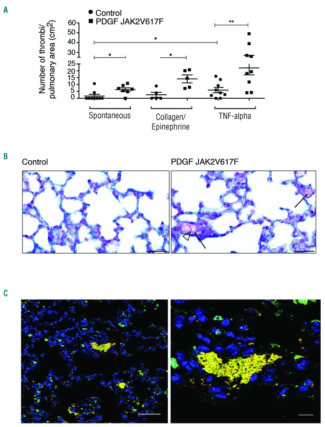
To investigate whether Pdgfb-iCreERT2;JAK2V617F/WT mice displayed a greater propensity to thrombosis, we examined pulmonary thrombus formation using experimental conditions that allowed assessment of EC involvement, without exposure of the sub-endothelium: (i) spontaneous thrombosis under basal conditions, (ii) a model of mild thrombosis with administration of low doses of collagen together with epinephrine to induce weak activation of platelets and vasoconstriction and better demonstrate an intrinsic pro-thrombotic phenotype of JAK2V617F EC, and (iii) a weak inflammatory trigger of thrombosis with injection of a low dose of TNF-α (250 ng/mouse). A dose of 500 ng TNF-α is commonly used to trigger inflammation25–27 but we chose the lower dose to reveal a potential hypersensitivity to inflammation. To quantify thrombi, we performed Carstairs staining which allows visualization of platelets, red blood cells, and fibrin.28 Small, spontaneously formed thrombi were observed in the lungs of Pdgfb-iCreERT2;JAK2V617F/WT mice, but not in littermate JAK2V617F/WT control mice (Figure 1A). In both models of mild induction of thrombosis, we observed a significant increase in thrombus formation in Pdgfb-iCreERT2;JAK2V617F/WT mice compared with that in controls (Figure 1A-C). Together, these results demonstrate that JAK2V617F EC have a pro-thrombotic phenotype.
Figure 1.
The presence of JAK2V617F in endothelial cells promotes thrombus formation. (A) In Pdgfb-iCreERT2;JAK2V617F/WT mice, thrombus formation occurs spontaneously and is increased after weak platelet activation by low-dose collagen plus epinephrine, or injection of tumor necrosis factor (TNF)-alpha. (B) Carstairs staining of pulmonary thrombi in control mice (left) and Pdgfb-iCreERT2;JAK2V617F/WT mice (right) injected with TNF-alpha. Black arrows indicate thrombi. The clear arrow head indicates fibrin deposition. Scale bar: 500 mm. (C) Representative image of a thrombus formed by neutrophils (green) and platelets (yellow) in Pdgfb-iCreERT2;JAK2V617F/WT mice. Scale bar in the left image: 50 μM. Scale bar in the right image: 10 μM. Results are expressed as mean value ± SEM. Statistical significance was determined by the Mann-Whitney test. *P<0.05; **P<0.01.
JAK2V617F endothelial cells have normal anticoagulant activity
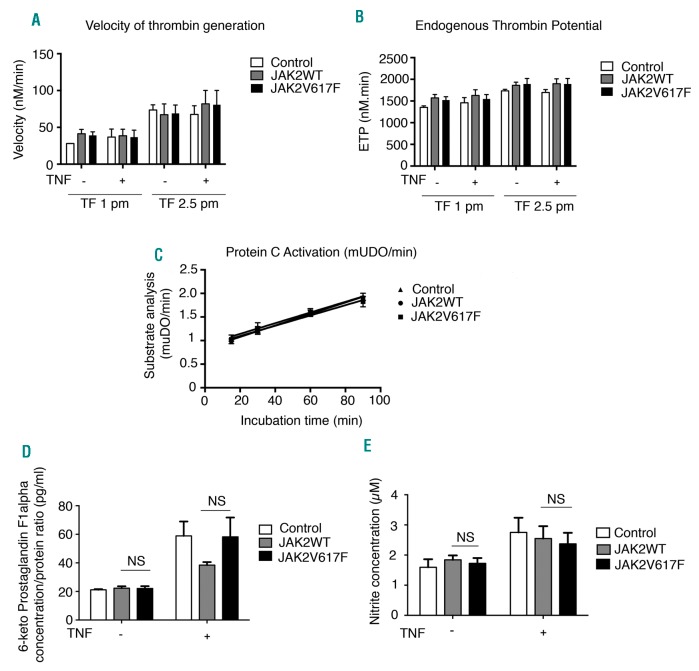
Next, we sought to decipher the mechanisms by which JAK2V617F expression in EC leads to a pro-thrombotic phenotype. To assess whether the expression of JAK2V617F by EC decreased these cells’ anticoagulant properties or even triggered pro-coagulant properties, we measured thrombin generation at the surface of HUVEC transduced with lentivirus encoding for either human JAK2V617F or wildtype JAK2 (JAK2WT), or with an empty lentivirus as a control. Our data show that HUVEC were efficiently transduced as: (i) 95% of JAK2V617F or JAK2WT HUVEC were GFP-positive as assessed by flow cytometry (Online Supplementary Figure S2A) and (ii) the mRNA JAK2V617F mutational burden was 99.5% in JAK2V617F HUVEC (Online Supplementary Figure S2B). Western blot analysis revealed induced protein expression of JAK2 in JAK2V617F- and JAK2WT-transduced cells. The introduction of JAK2V617F led to increased phosphorylation of JAK2, STAT3 and AKT, in agreement with hyperactivation of the JAK/STAT pathway (Online Supplementary Figure S2C). Under resting conditions, measurement of thrombin generation at the surface of control, JAK2WT-or JAK2V617F-lentivirus infected HUVEC did not reveal any significant differences in the kinetics or extent of thrombin generation. These results indicate that there is not a significant gain of pro-coagulant activity in response to JAK2WT- or JAK2V617F-induced HUVEC expression (Figure 2A,B). We next hypothesized that JAK2V617F HUVEC might acquire a procoagulant phenotype due to exposure to circulating inflammatory stimuli. We thus repeated the experiments after overnight activation with 10 ng/mL TNF-α, but did not observe any difference (Figure 2A,B). We also measured the rate of thrombin-triggered protein C activation and did not observe any difference between the cells (Figure 2C). Finally, we quantified the production of nitrite and prostaglandin (6-keto-prostaglandin 1-α) and did not find any difference between JAK2WT and JAK2V617F HUVEC which were or were not stimulated by TNF-α (Figure 2D,E). To assess whether JAK2V617F EC increase hemostatic potential in vivo, we used whole blood thromboelastometry in Pdgfb-iCreERT2;JAK2V617F/WT mice. No difference was observed in clotting time, clot formation time, 10-minute amplitude or alpha angle after stimulation of the extrinsic pathway (Online Supplementary Figure S3).
Figure 2.
JAK2V617F-expressing endothelial cells have normal anticoagulant activity. (A,B) There was no statistical difference in either (A) the rate or (B) the extent of tissue factor (TF) -triggered thrombin generation at the surface of endothelial cells which were or were not activated with tissue necrosis factor (TNF)-alpha (n=3 for all conditions). (C-E) There was no statistical difference in (C) the rate of thrombin-triggered protein C activation at the surface of endothelial cells (n=3 for all conditions), (D) the production of nitrite or (E) the production of 6-keto prostaglandin F1a (n=3 for all conditions). Results are expressed as mean value ± SEM from three experiments. Statistical significance was determined by the Mann-Whitney test.
JAK2V617F endothelial cells have a pro-adhesive phenotype in vitro and in vivo
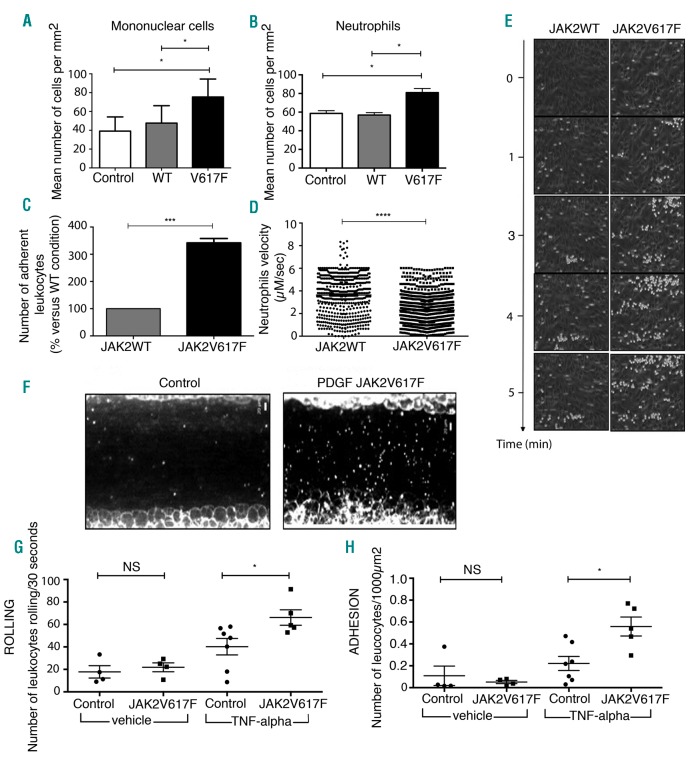
Exposing EC to inflammatory stimuli leads to expression of adhesion molecules that allow rolling and adhesion of leukocytes, a phenomenon that is thought to participate in the pathogenesis of thrombosis. Using JAK2V617F-transduced HUVEC, we observed an increase in static adhesion of normal mononuclear cells (Figure 3A) and normal polymorphonuclear neutrophils isolated from healthy subjects (Figure 3B), as previously reported with JAK2V617F EC from patients. Under flow conditions, we also observed that more normal polymorphonuclear neutrophils rolled and stably adhered to JAK2V617F HUVEC than to JAK2WT HUVEC (Figure 3C-E). To assess whether this pro-adhesive phenotype was also present in vivo, we injected Pdgfb-iCreERT2;JAK2V617F/WT mice with rhodamine to track leukocytes. Using intravital microscopy, we observed mesenteric venules and quantified leukocyte adhesion and rolling. We observed that both leukocyte rolling and adhesion were significantly increased in Pdgfb-iCreERT2;JAK2V617F/WTmice previously exposed to low-dose TNF-α (Figure 3F-H).
Figure 3.
JAK2V617F-expressing endothelial cells have a pro-adhesive phenotype in vitro and in vivo. (A,B) Under static conditions (A) normal mononuclear cells and (B) neutrophils adhere more to human umbilical vein endothelial cells (HUVEC) expressing JAK2V617F than to JAK2WT HUVEC. HUVEC transduced with lentiviruses containing green fluorescent protein alone were used as negative controls. Statistical significance determined by one-way analysis of variance and the post-hoc Tukey test. Results are expressed as mean value ± SEM from three experiments. (C,D) Under flow conditions (C) adhesion of neutrophils is increased in the presence of JAK2V617F HUVEC and (D) neutrophil velocity is reduced. Each dot represents one cell. Statistical significance was determined by the Student t-test. Results are expressed as mean value ± SEM from three experiments. (E) Representative images of adhesion of neutrophils on JAK2WT and JAK2V617F HUVEC under flow conditions. (F) Representative images of leukocyte adhesion in mesenteric venules of control and Pdgfb-iCreERT2;JAK2V617F/WT mice treated with tumor necrosis factor (TNF). (G) Leukocyte rolling and (H) adhesion are increased in mesenteric venules of Pdgfb-iCreERT2;JAK2V617F/WT mice treated with TNF. Results are expressed as mean value ± SEM. Statistical significance was determined by the Mann-Whitney test. *P<0.05; ***P<0.001. ****P<0.0001.
P-selectin expression is increased in JAK2V617F endothelial cells
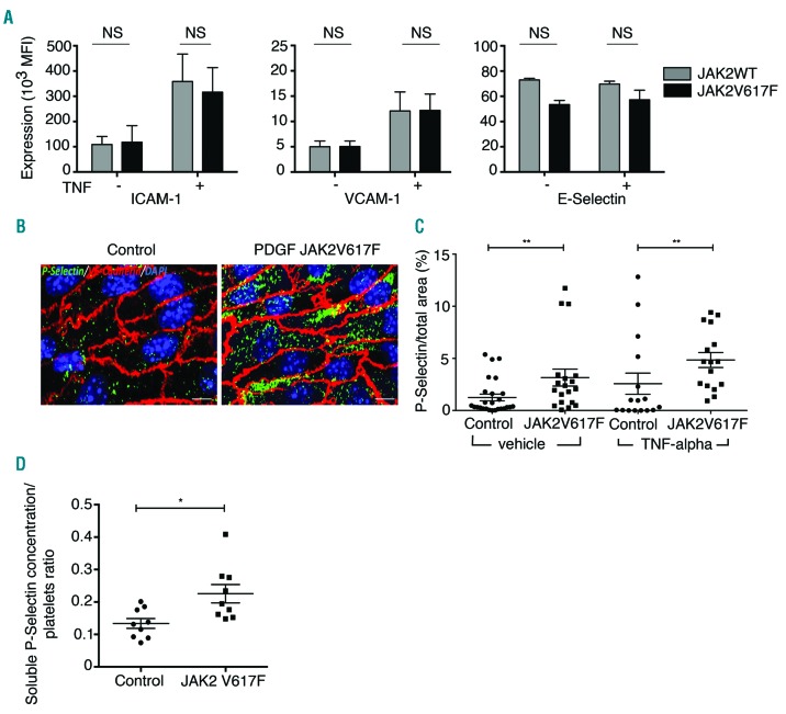
The adhesion of leukocytes to EC is mediated by cell adhesion molecules and selectins.29 Flow cytometry analysis demonstrated that JAK2V617F HUVEC expressed intercellular adhesion molecule (ICAM-1), vascular cell adhesion molecule-1 (VCAM-1) and E-selectin at the same level as JAK2WT HUVEC, whether or not they were previously activated with TNF-α (Figure 4A and Online Supplementary Figure S4). Immunostaining of non-permeabilized carotid arteries from Pdgfb-iCreERT2;JAK2V617F/WT mice showed an increased exposure of P-selectin at the EC surface in vivo, independently of prior administration of TNF-α (Figure 4B,C). Conversely we observed increased levels of soluble P-selectin in the plasma of Pdgfb-iCreERT2;JAK2V617F/WT mice (Online Supplementary Figure S5A). As most soluble P-selectin is of platelet origin, we ruled out an increase of soluble P-selectin due to increased platelet count (Figure 4D). Finally, we excluded increased platelet activation in Pdgfb-iCreERT2;JAK2V617F/WT mice by quantifying soluble platelet factor 4 (Online Supplementary Figure S5B). Collectively, our results support the notion of increased membrane-attached and plasma soluble P-selectin of endothelial origin without an increase in EC expression of ICAM-1, VCAM-1 or E-selectin.
Figure 4.
P-selectin expression is increased in JAK2V617F-expressing endothelial cells. (A) There was no modification of cell surface expression of the adhesion molecules, ICAM-1, VCAM-1, and E-selectin on JAK2V617F human umbilical vein endothelial cells (HUVEC). Statistical significance was determined by the Student t-test. Results are expressed as mean value ± SEM from three experiments. (B) Representative images of P-selectin staining (green) in carotid endothelial cells. Nuclei are stained with DAPI (blue) and VE-cadherin (red). Scale bar: 50 mm. (C) Cell surface expression of mouse P-selectin is increased in carotid endothelial cells from Pdgfb-iCreERT2;JAK2V617F/WTmice whether or not they received tumor necrosis factor (TNF)-alpha. Each dot represents one image (4 images per mouse). Results are expressed as mean value ± SEM. Statistical significance was determined by the Mann-Whitney test. **P<0.01. (D) The ratio between soluble P-selectin concentration and platelet count is significantly increased in Pdgfb-iCreERT2;JAK2V617F/WT mice. Results are expressed as mean value ± SEM. Statistical significance was determined by the Mann-Whitney test. *P<0.05.
Endothelial expression and release of Von Willebrand factor is increased in JAK2V617F endothelial cells in vitro and in vivo
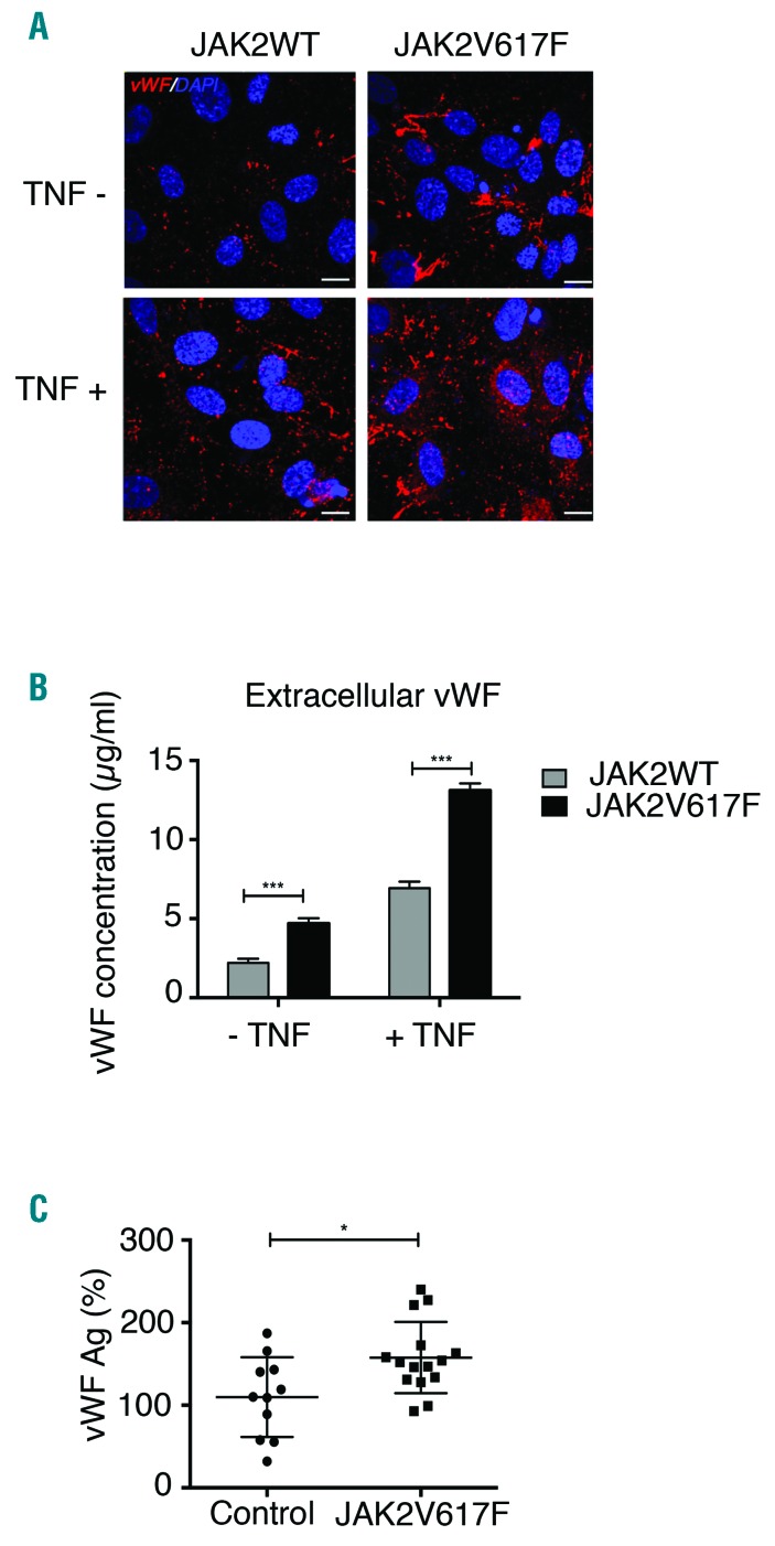
P-selectin is stored within EC in exocytotic organelles called Weibel-Palade bodies together with von Willebrand factor (vWF). Exocytosis of Weibel-Palade bodies leads to cell surface expression of vWF and P-selectin. Since we had observed an increase in endothelial P-selectin expression, we next investigated whether vWF was also increased. In vitro, using immunostaining on non-permeabilized HUVEC, we observed increased vWF expression at the surface of JAK2V617F HUVEC, spontaneously and after overnight activation with 10 ng/mL TNF-α (Figure 5A). Furthermore, quantification of vWF in the conditioned media revealed higher amounts of vWF released by JAK2V617F HUVEC (Figure 5B), a difference that was even greater when the cells had been treated with TNF-α. These results were confirmed in vivo with higher levels of vWF antigen in Pdgfb-iCreERT2;JAK2V617F/WT mice than in control mice (Figure 5C). There was no difference in the distribution of vWF multimers between Pdgfb-iCreERT2;JAK2V617F/WT mice and control animals. These data demonstrate that endothelial JAK2V617F increased the levels of vWF protein and the release of soluble vWF, in association with increased P-selectin expression at the cell surface, as a consequence of increased degranulation of Weibel-Palade bodies.
Figure 5.
von Willebrand factor expression is increased in JAK2V617F-expressing endothelial cells. (A) Cell surface expression of von Willebrand factor (vWF) (red) is higher in JAK2V617F human umbilical vein endothelial cells (HUVEC) (right) than in JAK2WT HUVEC (left). Nuclei are stained by DAPI (blue). Scale bar: 20 mm. (B) JAK2V617F HUVEC secrete more vWF than JAK2WT HUVEC in the absence or presence of tumor necrosis factor (TNF)-alpha. Statistical significance was determined by Mann-Whitney test. Results are mean value ± SEM from three experiments. (C) Plasma level of vWF antigen (Ag) is increased in Pdgfb-iCreERT2;JAK2V617F/WT mice. Results are expressed as mean value ± SEM. Statistical significance was determined by the Student t-test. *P<0.05;***P<0.001.
Increased P-selectin exposure is involved in the pro-adhesive and pro-thrombotic phenotype of JAK2V617F endothelial cells
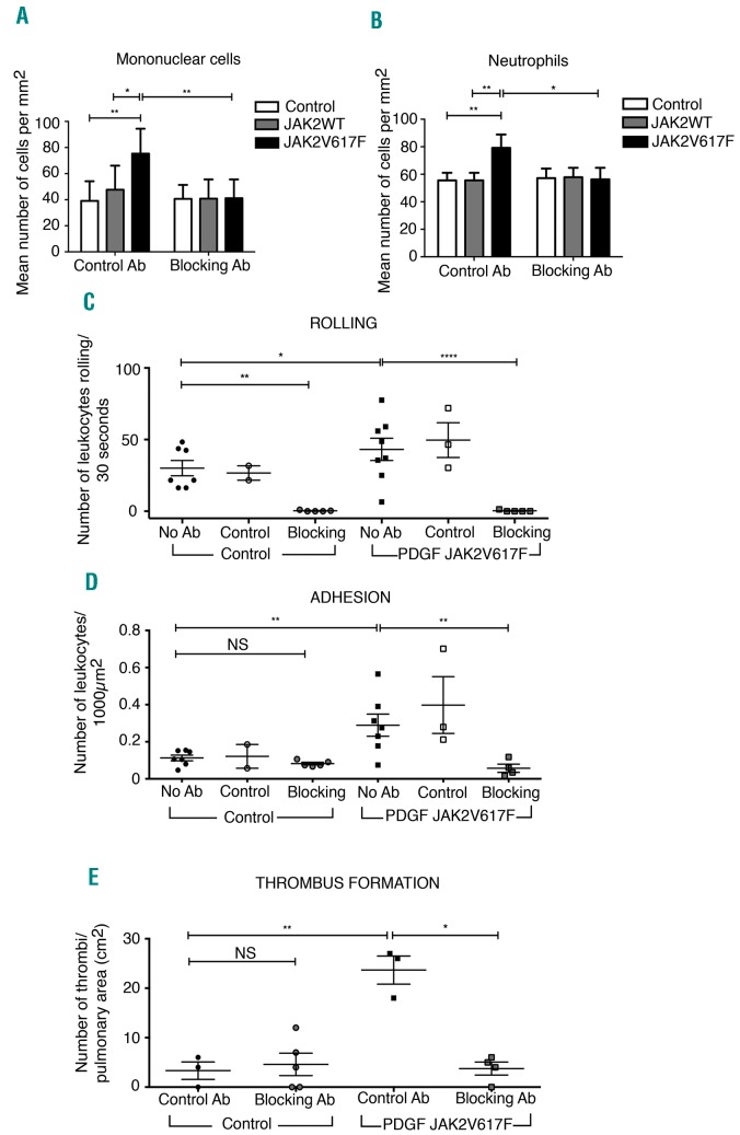
To investigate a potential causal link between increased P-selectin expression at the EC surface and the pro-adhesive phenotype of JAK2V617F EC, we reproduced the same experiments as previously described, but in the presence of a P-selectin blocking antibody. Our in vitro approach with JAK2V617F HUVEC showed a complete reversion of the hyper-adhesive properties of JAK2V617F HUVEC when these cells had been exposed for 30 min to the P-selectin blocking antibody (Figure 6A,B). Quantification of leukocytes in Pdgfb-iCreERT2;JAK2V617F/WT mice treated with TNF-α and the P-selectin blocking antibody revealed a complete inhibition of leukocyte rolling in control mice, which was expected given the well-established role of P-selectin in leukocyte rolling (Figure 6C). In mutant mice, administration of P-selectin blocking antibody completely reversed the pathologically increased leukocyte adhesion (Figure 6C,D). To examine whether increased P-selectin was also responsible for thrombus formation, we used the model of low-dose TNF-α-induced lung thrombus formation (as shown in Figure 1). We observed that pre-treatment of the mice with the P-selectin blocking antibody completely abrogated thrombus formation in Pdgfb-iCreERT2;JAK2V617F/WT mice, but had no effect in control mice (Figure 6E). These results demonstrate that the pro-thrombotic phenotype of JAK2V617F EC is primarily the consequence of increased adhesive properties, due to overex-pression of membrane P-selectin.
Figure 6.
Increased endothelial P-selectin expression is responsible for the pro-adhesive phenotype of JAK2V617F-expressing endothelial cells. (A,B) Under static conditions, increased adhesion of (A) normal mononuclear cells and (B) neutrophils on JAK2V617F human umbilical vein endothelial cells is reversed in the presence of a P-selectin blocking antibody (Ab). Results are mean value ± SEM from three experiments. In Pdgfb-iCreERT2;JAK2V617F/WT mice, increased (C) rolling and (D) adhesion of leukocytes is abolished in the presence of a P-selectin blocking antibody. (E) Increased thrombus formation in Pdgfb-iCreERT2; JAK2V617F/WT mice is abrogated in the presence of a P-selectin blocking antibody. Results are expressed as mean value ± SEM. Statistical significance was determined by two-way analysis of variance and a Sidak post-hoc test. *P<0.05; **P<0.01; ****P<0.0001.
Treatment with hydroxyurea abrogates tumor necrosis factor-induced thrombosis in Pdgfb-iCreERT2;JAK2V617F/WT mice through decreased P-selectin expression
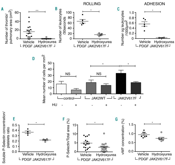
Hydroxyurea is an anti-metabolite frequently used in MPN to reduce the occurrence of thrombosis. Its anti-thrombotic effect is reported to occur via the reduction of blood cell counts. Hydroxyurea is also used in sickle cell disease to reduce vaso-occlusive crises, and its beneficial effect is in part mediated by a direct effect on EC, with a reduction in leukocyte adhesion.30 We hypothesized that hydroxyurea might be capable of reducing the pro-thrombotic effect of JAK2V617F EC. To test this, we treated Pdgfb-iCreERT2;JAK2V617F/WT and control mice with hydroxyurea for 10 days, injected a low dose of TNF-α and quantified thrombus formation in the lungs after 4 h. We observed a significant inhibition of thrombus formation in Pdgfb-iCreERT2;JAK2V617F/WT mice (Figure 7A). We then investigated whether hydroxyurea had modified the adhesiveness of the EC. We observed a reduction in leukocyte rolling and adhesion in mesenteric venules 4 h after TNF-α administration in Pdgfb-iCreERT2;JAK2V617F/WT mice treated with hydroxyurea (Figure 7B,C). To further determine whether this was due to a direct effect on EC, we treated JAK2V617F HUVEC with 100 mM hydroxyurea for 24 h, washed them and analyzed neutrophil adhesion in vitro. Interestingly, there was a significant reduction of neutrophil adhesion on JAK2V617F HUVEC pretreated with hydroxyurea (Figure 7D). Having previously demonstrated that the pro-adhesive phenotype of JAK2V617F EC was mediated by P-selectin, we examined whether the protective effect of hydroxyurea acted through reduction of endothelial P-selectin. As we had previously shown that soluble P-selectin was a reflection of endothelial P-selectin in Pdgfb-iCreERT2;JAK2V617F/WT mice, we measured levels of soluble P-selectin after treatment with hydroxyurea. We observed that the P-selectin:platelet ratio was significantly lower in hydroxyurea-treated Pdgfb-iCreERT2;JAK2V617F/WT mice than in untreated mice (Figure 7E). In these same mice, we used immunostaining to quantify P-selectin expression at the surface of carotid EC. We observed a sig nificant decrease of endothelial membrane P-selectin expression, confirming a direct effect of hydroxyurea on JAK2V617F EC (Figure 7F). This effect is in part secondary to JAK/STAT pathway inhibition, as hydroxyurea administration reduces the level of STAT3 phosphorylation in JAK2V617F HUVEC (Online Supplementary Figure S6). Finally, we observed decreased vWF concentrations in the supernatant of JAK2V617F HUVEC treated with hydroxyurea, indicating a reduced release of Weibel Palade bodies (Figure 7G). Collectively, in vitro and in vivo results demonstrate that hydroxyurea has a direct effect on JAK2V617F EC, decreasing endothelial P-selectin release and surface expression, thus decreasing the pro-thrombotic phenotype of the EC.
Figure 7.
Treatment with hydroxyurea decreases the pro-thrombotic and pro-adhesive phenotype of JAK2V617F-expressing endothelial cells in vitro and in vivo via inhibiting P-selectin and von Willebrand factor expression. (A) Treatment with hydroxyurea for 15 days decreases tumor necrosis factor-induced thrombosis in the lungs of Pdgfb-iCreERT2;JAK2V617F/WT mice. Treatment with hydroxyurea decreases (B) rolling and (C) adhesion of leukocytes on mesenteric venules in Pdgfb-iCreERT2;JAK2V617F/WT mice treated with tumor necrosis factor. Results are expressed as mean value ± SEM. Statistical significance was determined by the Mann-Whitney test. *P<0.05, ***P<0.001. (D) Pre-treatment of JAK2V617F HUVEC with hydroxurea (HU) decreases static adhesion of neutrophils. Results are expressed as mean value ± SEM. Statistical significance determined by two-way ANOVA analysis of variance and the Sidak post-hoc test. (E) Treatment with hydroxyurea for 15 days led to a decrease of the ratio of soluble P-selectin: number of platelets in plasma of Pdgfb-iCreERT2;JAK2V617F/WT mice treated with tumor necrosis factor. (F) Hydroxyurea decreases the expression of P-selectin at the surface of carotid JAK2V617F endothelial cells. Each dot represents one image with four images per mouse (n=4). (G) Treatment of JAK2V617F HUVEC with hydroxyurea decreases secretion of von Willebrand factor (vWF). Results are expressed as mean value ± SEM. Statistical significance was determined by the Mann-Whitney test. *P<0.05.
Discussion
Despite significant advances in deciphering the molecular mechanisms responsible for the occurrence and transformation of MPN, the mechanisms that lead to thrombosis, the main cause of morbidity and mortality, remain largely elusive. Recent identification of the JAK2V617F mutation in EC of patients with MPN13–15 opened new perspectives in the pathogenesis of thrombosis in MPN. Here we demonstrate that JAK2V617F-positive EC promote spontaneous thrombosis under basal conditions and have an increased thrombotic response to weak inflammatory stimuli. We describe that the mechanism that leads to thrombosis involves endothelial P-selectin release and cell surface exposure and subsequent leukocyte rolling and adhesion. We also describe that treatment with hydroxy-urea decreases P-selectin endothelial expression and thrombus formation in mice expressing JAK2V617F only in EC.
We used a mouse model that allows expression of JAK2V617F only in EC to ensure that the phenotype observed in these mice was solely due to the presence of JAK2V617F EC. We are aware that there are differences between our model and patients with MPN, who often have JAK2V617F -positive blood cells together with some JAK2V617F EC. Moreover, not all patients have JAK2V617F EC and, for those that do, it is currently not known where they are located. Our mouse model is not, therefore, representative of the human situation and is not meant to be a model of MPN; however, it does allow precise characterization of the properties of JAK2V617F EC.
The link between P-selectin expression and thrombosis has been described previously, and in most cases P-selectin originates from platelets.28,31,32 The mechanism of P-selectin-mediated thrombosis involves neutrophil activation, either through tissue factor expression and activation of the extrinsic coagulation pathway,28 or through priming for neutrophil extracellular trap formation,33 a process that is now well-recognized to participate in thrombus formation.34 An increase in endothelial P-selectin and subsequent thrombosis were seen in response to venous flow reduction and local hypoxia.28 Here, we show for the first time that EC can have constitutively increased expression of P-selectin, even without any hypoxic or inflammatory stimuli. Further studies are now required to decipher the specific intracellular mechanism responsible for increased P-selectin expression in JAK2V617F-expressing EC. One possible mechanism could be increased STAT3 phosphorylation, as STAT3 activation has been shown to upregulate expression of P-selectin in EC35 (Online Supplementary Figure S7).
Given that MPN are acquired hematologic malignancies, the description of pro-thrombotic JAK2V617F EC raises the question of their origin. JAK2V617F EC have been found in microdissected vessels13,14 and after culture of circulating endothelial progenitors, both in colony-forming unit -endothelial cell (CFU-EC)15,36–38 and endothelial colony-forming cells (E-CFC).15,38 The finding that JAK2V617F CFU-EC are present in all MPN patients was not surprising given that CFU-EC are of hematopoietic origin. However, E-CFC are not of hematopoietic origin and the presence of JAK2V617F EC thus suggests the existence of a progenitor cell of hematopoietic and endothelial lineages. Such cells certainly exist in the embryo but their existence in adults is a matter of debate. In the case of microdissection experiments,13,14 it is possible that the JAK2V617F EC that have been microdissected derive from CFU-EC.39 The presence of JAK2V617F EC of real endothelial origin in MPN is probably rare, but the presence of JAK2V617F EC of hematopoietic origin is common. Demonstrating that JAK2V617F EC have a pro-thrombotic phenotype is thus particularly relevant to our understanding of the pathogenesis of thrombosis in MPN.
Our study has important therapeutic implications. We demonstrate that treatment with hydroxyurea inhibits the pathological hyper-adhesive phenotype of JAK2V617F EC. The results presented here challenge current thinking, according to which the anti-thrombotic effect of hydroxyurea in MPN is only mediated by lowering the blood cell count. However, a direct effect of hydroxyurea on EC has been reported40,41 and is thought to occur via stimulation of the nitric oxide-cyclic GMP pathway in EC, and reduction of leukocyte rolling.30 Moreover, in patients with sickle cell disease, treatment with hydroxyurea efficiently decreases the frequency of vaso-occlusive crises.42 These findings suggest that hydroxyurea should be considered for patients with MPN and a history of thrombosis. This is often the case, as hydroxyurea is the first-line therapy in high-risk patients. However, in young patients, hydroxyurea is usually avoided because of its potential leukemic effect, even if large, retrospective studies have not confirmed this effect.43–45 The antithrombotic and leukemic effects of hydroxyurea should thus be addressed in a prospective cohort of young MPN patients with thrombosis.
Another therapeutic implication of our work comes from the demonstration that increased endothelial P-selectin favors thrombosis in MPN, as was shown in sickle cell disease.46 Very recently, a clinical trial demonstrated that treatment with an anti-P-selectin antibody, crizanlizumab, efficiently prevented pain crises in patients with sickle cell disease.47 It is thus tempting to speculate that such treatment could have therapeutic benefits in MPN patients at high risk of thrombosis, who need to receive anticoagulant treatment for a MPN-related thrombosis (such as splanchnic thrombosis).
Our description of the pro-thrombotic profile of JAK2V617F EC may appear to contradict with the results of Etheridge et al.48 These authors used Tie2-Cre/FF1 mice, which allow constitutive expression of human JAK2V617F in blood and EC. These mice were irradiated then transplanted with the bone marrow of wild-type mice to generate mice in which only the EC express JAK2V617F. Etheridge et al. reported a hemorrhagic diathesis due to acquired von Willebrand disease. There are several possible reasons for the apparently contradictory results. Studies have shown the importance of the JAK2V617F:JAK2WT ratio for the MPN phenotype49 and Etheridge et al. used transplanted mice with a human JAK2V617F:mouse JAK2WT ratio of 1:5 in EC, whereas we used a tamoxifen-inducible mouse model that allows heterozygous expression of mouse JAK2V617F/JAK2WT specifically in EC. Another major difference lies in the models of thrombosis, as Etheridge et al. used models in which the endothelial layer is disrupted whereas we chose three models in which the endothelial barrier is maintained intact. Of note, Etheridge et al. reported an overall increase in the content of vWF in EC, which is in accordance with our results.
Altogether, we believe that our work is clinically relevant for three reasons; (i) it suggests that specific biomarkers of EC activation should be intensively investigated to determine which MPN patients are at high thrombotic risk; (ii) it opens the way to new therapeutic options in MPN, such as hydroxyurea in patients at high risk of thrombosis or with raised levels of markers of endothelial activation, as well as anti-P-selectin antibody instead of or in addition to the standard of care to prevent thrombosis in high-risk MPN patients; and (iii) it provides the proof of concept that an acquired genetic mutation can alter the phenotype of EC, as shown for the pro-thrombotic phenotype acquired upon expression of the JAK2 mutation. This suggests that other activating mutations in EC could be causal in thrombotic disorders of unknown cause, which account for 50% of recurrent venous thromboses.
Supplementary Material
Acknowledgments
The authors wish to thank Myriam Petit and Annabel Reynaud and Beatrice Jaspard-Vinassa for their assistance in histology, Beatrice Jaspard-Vinassa for her help with western blots, Jeremie Teillon for his help in image quantification, Veronique Ollivier for her assistance with in vitro flow experiments, Sylvain Grolleau for his help in mice genotyping, Olivier Mansier for his help with the quantitative polymerase chain reaction, Jerome Guignard for his help with mice and the vectorology platform for lentiviruses production (Inserm U1035). We are grateful to William Vainchenker (Institut Gustave Roussy, Inserm U1009), Pierre-Emmanuel Rautou (Inserm U970) and Jean-François Viallard (Inserm U1034) for their helpful discussions. We are also grateful to the French Intergroup Myeloproliferative (FIM).
Footnotes
Check the online version for the most updated information on this article, online supplements, and information on authorship & disclosures: www.haematologica.org/content/104/1/70
Funding
This study was supported by research grants from ANR-DFG JAKPOT (N° ANR-14-CE35-0022-02), INSERM, The Fondation Bettencourt Schueller and the Aquitaine Region. AG was supported by a research grant from INSERM (Poste Accueil INSERM) and VGL was supported by ANR-DFG JAKPOT.
References
- 1.Arber DA, Orazi A, Hasserjian R, et al. The 2016 revision to the World Health Organization classification of myeloid neoplasms and acute leukemia. Blood. 2016; 127(20):2391–2405. [DOI] [PubMed] [Google Scholar]
- 2.Baxter EJ, Scott LM, Campbell PJ, et al. Acquired mutation of the tyrosine kinase JAK2 in human myeloproliferative disorders. Lancet. 2005;365(9464):1054–1061. [DOI] [PubMed] [Google Scholar]
- 3.James C, Ugo V, Le Couedic JP, et al. A unique clonal JAK2 mutation leading to constitutive signalling causes poly-cythaemia vera. Nature. 2005;434(7037): 1144–1148. [DOI] [PubMed] [Google Scholar]
- 4.Kralovics R, Passamonti F, Buser AS, et al. A gain-of-function mutation of JAK2 in myeloproliferative disorders. N Engl J Med. 2005;352(17):1779–1790. [DOI] [PubMed] [Google Scholar]
- 5.Levine RL, Wadleigh M, Cools J, et al. Activating mutation in the tyrosine kinase JAK2 in polycythemia vera, essential thrombocythemia, and myeloid metaplasia with myelofibrosis. Cancer Cell. 2005;7(4):387–397. [DOI] [PubMed] [Google Scholar]
- 6.Lu X, Huang LJ, Lodish HF. Dimerization by a cytokine receptor is necessary for constitutive activation of JAK2V617F. J Biol Chem. 2008;283(9):5258–5266. [DOI] [PubMed] [Google Scholar]
- 7.Elliott MA, Tefferi A. Pathogenesis and management of bleeding in essential thrombocythemia and polycythemia vera. Curr Hematol Rep. 2004;3(5):344–351. [PubMed] [Google Scholar]
- 8.Barbui T, Finazzi G, Falanga A. Myeloproliferative neoplasms and thrombosis. Blood. 2013;122(13):2176–2184. [DOI] [PubMed] [Google Scholar]
- 9.Falanga A, Marchetti M, Vignoli A, Balducci D, Barbui T. Leukocyte-platelet interaction in patients with essential thrombocythemia and polycythemia vera. Exp Hematol. 2005;33(5):523–530. [DOI] [PubMed] [Google Scholar]
- 10.Barbui T, Carobbio A, Rambaldi A, Finazzi G. Perspectives on thrombosis in essential thrombocythemia and polycythemia vera: is leukocytosis a causative factor? Blood. 2009;114(4):759–763. [DOI] [PMC free article] [PubMed] [Google Scholar]
- 11.Landolfi R, Di Gennaro L, Barbui T, et al. Leukocytosis as a major thrombotic risk factor in patients with polycythemia vera. Blood. 2007;109(6):2446–2452. [DOI] [PubMed] [Google Scholar]
- 12.De Grandis M, Cambot M, Wautier MP, et al. JAK2V617F activates Lu/BCAM-mediated red cell adhesion in polycythemia vera through an EpoR-independent Rap1/Akt pathway. Blood. 2013;121(4):658–665. [DOI] [PubMed] [Google Scholar]
- 13.Rosti V, Villani L, Riboni R, et al. Spleen endothelial cells from patients with myelofibrosis harbor the JAK2V617F mutation. Blood. 2012;121(2):360–368. [DOI] [PubMed] [Google Scholar]
- 14.Sozer S, Fiel MI, Schiano T, Xu M, Mascarenhas J, Hoffman R. The presence of JAK2V617F mutation in the liver endothelial cells of patients with Budd-Chiari syndrome. Blood. 2009;113(21): 5246–5249. [DOI] [PMC free article] [PubMed] [Google Scholar]
- 15.Teofili L, Martini M, Iachininoto MG, et al. Endothelial progenitor cells are clonal and exhibit the JAK2(V617F) mutation in a subset of thrombotic patients with Ph-negative myeloproliferative neoplasms. Blood. 2011; 117(9):2700–2707. [DOI] [PubMed] [Google Scholar]
- 16.Wakefield TW, Myers DD, Henke PK. Mechanisms of venous thrombosis and resolution. Arterioscler Thromb Vasc Biol. 2008;28(3):387–391. [DOI] [PubMed] [Google Scholar]
- 17.Kaneider NC, Forster E, Mosheimer B, Sturn DH, Wiedermann CJ. Syndecan-4-dependent signaling in the inhibition of endotoxin-induced endothelial adherence of neutrophils by antithrombin. Thromb Haemost. 2003;90(6):1150–1157. [DOI] [PubMed] [Google Scholar]
- 18.Burns MP, DePaola N. Flow-conditioned HUVECs support clustered leukocyte adhesion by coexpressing ICAM-1 and E-selectin. Am J Physiol Heart Circ Physiol. 2005;288(1):H194–204. [DOI] [PubMed] [Google Scholar]
- 19.Miszti-Blasius K, Debreceni IB, Felszeghy S, Dezso B, Kappelmayer J. Lack of P-selectin glycoprotein ligand-1 protects mice from thrombosis after collagen/epinephrine challenge. Thromb Res. 2011;127(3):228–234. [DOI] [PubMed] [Google Scholar]
- 20.Severin S, Gratacap MP, Lenain N, et al. Deficiency of Src homology 2 domain-containing inositol 5-phosphatase 1 affects platelet responses and thrombus growth. J Clin Invest. 2007;117(4):944–952. [DOI] [PMC free article] [PubMed] [Google Scholar]
- 21.Hasan S, Lacout C, Marty C, et al. JAK2V617F expression in mice amplifies early hematopoietic cells and gives them a competitive advantage that is hampered by IFNalpha. Blood. 2013;122(8):1464–1477. [DOI] [PubMed] [Google Scholar]
- 22.Claxton S, Kostourou V, Jadeja S, Chambon P, Hodivala-Dilke K, Fruttiger M. Efficient, inducible Cre-recombinase activation in vascular endothelium. Genesis. 2008; 46(2):74–80. [DOI] [PubMed] [Google Scholar]
- 23.Thambyrajah R, Mazan M, Patel R, et al. GFI1 proteins orchestrate the emergence of haematopoietic stem cells through recruitment of LSD1. Nat Cell Biol. 2016;18(1):21–32. [DOI] [PubMed] [Google Scholar]
- 24.Wang L, Benedito R, Bixel MG, et al. Identification of a clonally expanding haematopoietic compartment in bone marrow. EMBO J. 2013;32(2):219–230. [DOI] [PMC free article] [PubMed] [Google Scholar]
- 25.Chauhan AK, Kisucka J, Brill A, Walsh MT, Scheiflinger F, Wagner DD. ADAMTS13: a new link between thrombosis and inflammation. J Exp Med. 2008;205(9):2065–2074. [DOI] [PMC free article] [PubMed] [Google Scholar]
- 26.Wegmann F, Petri B, Khandoga AG, et al. ESAM supports neutrophil extravasation, activation of Rho, and VEGF-induced vascular permeability. J Exp Med. 2006; 203(7):1671–1677. [DOI] [PMC free article] [PubMed] [Google Scholar]
- 27.Yago T, Tsukamoto H, Liu Z, Wang Y, Thompson LF, McEver RP. Multi-inhibitory effects of A2A adenosine receptor signaling on neutrophil adhesion under flow. J Immunol. 2015;195(8):3880–3889. [DOI] [PMC free article] [PubMed] [Google Scholar]
- 28.von Bruhl ML, Stark K, Steinhart A, et al. Monocytes, neutrophils, and platelets cooperate to initiate and propagate venous thrombosis in mice in vivo. J Exp Med. 2012;209(4):819–835. [DOI] [PMC free article] [PubMed] [Google Scholar]
- 29.Ley K, Laudanna C, Cybulsky MI, Nourshargh S. Getting to the site of inflammation: the leukocyte adhesion cascade updated. Nat Rev Immunol. 2007;7(9):678–689. [DOI] [PubMed] [Google Scholar]
- 30.Almeida CB, Scheiermann C, Jang JE, et al. Hydroxyurea and a cGMP-amplifying agent have immediate benefits on acute vaso-occlusive events in sickle cell disease mice. Blood. 2012;120(14):2879–2888. [DOI] [PMC free article] [PubMed] [Google Scholar]
- 31.Andre P, Hartwell D, Hrachovinova I, Saffaripour S, Wagner DD. Pro-coagulant state resulting from high levels of soluble P-selectin in blood. Proc Natl Acad Sci USA. 2000; 97(25):13835–13840. [DOI] [PMC free article] [PubMed] [Google Scholar]
- 32.Zetterberg E, Verrucci M, Martelli F, et al. Abnormal P-selectin localization during megakaryocyte development determines thrombosis in the gata1low model of myelofibrosis. Platelets. 2014;25(7):539–547. [DOI] [PMC free article] [PubMed] [Google Scholar]
- 33.Etulain J, Martinod K, Wong SL, Cifuni SM, Schattner M, Wagner DD. P-selectin promotes neutrophil extracellular trap formation in mice. Blood. 2015;126(2):242–246. [DOI] [PMC free article] [PubMed] [Google Scholar]
- 34.Martinod K, Wagner DD. Thrombosis: tangled up in NETs. Blood. 2014;123(18):2768–2776. [DOI] [PMC free article] [PubMed] [Google Scholar]
- 35.Kim KJ, Kwon SH, Yun JH, et al. STAT3 activation in endothelial cells is important for tumor metastasis via increased cell adhesion molecule expression. Oncogene. 2017;36(39):5445–5459. [DOI] [PubMed] [Google Scholar]
- 36.Helman R, Pereira WO, Marti LC, et al. Granulocyte whole exome sequencing and endothelial JAK2V617F in patients with JAK2V617F positive Budd-Chiari syndrome without myeloproliferative neoplasm. Br J Haematol. 2018;180(3):443–445. [DOI] [PubMed] [Google Scholar]
- 37.Piaggio G, Rosti V, Corselli M, et al. Endothelial colony-forming cells from patients with chronic myeloproliferative disorders lack the disease-specific molecular clonality marker. Blood. 2009;114(14):3127–3130. [DOI] [PubMed] [Google Scholar]
- 38.Yoder MC, Mead LE, Prater D, et al. Redefining endothelial progenitor cells via clonal analysis and hematopoietic stem/progenitor cell principals. Blood. 2007;109(5):1801–1809. [DOI] [PMC free article] [PubMed] [Google Scholar]
- 39.Sozer S, Ishii T, Fiel MI, et al. Human CD34+ cells are capable of generating normal and JAK2V617F positive endothelial like cells in vivo. Blood Cells Mol Dis. 2009;43(3):304–312. [DOI] [PubMed] [Google Scholar]
- 40.Cokic VP, Beleslin-Cokic BB, Tomic M, Stojilkovic SS, Noguchi CT, Schechter AN. Hydroxyurea induces the eNOS-cGMP pathway in endothelial cells. Blood. 2006;108(1):184–191. [DOI] [PubMed] [Google Scholar]
- 41.Cokic VP, Beleslin-Cokic BB, Noguchi CT, Schechter AN. Hydroxyurea increases eNOS protein levels through inhibition of proteasome activity. Nitric Oxide. 2007;16(3):371–378. [DOI] [PubMed] [Google Scholar]
- 42.Charache S, Terrin ML, Moore RD, et al. Effect of hydroxyurea on the frequency of painful crises in sickle cell anemia. Investigators of the multicenter study of hydroxyurea in sickle cell anemia. N Engl J Med. 1995;332(20):1317–1322. [DOI] [PubMed] [Google Scholar]
- 43.Gangat N, Wolanskyj AP, McClure RF, et al. Risk stratification for survival and leukemic transformation in essential thrombocythemia: a single institutional study of 605 patients. Leukemia. 2007;21(2):270–276. [DOI] [PubMed] [Google Scholar]
- 44.Finazzi G, Caruso V, Marchioli R, et al. Acute leukemia in polycythemia vera: an analysis of 1638 patients enrolled in a prospective observational study. Blood. 2005;105(7):2664–2670. [DOI] [PubMed] [Google Scholar]
- 45.Tefferi A, Rumi E, Finazzi G, et al. Survival and prognosis among 1545 patients with contemporary polycythemia vera: an international study. Leukemia. 2013;27(9):1874–1881. [DOI] [PMC free article] [PubMed] [Google Scholar]
- 46.Matsui NM, Borsig L, Rosen SD, Yaghmai M, Varki A, Embury SH. P-selectin mediates the adhesion of sickle erythrocytes to the endothelium. Blood. 2001;98(6):1955–1962. [DOI] [PubMed] [Google Scholar]
- 47.Ataga KI, Kutlar A, Kanter J, et al. Crizanlizumab for the prevention of pain crises in sickle cell disease. N Engl J Med. 2017;376(5):429–439. [DOI] [PMC free article] [PubMed] [Google Scholar]
- 48.Etheridge SL, Roh ME, Cosgrove ME, et al. JAK2V617F-positive endothelial cells contribute to clotting abnormalities in myelo-proliferative neoplasms. Proc Natl Acad Sci USA. 2014;111(6):2295–2300. [DOI] [PMC free article] [PubMed] [Google Scholar]
- 49.Tiedt R, Hao-Shen H, Sobas MA, et al. Ratio of mutant JAK2-V617F to wild-type Jak2 determines the MPD phenotypes in transgenic mice. Blood. 2008;111(8):3931–3940. [DOI] [PubMed] [Google Scholar]
Associated Data
This section collects any data citations, data availability statements, or supplementary materials included in this article.


