Figure 1.
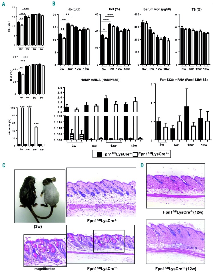
Transient alopecia and anemia are present in Fpn1fl/flLysCre+/− mice. (A) Hemoglobin (Hb) levels and hematocrit (Hct) in 3- to 6-week (w) old mice (mean ± SEM of 50 mice for each group; ***P<0.0001, **P<0.001). The histogram at the bottom shows the percentage alopecia at different time-points (mean ± SEM of 50 mice for each group; ***P<0.0001, **P<0.001 versus Fpn1fl/flLysCre−/−). (B) Top: Hb levels, Hct, serum iron and transferrin saturation (TS) in 3-, 6-, 12-, and 18-week old mice (mean ± SEM of 50 mice for each group; ***P<0.0001, **P<0.001, *P<0.01). Bottom: hepcidin (HAMP) expression in the liver (solid bars) and skin (striped bars) and spleen erythroferrone (Fam132b) mRNA levels of 3-, 6-, 12-, and 18-week old mice. mRNA levels were measured by quantitative real time polymerase chain reaction and normalized to the housekeeping gene 18S RNA. Data are presented as mean ± SEM of 10 mice for each group. (C) Representative appearance of 3-week old Fpn1fl/flLysCre−/− (left) and Fpn1fl/flLysCre+/− (right) mice and representative histology (dorsal area) of the same mice. Magnification 10X, in the inset 20X. (D) Representative histology of the skin (dorsal area) of adult (12-week old) mice. Tissue sections were stained with hematoxylin and eosin. Magnification: 10X.

