Abstract
The direct anterior approach to the hip for total joint arthroplasty has been suggested to have several advantages compared to other popular approaches through its use of a natural intramuscular and intra-nervous interval. Recent emphasis on tissue sparing and minimally invasive outpatient joint replacements has given rise to a significant increase in the utilization of direct anterior total hip arthroplasty (DAA). Proponents of this approach cite improved recovery times, lower pain levels, improved patient satisfaction as well as improved accuracy on both implant placement/alignment and leg length restoration. A number of variations of the procedure have been described and many authors have published their experiences and technical keys to successfully accomplishing this procedure. Described techniques have been performed using specifically designed instruments and specific fracture tables and intra-operative flouroscopy, however this approach may be performed using a regular table with standard arthroplasty tools with alternative patient positioning and without intraoperative imaging. This review summarizes several aspects of the direct anterior approach for total hip arthroplasty and its comparison to other popular approaches to modern hip replacement.
Introduction
There are several approaches to the hip joint that can be utilized for total hip replacement and these include with some variations the posterior approach (Moore or Southern), the lateral approach (Hardinge), the anterolateral approach (Watson Jones), and the direct anterior approach (Smith-Peterson). Although more recently popularized for hip arthroplasty, the direct anterior approach to the hip was originally described by Carl Heuter in 1881. Smith-Peterson is frequently credited with popularizing this technique through prolific use of this technique during his career after publishing his first description of the approach in 1917.1 The anterior-based incision utilizes the interval to the hip joint between the tensor fascia lata and the sartorius muscles. Light and Keggi published their experience using this approach for hip arthroplasty in 1980, and Judet described the procedure with the use of a fracture table in 1985.2 Recent desire to perform hip reconstruction through less invasive and tissue sparing as well as in an outpatient setting have been key factors in the newfound interest of this approach. This has led to a surge in its use for hip replacement over the last 10–15 years. During this time, many authors have described variations of the technique and important pearls to safe and successful performance of anterior hip arthroplasty.3 Although not as common, several have noted routine use of this technique for revision total arthroplasty as well as hemiarthroplasty for fractures.4
The direct anterior approach (DAA) can be used for patients of nearly all body habitus and hip conditions. Some anatomic features of the native hip and pelvis are recognized to make a direct anterior approach more difficult. Acetabular protrusio brings the femoral canal closer to the pelvis and can limit the access to the femur. Neck shaft angle with decreased offset positions the femoral canal deeper in the thigh, and factors associated with obese muscular males can limit the exposure. A potential disadvantage of the anterior approach is diminished access to the posterior column. If a patient has retained posterior acetabular hardware or posterior wall deficiency with augmentation contemplated, the anterior exposure might prove unsuitable.5
Surgical Technique
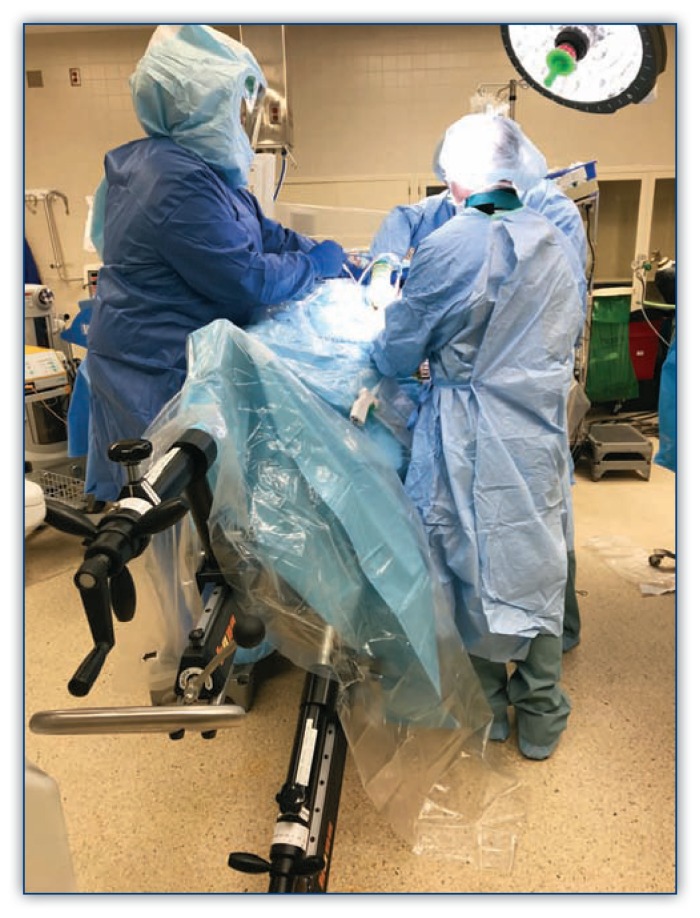
The vast majority of authors describing direct anterior approach position the patient supine on a fracture table or regular table. Michel et al. also reported performing anterior total hip arthroplasty in lateral decubitus position.6 When using a regular operating room table, the patient is positioned with the hip located over the table break which can be reflexed to allow hyperextension of the hip joint. The contralateral leg is typically draped into the field and a mayo stand placed alongside to allow for figure-four adduction during the femoral exposure. The author’s preferred method includes the use of a HANA or fracture table and a shower curtain laid across the patient or other draping system can be utilized. (Figure 1.)
Figure 1.
Surgical table set up with author’s preferred draping technique on HANA table. Operative leg shown extended and adducted.
Obese patients should have the pannus retracted with adhesive tape to avoid any possible interference with the exposure. Incisions vary by surgeon, however most authors rely on the ASIS and greater trochanter as anatomic landmarks for reference. An oblique incision is made originating 2–4 cm distal and lateral to the ASIS to a point a few fingerbreadths anterior to the greater trochanter. Dissection is taken deep to expose the overlying fascia of the tensor fascia lata (TFL) which is then incised along its fibers. Blunt finger dissection is utilized under the medial fascia and the interval is developed between the sartorius and the TFL. A wound protector can help minimize soft tissue, muscle, and skin damage caused by retraction.
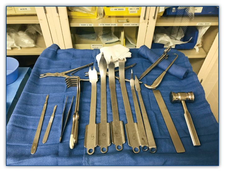
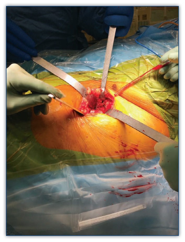
The ascending branch of the lateral circumflex femoral artery and vein are identified over the intertrochanteric line and cauterized and the anterior hip capsule is exposed. Specific DAA retractors are useful for exposure. (Figure 2) A cobra type retractor is placed superior to the lateral capsule to retract the abductors and a second large Hohmann type retractor is placed inferior to the femoral neck. A curved third retractor maybe useful proximally to elevate the rectus tendon, especially in heavier patients. The anterior capsule can be partially excised, fully excised, or incised and tagged for retention and later repair. The femoral neck osteotomy can then be made either with a single cut or some prefer a napkin ring type parallel two cut technique to facilitate removal of the femoral head. An osteotome can be used to complete the cuts (Figure 3). Flouroscopy at this point can be useful to confirm the accuracy of the planned cuts. A corkscrew inserted through the cortical side of the femoral head or through the femoral neck cut will aid to spin the head and rupture the ligamentum flavum and assist with head removal.
Figure 2.
Direct anterior approach specific retractors and instruments which are used to facilitate the approach.
Figure 3.
Hip exposure with retractors and osteotome for femoral neck cut completion in minimally invasive direct anterior approach (DAA).
Excellent exposure and visualization of the acetabulum can be achieved with the use of two or three retractors. A Mueller type retractor can be placed inferiorly on the posterior border of the acetabulum. Downward pressure on this retractor will bring the femur posterior. One or two retractors can be placed along the anterior acetabulum. Rim osteophytes and labrum are removed as well as the ligamentum flavum stump and soft tissue. An inferior capsular release may be useful for exposure. Reaming proceeds as in other approaches medially at first to reach the true floor then to anatomic position. Flouroscopy can be used to check the size and as well as inclination and version of the reaming and acetabular impaction seating. Screw fixation of the acetabular component may be added pending surgeon preference followed by liner impaction.
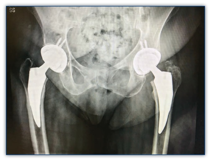
Femoral exposure on a HANA/fracture table is initiated then by external rotation of the femur typically over 90 degrees. A proximal femoral hook is placed for elevating the proximal femur to facilitate exposure. The leg is extended by lowering the leg to the floor followed by adduction of the extremity. For DAA arthroplasty on a standard table femoral exposure is accomplished by reflexing the table to extend the extremity and placing the leg figure-four under the opposite leg and knee. A cobra type retractor is placed on the femur medial to the neck cut and a double-pronged retractor is placed under the greater trochanter. Soft tissue releases are performed in the piriformis fossa and greater trochanter along while elevating the proximal femur with the hook device to visualize the osteotomy plane and allow for broaching and placement of a femoral trial component. A key advantage of DAA arthroplasty is the ability to evaluate leg length at this point in the procedure prior to final implant placement. The image of the trial components can compared to the preop plan, a preop image and/or the contralateral hip to confirm alignment, version, fit and leg length. (Figure 4)
Figure 4.
Radiographic image showing comparison of hips for leg length, fit and alignment.
Closure of the wound is initiated with repair of the capsule, if preserved, followed by closure of the TFL fascia with either running or interrupted suture. The use of a drain is optional. The subcutaneous tissue is closed with resorbable suture and skin is closed with technique of choice but glue technique with a long term adhesive dressing continued for two weeks may be useful in patients with a large pannus which can overly the incision.
Postoperative care is frequently initiated with the use of an abduction pillow along with anterior hip precaution instruction. Immediate weight bearing is allowed and the patient can be discharged when the patient is safely ambulating and can manage stairs. This is frequently within 23 hours and can occur on the same day of surgery as a true outpatient procedure.
Discussion
As with any surgical procedure there is a significant learning curve associated with the adoption of the anterior approach to the hip for arthroplasty. Outcomes related to anterior hip arthroplasty have been described in a number of studies, though the majority are retrospective with smaller samples sizes. A meta-analysis comparing the anterior and posterior approaches showed the anterior approach (DAA) may have potential advantages in patient reported pain and functional outcomes, post-operative length of stay, dislocations and postoperative narcotic requirements. It additionally noted that the DAA trended to higher rates of patients discharged to home versus a skilled facility and a larger percentage of acetabular cups placed within the “safe” zone of alignment due to the use of fluoroscopy.6
One retrospective study compared 41 DAA and 47 posterior approaches and found shorter hospital stay and fewer days to mobilization with the DAA. Incision length was shorter in the DAA, however lateral femoral cutaneous nerve injury and femoral fractures were more common and operative time was 20% longer.7 Another report compared 419 patients receiving either a lateral approach or DAA and found similar operative times and blood loss, but less pain, shorter time to discharge and greater percentage of patients discharged directly home with the DAA.8
Another aspect of comparison in approaches to the hip involves functional capacity in the early and long term postoperative period. One prospective, randomized single surgeon study compared 43 DAA to 44 posterior approaches with endpoint analysis of normal ability to climb stairs and walk unlimited distances. The study revealed the DAA cohort performed better in the immediate post-op period with lower VAS pain scores on post-op day one, more patients climbing stairs and walking unlimited distances at six weeks and higher HOOS Symptom scores at three months. However, there were no significant differences at later time end points.9 Other studies have found similar results that most advantages in functional outcome with DAA may be largely limited to the early postoperative recovery period of six to twelve weeks.10–14
A recent prospective, randomized Mayo clinic study compared 101 patients who were randomized to four surgeons. Two specialists were experts in DAA and the other two surgeons were experts on the “mini” posterior approach (MPA). The study documented quicker recovery by DAA subjects compared to the MPA patients. Specifically, advantages included discontinuing use of a walker (10 days post-op versus 14.5 days), discontinuing all walking aids (17.3 days versus 23.6 days), discontinuing narcotics (9.1 versus 14 days), ascending stairs with gait aid (5.4 days versus 10.3 days), and walking six blocks (20 days versus 26 days). There was no difference in activity levels between the groups pre-op, and at two months and one year post op.15
Many studies have examined the relative complication rates associated with the DAA compared to other approaches to the hip. Some have noted as with all surgical procedures that complication rates correlate with surgeon experience and significantly drop after the first 40–100 cases.16,17 Femoral fractures have been found to be of high incidence in initial experience with DAA likely due to excessive force for elevating the femur into the wound and poor exposure limiting access to the femur resulting in poor orientation for broaching and implant placement. Dislocation rates have been shown to be significantly lower with the DAA and several authors do not restrict patients with anterior hip precautions or abduction pillows postoperatively.19–22 Lateral femoral cutaneous nerve injury and wound complications in some studies have been shown to be slightly higher with the DAA likely due to excessive traction on the soft tissues for exposure as well as the effect of a pannus resting on the incision in larger patients. Ring tissue protectors and extended sterile dressings have been suggested to improve this complication.23–24
Conclusion
Most standard approaches to hip arthroplasty have been shown to be safe and effective in the management of arthritis and with proper training and meticulous technique can be successful with exceptional patient satisfaction. There remains a significant learning curve associated with the DAA approach due to its recent popularization and lack of significant experience with this technique in residency training programs until the last decade. The growing emphasis for minimally invasive arthroplasty and improved and expedited functional results make this approach an attractive choice. As hospital associated costs and consumer driven healthcare become more important in the practice of total joint arthroplasty, more surgeons will continue examine surgical approaches/techniques along with other factors in order to drive improved patient outcomes.
Footnotes
Gregory R. Galakatos, MD, MSMA member since 1997, is the Missouri Medicine Editorial Board member for Orthopaedic Surgery. He practices at the Mercy Clinic in Orthopaedic Surgery in St. Louis, Missouri.
Contact: Gregory.Galakatos@Mercy.Net
Disclosure
None reported.
References
- 1.Rachbauer F, Kain MS, Leunig M. The History of the Anterior Approach to the Hip. Orthop Clin North Am. 2009;40:311–320. doi: 10.1016/j.ocl.2009.02.007. [DOI] [PubMed] [Google Scholar]
- 2.Light TR, Keggi KJ. Anterior Approach to Hip Arthroplasty. Clin Orthop Relat Res. 1980;(152):255–260. [PubMed] [Google Scholar]
- 3.Post ZD. Direct Anterior Approach for Total Hip Arthroplasty: Indications, Technique, and Results. J Am Acad Orthop Surg. 2014;22:595–603. doi: 10.5435/JAAOS-22-09-595. [DOI] [PubMed] [Google Scholar]
- 4.Mast NH, Laude F. Revision Total Hip Arthroplasty Performed Through the Hueter Interval. J Bone Joint Surg Am. 2011;93(Suppl 2):143–148. doi: 10.2106/JBJS.J.01736. [DOI] [PubMed] [Google Scholar]
- 5.Berend KR, Lombardi AV, Seng BE, Adams JB. Enhanced early outcomes with the anterior supine intermuscular approach in primary total hip arthroplasty. J Bone Joint Surg Am. 2009;91(Suppl 6):107–120. doi: 10.2106/JBJS.I.00525. [DOI] [PubMed] [Google Scholar]
- 6.Higgins BT, Barlow DR, Heagerty NE, Lin TJ. Anterior vs. posterior approach for total hip arthroplasty, a systematic review and meta-analysis. J Arthroplasty. 2015;30:419–434. doi: 10.1016/j.arth.2014.10.020. [DOI] [PubMed] [Google Scholar]
- 7.Martin CT, Pugely AJ, Gao Y, Clark CR. A Comparison of Hospital Length of Stay and Short-Term Morbidity Between the Anterior and the Posterior Approaches to Total Hip Arthroplasty. J Arthroplasty. 2013;28:849–854. doi: 10.1016/j.arth.2012.10.029. [DOI] [PubMed] [Google Scholar]
- 8.Alecci V, Valente M, Crucil M, Minerva M, Pellegrino CM, Sabbadini DD. Comparison of Primary Total Hip Replacements Performed with a Direct Anterior Approach versus the Standard Lateral Approach: Perioperative Findings. J Orthop Traumatol. 2011;12:123–129. doi: 10.1007/s10195-011-0144-0. [DOI] [PMC free article] [PubMed] [Google Scholar]
- 9.Barrett WP, Turner SE, Leopold JP. Prospective Randomized Study of Direct Anterior vs Postero-lateral Approach for Total Hip Arthroplasty. J Arthroplasty. 2013;28:1634–1638. doi: 10.1016/j.arth.2013.01.034. [DOI] [PubMed] [Google Scholar]
- 10.Lamontagne M, Varin D, Beaulé PE. Does the anterior approach for total hip arthroplasty better restore stair climbing gait mechanics? J Orthop Res. 2011;29:1412–1417. doi: 10.1002/jor.21392. [DOI] [PubMed] [Google Scholar]
- 11.Mayr E, Nogler M, Benedetti MG, Kessler O, Reinthaler A, Krismer M, Leardini A. A prospective randomized assessment of earlier functional recovery in THA patients treated by minimally invasive direct anterior approach: a gait analysis study. Clin Biomech. 2009;24:812–818. doi: 10.1016/j.clinbiomech.2009.07.010. [DOI] [PubMed] [Google Scholar]
- 12.Rathod PA, Orishimo KF, Kremenic IJ, Deshmukh AJ, Rodriguez JA. Similar improvement in gait parameters following direct anterior & posterior approach total hip arthroplasty. J Arthroplasty. 2014;29:1261–1264. doi: 10.1016/j.arth.2013.11.021. [DOI] [PubMed] [Google Scholar]
- 13.Klausmeier V, Lugade V, Jewett BA, Collis DK, Chou LS. Is there faster recovery with an anterior or anterolateral THA? A pilot study. Clin Orthop Relat Res. 2010;468:533–541. doi: 10.1007/s11999-009-1075-4. [DOI] [PMC free article] [PubMed] [Google Scholar]
- 14.Restrepo C, Parvizi J, Pour AE, Hozack WJ. Prospective randomized study of two surgical approaches for total hip arthroplasty. J Arthroplasty. 2010;25:671–679.e1. doi: 10.1016/j.arth.2010.02.002. [DOI] [PubMed] [Google Scholar]
- 15.Taunton MJ. Direct Anterior Hip Arthroplasty. Mayo Clin Surg Update. 2017;11(2):2–3. [Google Scholar]
- 16.Moskal JT, Capps SG, Scanelli JA. Anterior muscle sparing approach for total hip arthroplasty. World J Orthop. 2013;4:12–18. doi: 10.5312/wjo.v4.i1.12. [DOI] [PMC free article] [PubMed] [Google Scholar]
- 17.Alexandrov T, Ahlmann ER, Menendez LR. Early clinical and radiographic results of minimally invasive anterior approach hip arthroplasty. Adv Orthop. 2014;2014:954208. doi: 10.1155/2014/954208. [DOI] [PMC free article] [PubMed] [Google Scholar]
- 18.Jewett BA, Collis DK. High complication rate with anterior total hip arthroplasties on a fracture table. Clin Orthop Relat Res. 2011;469:503–507. doi: 10.1007/s11999-010-1568-1. [DOI] [PMC free article] [PubMed] [Google Scholar]
- 19.Barton C, Kim PR. Complications of the direct anterior approach for total hip arthroplasty. Orthop Clin North Am. 2009;40:371–375. doi: 10.1016/j.ocl.2009.04.004. [DOI] [PubMed] [Google Scholar]
- 20.Siguier T, Siguier M, Brumpt B. Mini-incision anterior approach does not increase dislocation rate: a study of 1037 total hip replacements. Clin Orthop Relat Res. 2004;426:164–173. doi: 10.1097/01.blo.0000136651.21191.9f. [DOI] [PubMed] [Google Scholar]
- 21.Sheth D, Cafri G, Inacio MC, Paxton EW, Namba RS. Anterior and Anterolateral Approaches for THA Are Associated With Lower Dislocation Risk Without Higher Revision Risk. Clin Orthop Relat Res. 2015;473:3401–3408. doi: 10.1007/s11999-015-4230-0. [DOI] [PMC free article] [PubMed] [Google Scholar]
- 22.Sariali E, Leonard P, Mamoudy P. Dislocation after total hip arthroplasty using Hueter anterior approach. J Arthroplasty. 2008;23:266–272. doi: 10.1016/j.arth.2007.04.003. [DOI] [PubMed] [Google Scholar]
- 23.Christensen CP, Karthikeyan T, Jacobs CA. Greater prevalence of wound complications requiring reoperation with direct anterior approach total hip arthroplasty. J Arthroplasty. 2014;29:1839–1841. doi: 10.1016/j.arth.2014.04.036. [DOI] [PubMed] [Google Scholar]
- 24.Alvarez-Pinzon AM, Mutnal A, Suarez JC, Jack M, Friedman D, Barsoum WK, Patel PD. Evaluation of wound healing after direct anterior total hip arthroplasty with use of a novel retraction device. Am J Orthop. 2015;44:E17–E24. [PubMed] [Google Scholar]


