Abstract
Rhizophagus irregularis, an arbuscular mycorrhizal fungus, and Bacillus amyloliquefaciens, a bacterium, are microorganisms that promote plant growth. They associate with plant roots and facilitate nutrient absorption by their hosts, increase resistance against pathogens and pests, and regulate plant growth through phytohormones. In this study, eight local plant species in Finland (Antennaria dioica, Campanula rotundifolia, Fragaria vesca, Geranium sanguineum, Lotus corniculatus, Thymus serpyllum, Trifolium repens, and Viola tricolor) were inoculated with R. irregularis and/or B. amyloliquefaciens in autoclaved substrates to evaluate the plant growth−promoting effects of different plant/microbe combinations under controlled conditions. The eight plant species were inoculated with R. irregularis, B. amyloliquefaciens, or both microbes or were not inoculated as a control. The impact of the microbes on the plants was evaluated by measuring dry shoot weight, colonization rate by the arbuscular mycorrhizal fungus, bacterial population density, and chlorophyll fluorescence using a plant phenotyping facility. Under dual inoculation conditions, B. amyloliquefaciens acted as a “mycorrhiza helper bacterium” to facilitate arbuscular mycorrhizal fungus colonization in all tested plants. In contrast, R. irregularis did not demonstrate reciprocal facilitation of the population density of B. amyloliquefaciens. Dual inoculation with B. amyloliquefaciens and R. irregularis resulted in the greatest increase in shoot weight and photosynthetic efficiency in T. repens and F. vesca.
Introduction
Countless microorganisms reside and propagate in the rhizosphere where plant roots and soil meet. Some of the microbes have neutral or lethal effects on the growth and survival of plants, whereas others support their host plants via various mechanisms [1]. Because of their plant growth−promoting attributes, some of the microorganisms are used as commercial soil additives. Rhizophagus irregularis (Schenck and Smith) (formerly Glomus intraradices) and Bacillus amyloliquefaciens (Fukumoto) are among the most commonly applied plant growth−promoting microorganisms.
R. irregularis is an arbuscular mycorrhizal fungus (AMF) that is found in nearly all soil ecosystems and types [2,3]. It colonizes plants by forming intraradical hyphae and arbuscules as well as vesicles inside roots. Arbuscules are tree-like structures that function as carbohydrate/mineral/lipids exchange systems between host plants and the fungus. Vesicles are oval structures that act as nutrient reservoirs and, in some cases, as propagules [4–7]. When the arbuscules and vesicles appear, the hyphae function as nutrient transportation ducts [2]. R. irregularis facilitates plant growth by direct and indirect means, such as nutrient uptake and transportation [8,9], water absorption [10], salinity resistance [11,12], heavy-metal detoxification [13,14], and pathogen resistance [15,16].
B. amyloliquefaciens is a Gram-positive, spore-forming bacterium closely related to Bacillus subtilis [17]. It was given its name because it produces α-amylase and protease [18]. B. amyloliquefaciens is attracted by root exudates and lives on root surfaces. Successful colonization and effective plant growth promotion occur when a layer of bacterial cells, known as a biofilm, is formed on the surface of seeds, roots, or root hairs, as it prevents competition by other microorganisms [19–21]. Similar to R. irregularis, B. amyloliquefaciens facilitates the growth of its host plant in several ways: salt tolerance [22], drought tolerance [23], nutrient uptake [24,25], and pathogen resistance [26–28].
The use of plant growth−promoting microorganisms has increased at a rate of 10% annually in crop production worldwide over the last decade [29]. In particular, they have the potential to be used in green roofs (rooftops covered with vegetation), which are especially desirable in cities where they can provide multiple ecosystem services to urban residents, including mitigating air pollution, relieving the urban heat island effect, saving energy, and retaining stormwater [30–35]. Green roof applications are, however, limited by three major challenges: extreme weather conditions, the choice of suitable plants, and high installation and maintenance costs [36,37]. McGuire et al. [38] found that green roof soils support microbial communities that are distinct from city park soils and suggested this may be due to differences in soil depth, plant species, proximity to parks, and the conditions on the roofs. Studies have thus been conducted on green roofs to manipulate soil microbial communities [39], study AMF colonization patterns [40], and improve soil nutritional status by using AMF inoculum [41]. Nevertheless, more attention should be paid to microbe-plant interactions under green roof conditions to understand how the microbial community functions and whether microbial manipulation benefits green roof applications.
In 2012, a green roof experiment was conducted in Vantaa, Finland, led by the Fifth Dimension Green Roof Research Group. R. irregularis and B. amyloliquefaciens were added separately to the green roof plots to study microbial survival and growth. The preliminary findings suggested that the density of B. amyloliquefaciens in the rhizosphere was likely to be enhanced by R. irregularis co-inoculated in the soil. Interactions between AMF and bacteria have been reported in the literature, and these interactions can be commensalistic as well as amensalistic [42]. Mansfeld-Giese et al. [43] found that the presence of R. irregularis either promoted or suppressed the population density for 14 bacterial species. In another study using Medicago sativa (L.) as a host plant, co-inoculation with Glomus deserticola (Trappe, Bloss & Menge) and Bacillus pumilus (Meyer & Gottheil) increased shoot biomass and root length compared with inoculation with either of the two microbes alone [44]. Toro et al. [45] concluded that co-inoculation with B. subtilis and R. irregularis significantly increased biomass and nitrogen/phosphorous (N/P) accumulation in tissues of onion plants. If synergy between R. irregularis and B. amyloliquefaciens could be achieved in plants suitable to be grown on green roofs, it would optimize ecosystem services provided by green roofs to urban areas via plant growth enhancement.
In this study, we determined whether synergistic interactions occur between R. irregularis and B. amyloliquefaciens by setting up greenhouse experiments under controlled conditions using eight local plant species from Finland as hosts (Antennaria dioica, Campanula rotundifolia, Fragaria vesca, Geranium sanguineum, Lotus corniculatus, Thymus serpyllum, Trifolium repens, and Viola tricolor). A few of these plants, such as T. serpyllum, T. repens, and L. croniculatus [46–48], are also stress resistant and hence potentially more suitable for green roofs. Four treatments were conducted for each plant species: inoculation with (i) R. irregularis, (ii) B. amyloliquefaciens, (iii) co-inoculation with both microbes, and (iv) no microbial amendment (control). The aim was to find out which plant-microbe combinations promote the physiological performance of plants. Shoot biomass and the photosynthetic efficiency were chosen as indicators of physiological performance, as they directly reflect plant physiology and are relatively easy to measure.
Materials and methods
Experimentation
Experiments were conducted in the Small Plant Unit of the National Plant Phenotyping Infrastructure (NaPPI) on the Viikki campus, University of Helsinki, Finland [49] (https://www.helsinki.fi/en/infrastructures/national-plant-phenotyping). NaPPI is an in-house facility that conducts high-throughput and high-precision plant phenotyping. It consists of programmable tools for weighing, watering, and imaging the plants—both for red-green-blue pictures (RGB Camera) and chlorophyll fluorescence (Fluorecam). These tools allow comprehensive analysis of plant growth and physiology. Light-emitting diode lamps were used to avoid heating the plants by illumination.
Seed of the plant species were purchased from Suomen Niittysiemen (Jyväskylä, Finland) and stored at 4°C for 2 weeks before sowing. The inoculant products MYC4000 (4000 fungal spores of R. irregularis strain DAOM 181602 per gram) and Rhizocell (>109 CFU endospores of B. amyloliquefaciens strain FZB42 per gram) were provided by Lallemand Plant Care (Castelmaurou, France). MYC4000 and Rhizocell are powdered products. They were dissolved in distilled water according to the manufacturer’s instructions before being sprayed onto the growth substrate.
Plant seeds were sown in autoclaved sand for germination. After 4 weeks, seedlings were individually transplanted into plastic pots (8 × 8 × 8 cm; VWR, Center Valley, PA, USA) filled with 450 cm3 autoclaved soil from a conifer forest (provided by Kekkilä Oy, Vantaa, Finland), a soil volume that is typically used for rooting plants [50–53]. The small plant unit at NaPPI is designed to fit only this particular pot size, and it is suitable for plants up to 50 cm in height. The substrates used in this study were low in nutrients, and no fertilizer was applied to keep the seedlings smaller and to more readily observe the effects of the inoculants on plant growth [52,54,55]. A shallow soil depth was used, as is typical for green roofs. The soil properties were pH 6.4; organic matter, 5.6%; soluble P, 2.2 mg/kg; and soluble N, 0.4 mg/kg. For each plant species, 24 seedlings were divided into four treatments: six seedlings were inoculated with R. irregularis (R), six seedlings with B. amyloliquefaciens (B), six seedlings were co-inoculated with both microbes (R+B), and six seedlings remained as uninoculated controls. Altogether, 192 transplanted seedlings were maintained in the NaPPI facility for 7 weeks, during which the day/night length was set to 16 h/8 h. Light intensity was 172 μmol m−2 s−1. Seedlings were irrigated daily to maintain a water-holding capacity of 65% [56]. Two independent, repeated NaPPI experiments were conducted in July 2015 and June 2016. During the first experiment, the NaPPI watering program was interrupted a few times.
Before sampling, each plant was measured for chlorophyll fluorescence activity with the Fluorecam in the NaPPI to determine the photosystem II photochemical capacity. This was calculated from the ratio of the variable fluorescence to the maximum chlorophyll fluorescence (Fv/Fm), which reflects the photosynthetic efficiency of tested plants [57]. The optimal value is 0.83 for most plant species, and a value lower than that indicates poor plant growth [58]. Plant shoots (including all aboveground biomass such as branches, leaves, and stems) were cut, desiccated at 70°C in an oven for 48 h, and weighed to measure their dry biomass. Soil adhering to roots was collected from six different places in each pot and transferred to screw cap tubes. Each soil sample was mixed by vigorous shaking and stored at 4°C. Roots were carefully brushed and stored in 70% ethanol at 4°C.
Detection of R. irregularis in root samples
The classical AMF detection method includes root staining, microscope slide preparation, and AMF quantification [59]. After 7 weeks of cultivation in NaPPI, a root sample was collected from each of three random plants for each species and treatment. Roots were washed and stored in 70% ethanol until staining with Trypan Blue. The staining protocol was adjusted for each plant species [60] (Table 1). In brief, the roots were first soaked and softened in KOH solution, which made the staining process more effective. Then the roots were immersed in 1.5% hydrogen peroxide containing 5 ml/l ammonia (H2O2+NH3) to remove the background color of the cells. Next, the roots were transferred into 1% HCl solution and held in Trypan Blue solution before being stored in clear glycerol (Table 1). Finally, the roots were mounted on microscope slides in polyvinyl-lacto-glycerol solution (10 ml/l water, 10 ml/l lactic acid, 1 ml/l glycerol, and 1.66 mg/l polyvinyl alcohol).
Table 1. Detailed staining protocol for eight plant species.
| Staining solutions | ||||
|---|---|---|---|---|
| Plant species | KOH | H2O2+NH31 | 1% HCl | Trypan Blue2 |
| C. rotundifolia | 60 min in 2.5% KOH at 80°C | 40 min | 30 min | 60 min at 80°C |
| L. corniculatus | 60 min in 2.5% KOH at 80°C | None | 30 min | 45 min at 95°C |
| T. repens | 60 min in 2.5% KOH at 80°C | None | 30 min | 90 min at 90°C |
| G. sanguineum | 30 min in 2.5% KOH at 121°C | None | 30 min | 75 min at 90°C |
| F. vesca | 48 h in 1.25% KOH at RT3 | None | 60 min | 60 min at 80°C |
| V. tricolor | 60 min in 2.5% KOH at 80°C | None | 30 min | 75 min at 95°C |
| T. serpyllum | 20 min in 2.5% KOH at 90°C | None | 60 min | 90 min at 80°C |
| A. dioica | 48 h in 2.5% KOH at RT3 | 30 min | 90 min | 90 min at 90°C |
1 1.5% hydrogen peroxide containing 5 ml/l ammonia.
2 Lactic acid containing 63 ml/l glycerol, 63 ml/l water, and 0.02% Trypan Blue.
3 Room temperature.
A modified gridline intersect method was applied for AMF quantification [61]. There was a fine crosshair pre-scored on the ocular, and intersections of root and the vertical crosshairs were made by vertically moving the slides. When the vertical hair intersected a root, AMF structure (arbuscule, vesicle, or hypha) that was intersected by the vertical hair was recorded. Observations were categorized into eight groups: hypha (H); arbuscule (A); vesicle (V); hypha+arbuscule (HA); hypha+vesicle (HV); arbuscule+vesicle (AV); hypha+arbuscule+vesicle (HAV); and no arbuscules, vesicles, or hyphae (negative, N). A total of 100 intersections were analyzed for each slide. For instance, when calculating arbuscule rate, observations of A, HA, AV, and HAV categories were summed and divided by 100. Three plants from each species were analyzed per treatment, and data are presented as the mean ± SE.
Detection of B. amyloliquefaciens in soil samples
The primer pair BaG3F (5´-GTCGACCACTCTTGACGTTACGGTT-3´) and BaG4R (5´-CGATCACTTCAAGATCGGCCACAG-3´), which amplifies a 94-bp fragment from gyrB was used to identify and quantify B. amyloliquefaciens in soil samples. Soil DNA extraction was carried out with the PowerSoil DNA Extraction Kit (MO BIO, Carlsbad, CA, USA). Genomic DNA from the Rhizocell powder was extracted with the DNeasy Plant Mini Kit (QIAGEN, Hilden, Germany). DNA concentrations were measured with a Nanodrop (Thermo Fisher, Waltham, MA, USA). PCR was carried out to amplify the target fragment from both soil DNA samples and Rhizocell DNA samples. The PCR products were sent to the Haartman Institute (Helsinki, Finland) for sequencing to verify the Bacillus species as the one in the Rhizocell product.
Before quantitative PCR (qPCR), soil DNA samples were diluted to 5 ng/μl, and Rhizocell DNA was diluted with Milli-Q water at ratios of 1:1, 1:10, 1:100, 1:1000, and 1:10000. The Rhizocell DNA dilutions were used to produce a standard curve and calculate amplification efficiency for each qPCR run. qPCR reactions were run with the following program: 5 min at 95°C; 45 cycles of 10 s at 95°C, 10 s at 62°C, and 10 s at 72°C; and 5 min at 72°C. B. amyloliquefaciens population densities from soil samples were calculated according to the standard curve equation [62]
where “Ct” is the cycle threshold value from the qPCR; “slope” and “m” are the slope value and intercept value of the standard curve, respectively; “wt” is the weight of the soil from which the DNA was extracted; and “n” is the dilution ratio of each soil DNA sample.
Statistical analysis
Mean values for the plant shoot dry weight, photosynthetic efficiency, and AMF colonization rate were compared using least significant difference analysis (LSD0.05,% confidence). Levels of significance for the treatments, host plant species, and their interactions were calculated by analysis of variance (ANOVA) using the SPSS software package (IBM SPSS Statistics 25, Armonk, NY, USA).
Results
R. irregularis is enhanced in R+B treatments
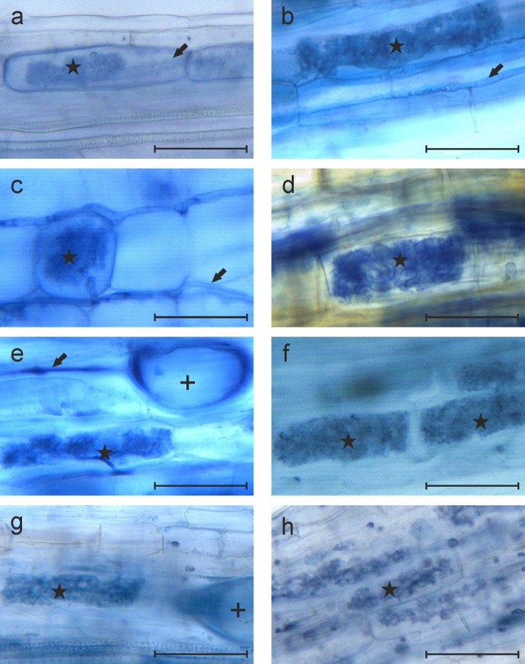
Eight selected plant species were inoculated with R. irregularis or B. amyloliquefaciens, were co-inoculated with both microbes, or were untreated as controls. Two repeated NaPPI experiments were conducted to study the effect of colonization on host plants. In the first experiment, AMF structures were not detected in plants from any of the four treatments. In the second experiment, hyphae of AMF were detected in five of eight plants from treatment R, whereas arbuscules and vesicles were rare or absent in all tested plants. In the R+B treatment, AMF structures such as arbuscules and hyphae were observed in all eight plant species (Fig 1) and vesicles were observed in all tested plants except C. rotundifolia. Even though hyphae and arbuscules were detected in roots of C. rotundifolia, the colonization rate was low (3% and 1%, respectively), and no vesicles were observed (Table 2). According to the LSD0.05 test, colonization by the specific fungal structures was significantly higher with the R+B treatment than with R alone (P < 0.05; Table 2).
Fig 1. Microscopic images of AMF structures in roots of eight plants co-inoculated with R. irregularis and B. amyloliquefaciens from the second experiment.
(a−h) Images from C. rotundifolia (a), L. corniculatus (b), T. repens (c), G. sanguineum (d), F. vesca (e), V. tricolor (f), T. serpyllum (g), and A. dioica (h). Scale bars represent 50 μm. A black arrow, a + symbol, and a star indicate a hypha, vesicle, and arbuscule, respectively.
Table 2. Colonization by AMF of roots of eight plant species following inoculation with R. irregularis and co-inoculation with R. irregularis and B. amyloliquefaciens from the second experiment.a.
| Plant species | Hyphae (%) | Arbuscules (%) | Vesicles (%) | |||
|---|---|---|---|---|---|---|
| R | R+B | R | R+B | R | R+B | |
| C. rotundifolia | 0 | 3.0 ± 1.0 | 0 | 1.0 ± 0.6 | 0 | 0 |
| L. corniculatus | 0 | 40.0 ± 3.6 | 0 | 30.7 ± 2.7 | 0 | 11.0 ± 1.5 |
| T. repens | 0 | 65.7 ± 2.2 | 0 | 58.0 ± 3.2 | 0 | 21.3 ± 1.2 |
| G. sanguineum | 3.0 ± 2.1 | 48.0 ± 2.5** | 0 | 38.3 ± 2.6 | 0 | 8.7 ± 1.9 |
| F. vesca | 6.7 ± 3.4 | 95.0 ± 2.0** | 2.7 ± 1.5 | 82.7 ± 3.2** | 0 | 14.0 ± 2.1 |
| V. tricolor | 17.7 ± 3.2 | 46.0 ± 1.0** | 11.7 ± 2.3 | 29.7 ± 4.2* | 0 | 1.3 ± 0.9 |
| T. serpyllum | 36.7 ± 2.3 | 87.7 ± 1.2** | 18.7 ± 0.9 | 47.0 ± 7.0* | 2.0 ± 0.6 | 8.0 ± 2.0* |
| A. dioica | 46.3 ± 1.2 | 82.0 ± 1.2** | 13.3 ± 1.5 | 63.3 ± 2.3** | 7.3 ± 0.7 | 14.0 ± 1.0** |
a Differences in the colonization rate of roots between the treatments R and R+B were tested by LSD0.05
*P < 0.05
**P < 0.01.
According to the AMF colonization response to co-inoculation with both microbes, the plant species could be divided into three groups: 1) plants that were not active R. irregularis host plants, regardless of the presence of B. amyloliquefaciens in the soil (C. rotundifolia); 2) plants that were R. irregularis host plants only when B. amyloliquefaciens was co-inoculated (L. corniculatus and T. repens); and 3) plants that were R. irregularis host plants when inoculated alone but became more efficiently colonized by R. irregularis when B. amyloliquefaciens was co-inoculated (G. sanguineum, F. vesca, V. tricolor, T. serpyllum, and A. dioica).
The rates of AMF structures in roots followed the pattern hyphae > arbuscules > vesicles in all the plant species, i.e., hyphae were the most prevalent AMF structure (Table 2).
B. amyloliquefaciens colonized all tested plant species
B. amyloliquefaciens was detected in varying amounts in the soil adhering to roots in nearly all plants inoculated with the bacteria. Extraction of the DNA from the soil and sequencing of the soil DNA confirmed that the gyrB sequence isolated from soil DNA matched that of the bacteria in the Rhizocell product. B. amyloliquefaciens was not detected by qPCR in the soil of non-treated controls nor in soil from R treatment. According to LSD0.05 analysis in both experiments, no statistically significant difference (i.e., P < 0.05) in B. amyloliquefaciens population density was detected between the treatments B and R+B (Table 3).
Table 3. Content of B. amyloliquefaciens in the soil adhering to roots in different plant species following different treatments, as measured by qPCR.
| Plant species | ||||||||||||||||
|---|---|---|---|---|---|---|---|---|---|---|---|---|---|---|---|---|
| C. rotundifolia | L. corniculatus | T. repens | G. sanguineum | F. vesca | V. tricolor | T. serpyllum | A. dioica | |||||||||
| Treatment | B | R+B | B | R+B | B | R+B | B | R+B | B | R+B | B | R+B | B | R+B | B | R+B |
|
Exp. 1 (ng/g) |
1.5 | 3.6 | 2.0 | 117.0 | 1.7 | 1.6 | 4.8 | 126.4 | 117.4 | 15.5 | 1.5 | 17.8 | 1.7 | 0.5 | 5.8 | 20.4 |
| 10.0 | 0 | 16.3 | 48.6 | 2.4 | 15.6 | 0 | 94.3 | 2.7 | 1.8 | 35.2 | 199.0 | 2.4 | 0.3 | 0.4 | 90 | |
| 1.4 | 0 | 1.4 | 3.5 | 0 | 0.5 | 0 | 0.5 | 2.6 | 0 | 3.2 | 3.2 | 1.7 | 2.8 | 0 | 41.9 | |
| P = 0.373 | P = 0.210 | P = 0.408 | P = 0.129 | P = 0.414 | P = 0.401 | P = 0.430 | P = 0.09 | |||||||||
|
Exp. 2 (ng/g) |
3.0 | 16.2 | 16.9 | 3.0 | 12.3 | 6.0 | 10.9 | 3.8 | 1.4 | 8.7 | 4.5 | 3.3 | 11.1 | 1.1 | 42.8 | 5.9 |
| 0.9 | 1.4 | 7.4 | 6.2 | 19.3 | 3.4 | 1.0 | 5.6 | 2.9 | 3.3 | 2.5 | 1.8 | 16.6 | 1.5 | 1.3 | 0.7 | |
| 3.9 | 2.3 | 10.3 | 3.8 | 2.5 | 2.2 | 2.6 | 2.2 | 0 | 0.8 | 8.8 | 0 | 0.3 | 0.7 | 15.1 | 2.5 | |
| P = 0.454 | P = 0.374 | P = 0.208 | P = 0.779 | P = 0.314 | P = 0.163 | P = 0.161 | P = 0.246 | |||||||||
No significant differences were observed between the treatments B and R+B (P < 0.05; LSD0.05).
Shoot weight of the plants
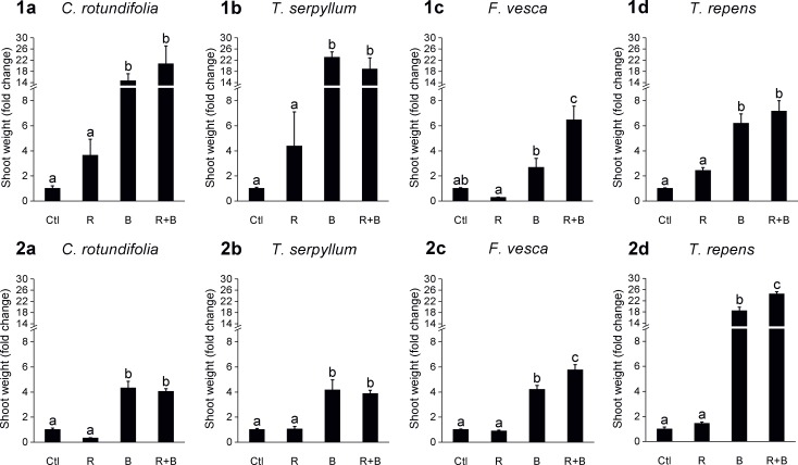
Differences in the plant shoot weight were studied in the two experiments by analyzing the fold change of the treated plants (R, B, and R+B) compared with untreated control plants. Plants were placed in two groups, namely those with consistent results (Fig 2) and those with less repeatable results (S1 Fig) obtained in the NaPPI experiments. The shoot weight of the four species with consistent results was significantly lower in untreated control plants and in R plants relative to B and R+B plants. For C. rotundifolia and T. serpyllum, no difference was detected between B and R+B, whereas for T. repens and F. vesca, plants in the R+B treatment group grew significantly larger than those in the B treatment group in at least one of the experiments.
Fig 2. Fold change in shoot weight of four plant species whose results were consistent across the two NaPPI experiments.
Bars (mean ± SE) represent fold changes in shoot weight of R, B, and R+B treated plants as compared with untreated control plants (Ctl). Graphs in the upper and lower row are from the first (1) and second (2) NaPPI experiment, respectively. (a−d) Data from C. rotundifolia (a), T. serpyllum (b), F. vesca (c), and T. repens (d). Different lowercase letters above the bars indicate statistical differences (LSD0.05) between the treatments at P < 0.05.
According to an ANOVA, there was a statistically significant interaction between the host plant species and treatment for shoot weight for all plant species (S1 Table, S2 Table).
Chlorophyll fluorescence
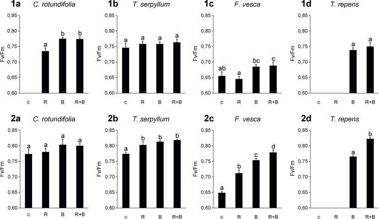
The four plant species that showed a consistent effect for shoot weight (Fig 2) also showed reproducible patterns in photosynthetic efficiency in the two NaPPI experiments (Fig 3). For C. rotundifolia and T. serpyllum, inoculation with the microbes showed little influence on photosynthetic efficiency. For T. repens and F. vesca, the Fv/Fm ratio was lowest in untreated control plants (for T. repens, the Fv/Fm ratio in the R treatment group and in the control was too low to be detected) and was higher after B and R+B treatments. Moreover, R+B treatment in T. repens and F. vesca produced the highest Fv/Fm ratio in at least one of the experiments (Fig 3). The Fv/Fm ratios for the four plant species that showed less-consistent results in the two experiments are presented in S2 Fig and S4 Table.
Fig 3. Quenching analysis of chlorophyll activities in leaves of plant species whose results were consistent in the two NaPPI experiments.
Bars (mean ± SE) represent the Fv/Fm ratio of chlorophyll fluorescence for treated and untreated plants. Bar charts in the upper and lower row are from the first and second NaPPI experiment, respectively. Different lowercase letters above the bars indicate statistical differences (LSD0.05) between the treatments. Missing data indicate that the Fv/Fm ratio was too low to be detected.
The host plant species, treatment, and their interaction had significant effects on photosynthetic efficiency in all plant species (S3 Table, S4 Table).
Discussion
Mycorrhiza helper bacterium (MHB) is a generic name for bacteria that can stimulate the formation of a mycorrhizal association with host plants [63]. In our study, the colonization rate of R. irregularis among the eight tested plant species was improved by co-inoculation with B. amyloliquefaciens. Moreover, for some plant species (L. corniculatus and T. repens), successful colonization by R. irregularis could only happen when B. amyloliquefaciens was co-inoculated. We can infer that AMF colonization in some plant species was strongly dependent on the presence of a MHB (B. amyloliquefaciens in this case).
Our findings are consistent with the studies of Yusran et al. [64], which indicated that different species of MHB promote mycorrhization to different levels and that co-inoculation with a MHB and an AMF has a positive impact on biomass and phytopathogen control in tomato plants. Our results show that the colonization-promoting effects of B. amyloliquefaciens on R. irregularis can occur across a broad range of host plants suggesting that such a promoting effect is fungus specific, rather than host plant specific [65,66]. Many MHB species have been identified, and it is predicted that more are likely to be discovered [44,67]. Finding such beneficial combinations would be promising for sustainable and cost-effective urban greening, and potential combinations should be extensively tested for their reliability and consistency under field conditions before wider exploitation.
The exact processes and compounds of microbes that are involved in stimulation of AMF colonization by MHB remain mostly unknown [68,69]. It does not necessarily require physical attachment but could happen through chemical signaling [63]. One possible mechanism behind these effects could be the release of gaseous volatiles, such as 2-methyl isoburneol, geosmin, and CO2, by the MHB that stimulates the growth of AMF. In addition to gaseous chemicals, other active metabolites produced by a MHB include vitamins, amino acids, and growth substances, any or all of which might directly stimulate the growth of an AMF [63,70].
The present study indicated that co-inoculation with R. irregularis and B. amyloliquefaciens led to increased plant biomass of at least T. repens and F. vesca in repeated experiments. A similar synergistic effect has been reported in a study that combined four Glomus species, namely G. aggregatum (Schenck and Smith), G. fasciculatum (Gerd & Trappe), G. intraradices, and G. mosseae (Nicolson & Gerd), with B. subtilis on Pelargonium graveolens (L'Her. ex Aiton). All four combinations (each of the four Glomus species inoculated separately with B. subtilis) resulted in synergistic effects to various degrees, as measured by increases in the plant dry weight of 25.7% to 74.3% as compared with that of control plants and of 7.8% to 14.5% as compared with inoculation with a single Glomus species [71].
We found that AMF colonization at a low level may not have any promoting effects on host plants. It is especially true if the internal hyphae are the only AMF structure present in the roots. However, little scientific data are available concerning the threshold level of arbuscules and vesicles needed for a significant promoting effect, and further studies on this topic are needed. C. rotundifolia hardly differed in biomass production between the two treatments R and R+B. A possible explanation could be that C. rotundifolia is highly tolerant to drought, soil pH, environmental variations, frost, and strong wind, so that it does not need symbiosis for further growth-promoting effects [72]. In contrast, Nuortila et al. [73] reported that, instead of increasing plant biomass, AMF colonization can significantly decrease the plant biomass of C. rotundifolia by 33% and can reduce flowers by 66%. Furthermore, in some specific AMF−host plant combinations, AMF can act as a “hitchhiker” that profits from the mycorrhizal-host network without returning benefits back to the host [74,75]. In the present study, T. serpyllum reached high levels of AMF colonization after the R+B treatment in the second experiment, but no significant difference in biomass production between R+B and B plants was observed. Similar results have been obtained using Thymus vulgaris (L.) as a host plant and G. mosseae as the AMF inoculant: a higher AMF colonization rate after AMF treatment did not increase plant biomass [76]. The phenomenon depends on the plant-AMF combinations, water availability, and soil types [77].
Enhanced photosynthetic efficiency is another benefit that AMF and plant growth−promoting rhizobacteria (PGPR) provide to some host plants, such as T. repens and F. vesca in the present study. A single inoculation with AMF or PGPR may significantly promote photosynthetic efficiency under suboptimal conditions such as high salinity and nutrient deficiency in the soil. For example, co-colonization by Glomus and Bacillus species enhances the photosynthetic efficiency of lettuce plants, compared with colonization by either of the two microbes. The extent of the synergistic effect is greatly dependent on the Glomus and Bacillus species combination [70].
Quantification of B. amyloliquefaciens revealed varying population densities between replicates in our study. Trevors et al. [78] also found large variation between replicates in their study of PGPR in the soil. It appears challenging to correlate PGPR population density with effects that promote plant growth [79], but the information would be useful, as beyond a certain PGPR population density no further enhancement may occur [79–82]. This scenario also seems possible in our study, as higher population densities of B. amyloliquefaciens did not lead to a higher biomass production or photosynthetic efficiency.
Our results suggest that neither a positive nor negative interaction was found between AMF colonization rate and the abundance of B. amyloliquefaciens in the soil in any of the eight tested plant species. These findings are supported by Kostoula et al. [83], who got similar results with Crithmum maritimum (L.) as a host plant and G. intraradices and B. amyloliquefaciens as inoculants. They found no significant effect of G. intraradices colonization on the population density of B. amyloliquefaciens in the soil. Likewise, Alam et al. [71] found no effect of four Glomus species on B. subtilis population density.
Interruptions in the NaPPI system during the first experiment made growth conditions unstable, which probably explains why the shoot weight and photosynthetic efficiency in half of the plant species were not reproducible across the two NaPPI experiments. Still, the other four plant species exhibited relatively consistent patterns in shoot weight and photosynthetic efficiency, suggesting that the plant species studied show different sensitivities to the stability of their growth environment.
In conclusion, for host plants T. repens and F. vesca, B. amyloliquefaciens could act as a MHB to facilitate colonization by R. irregularis, and dual inoculation with B. amyloliquefaciens and R. irregularis should result in higher shoot weight and photosynthetic efficiency. Such mycorrhiza-helping effects and dual-inoculation growth-promoting effects in other test plants were more conditional and dependent on the environment. Further field experiments to test these effects of co-infection on green roofs should focus on T. repens and F. vesca, as they showed consistent and promising results, and they are likely to produce better growth than other plants under such conditions.
Supporting information
(DOC)
(DOC)
(DOC)
(DOC)
(TIF)
(TIF)
Acknowledgments
We would like to extend our gratitude to Fifth Dimension Research Group, Plant Pathology Research Group, Daniel Richterich, Lasse Kuismanen, Kristiina Himanen, Katrin Artola, Linpin Wang and Delfia Marcenaro for valuable advice and assistance.
Data Availability
All relevant data are within the paper and its Supporting Information files.
Funding Statement
Long Xie was supported by China Scholarship Council (grant no. 20140796003); by the Finnish Cultural Foundation; and by Maiju and Yrjö Rikala Horticultural Foundation, Finland. Soil and microbial preparations were donated by Kekkilä Group and Verdera/Lallemand Inc., respectively. Dr. Susanna Lehvävirta obtained a grant from Helsinki-Uusimaa Regional Council, Finland. Dr. Susanna Lehvävirta obtained a grant from Helsinki University Center for Environment. The funders had no role in study design, data collection and analysis, decision to publish, or preparation of the manuscript.
References
- 1.Compant S, Clément C, Sessitsch A. Plant growth-promoting bacteria in the rhizo-and endosphere of plants: their role, colonization, mechanisms involved and prospects for utilization. Soil Biol Biochem. 2010; 42(5):669–678. [Google Scholar]
- 2.Strack D, Fester T, Hause B, Schliemann W, Walter MH. Arbuscular mycorrhiza: biological, chemical, and molecular aspects. J Chem Ecol. 2003; 29(9):1955–1979. [DOI] [PubMed] [Google Scholar]
- 3.Krüger M, Krüger C, Walker C, Stockinger H, Schüßler A. Phylogenetic reference data for systematics and phylotaxonomy of arbuscular mycorrhizal fungi from phylum to species level. New Phytol. 2012; 193(4):970–984. 10.1111/j.1469-8137.2011.03962.x [DOI] [PubMed] [Google Scholar]
- 4.Biermann B, Linderman RG. Use of vesicular-arbuscular mycorrhizal roots, intraradical vesicles and extraradical vesicles as inoculum. New Phytol. 1983; 95(1):97–105. [Google Scholar]
- 5.St-Arnaud M, Hamel C, Vimard B, Caron M, Fortin JA. Enhanced hyphal growth and spore production of the arbuscular mycorrhizal fungus Glomus intraradices in an in vitro system in the absence of host roots. Mycol Res. 1996; 100(3):328–332. [Google Scholar]
- 6.Pfeffer PE, Douds DD, Bécard G, Shachar-Hill Y. Carbon uptake and the metabolism and transport of lipids in an arbuscular mycorrhiza. Plant Physiol. 1999; 120(2):587–598. [DOI] [PMC free article] [PubMed] [Google Scholar]
- 7.Bago B, Pfeffer PE, Shachar-Hill Y. Carbon metabolism and transport in arbuscular mycorrhizas. Plant Physiol. 2000; 124(3):949–958. [DOI] [PMC free article] [PubMed] [Google Scholar]
- 8.Koide RT, Kabir Z. Extraradical hyphae of the mycorrhizal fungus Glomus intraradices can hydrolyse organic phosphate. New Phytol. 2000; 148(3):511–517. [DOI] [PubMed] [Google Scholar]
- 9.Bücking H, Shachar‐Hill Y. Phosphate uptake, transport and transfer by the arbuscular mycorrhizal fungus Glomus intraradices is stimulated by increased carbohydrate availability. New Phytol. 2005; 165(3):899–912. 10.1111/j.1469-8137.2004.01274.x [DOI] [PubMed] [Google Scholar]
- 10.Ruiz-Lozano JM, Azcón R, Gomez M. Effects of arbuscular-mycorrhizal glomus species on drought tolerance: physiological and nutritional plant responses. Appl Environ Microbiol. 1995; 61(2):456–460. [DOI] [PMC free article] [PubMed] [Google Scholar]
- 11.Ruiz‐Lozano JM, Azcon R, Gomez M. Alleviation of salt stress by arbuscular‐mycorrhizal Glomus species in Lactuca sativa plants. Physiol Plant. 1996; 98(4):767–772. [Google Scholar]
- 12.Aroca R, Porcel R, Ruiz‐Lozano JM. How does arbuscular mycorrhizal symbiosis regulate root hydraulic properties and plasma membrane aquaporins in Phaseolus vulgaris under drought, cold or salinity stresses? New Phytol. 2007; 173(4):808–816. 10.1111/j.1469-8137.2006.01961.x [DOI] [PubMed] [Google Scholar]
- 13.Hildebrandt U, Regvar M, Bothe H. Arbuscular mycorrhiza and heavy metal tolerance. Phytochem. 2007; 68(1):139–146. [DOI] [PubMed] [Google Scholar]
- 14.Gonzalez-Guerrero M, Melville LH, Ferrol N, Lott JN, Azcón-Aguilar C, Peterson RL. Ultrastructural localization of heavy metals in the extraradical mycelium and spores of the arbuscular mycorrhizal fungus Glomus intraradices. Can J Microbiol. 2008; 54(2):103–110. 10.1139/w07-119 [DOI] [PubMed] [Google Scholar]
- 15.Caron M, Fortin JA, Richard C. Effect of phosphorus concentration and Glomus intraradices on Fusarium crown and root rot of tomatoes. Phytopathology. 1986; 76(9):942–946. [Google Scholar]
- 16.St-Arnaud M, Hamel C, Fortin JA. Inhibition of Pythium ultimum in roots and growth substrate of mycorrhizal Tagetes patula colonized with Glomus intraradices. Can J Plant Pathol. 1994; 16(3):187–194. [Google Scholar]
- 17.Priest FG, Goodfellow M, Shute LA, Berkeley RC. Bacillus amyloliquefaciens sp. nov., nom. rev. Intern J Syst Evol Microbiol. 1987; 37(1):69–71. [Google Scholar]
- 18.Fukumoto J. Studies on the production of bacterial amylase. I. Isolation of bacteria secreting potent amylases and their distribution. Nippon Nogeikagaku Kaishi. 1943; 19:487–503. [Google Scholar]
- 19.Reva ON, Dixelius C, Meijer J, Priest FG. Taxonomic characterization and plant colonizing abilities of some bacteria related to Bacillus amyloliquefaciens and Bacillus subtilis. FEMS Microbiol Ecol. 2004; 48(2):249–259. 10.1016/j.femsec.2004.02.003 [DOI] [PubMed] [Google Scholar]
- 20.López D, Fischbach MA, Chu F, Losick R, Kolter R. Structurally diverse natural products that cause potassium leakage trigger multicellularity in Bacillus subtilis. Proc Natl Acad Sci. 2009; 106(1):280–285. 10.1073/pnas.0810940106 [DOI] [PMC free article] [PubMed] [Google Scholar]
- 21.Chen Y, Cao S, Chai Y, Clardy J, Kolter R, Guo JH, et al. A Bacillus subtilis sensor kinase involved in triggering biofilm formation on the roots of tomato plants. Mol Microbiol. 2012; 85(3):418–430. 10.1111/j.1365-2958.2012.08109.x [DOI] [PMC free article] [PubMed] [Google Scholar]
- 22.Nautiyal CS, Srivastava S, Chauhan PS, Seem K, Mishra A, Sopory SK. Plant growth-promoting bacteria Bacillus amyloliquefaciens NBRISN13 modulates gene expression profile of leaf and rhizosphere community in rice during salt stress. Plant Physiol Biochem. 2013; 66:1–9. 10.1016/j.plaphy.2013.01.020 [DOI] [PubMed] [Google Scholar]
- 23.Kasim WA, Osman ME, Omar MN, El-Daim IA, Bejai S, Meijer J. Control of drought stress in wheat using plant-growth-promoting bacteria. J Plant Growth Reg. 2013; 32(1):122–130. [Google Scholar]
- 24.Rodríguez H, Fraga R. Phosphate solubilizing bacteria and their role in plant growth promotion. Biotech Adv. 1999; 17(4–5):319–339. [DOI] [PubMed] [Google Scholar]
- 25.Idriss EE, Makarewicz O, Farouk A, Rosner K, Greiner R, Bochow H, et al. Extracellular phytase activity of Bacillus amyloliquefaciens FZB45 contributes to its plant-growth-promoting effect. Microbiology. 2002; 148(7):2097–2109. [DOI] [PubMed] [Google Scholar]
- 26.Li B, Xu LH, Lou MM, Li F, Zhang YD, Xie GL. Isolation and characterization of antagonistic bacteria against bacterial leaf spot of Euphorbia pulcherrima. Lett Appl Microb. 2008; 46(4):450–455. [DOI] [PubMed] [Google Scholar]
- 27.Hu HQ, Li XS, He H. Characterization of an antimicrobial material from a newly isolated Bacillus amyloliquefaciens from mangrove for biocontrol of Capsicum bacterial wilt. Biol Control. 2010; 54(3):359–365. [Google Scholar]
- 28.Benitez LB, Velho RV, da Motta AD, Segalin J, Brandelli A. Antimicrobial factor from Bacillus amyloliquefaciens inhibits Paenibacillus larvae, the causative agent of American foulbrood. Arch Microbiol. 2012; 194(3):177–185. 10.1007/s00203-011-0743-4 [DOI] [PubMed] [Google Scholar]
- 29.Berg G. Plant–microbe interactions promoting plant growth and health: perspectives for controlled use of microorganisms in agriculture. Appl Microbiol Biotech. 2009; 84(1):11–18. [DOI] [PubMed] [Google Scholar]
- 30.Peck SW, Callaghan C, Kuhn ME, Bass B. Greenbacks from green roofs: forging a new industry in Canada. Canada Mortgage and Housing Corporation (CMHC); 1999. http://publications.gc.ca/pub?id=9.615746&sl=0
- 31.DeNardo JC, Jarrett AR, Manbeck HB, Beattie DJ, Berghage RD. Stormwater mitigation and surface temperature reduction by green roofs. Transact ASAE. 2005; 48(4):1491–1496. [Google Scholar]
- 32.Oberndorfer E, Lundholm J, Bass B, Coffman RR, Doshi H, Dunnett N, et al. Green roofs as urban ecosystems: ecological structures, functions, and services. BioSci. 2007; 57(10):823–833. [Google Scholar]
- 33.Takebayashi H, Moriyama M. Surface heat budget on green roof and high reflection roof for mitigation of urban heat island. Build Environ. 2007; 42(8):2971–2979. [Google Scholar]
- 34.Yang J, Yu Q, Gong P. Quantifying air pollution removal by green roofs in Chicago. Atmosph Environ. 2008; 42(31):7266–7273. [Google Scholar]
- 35.Akita M, Lehtonen MT, Koponen H, Marttinen EM, Valkonen JPT. Infection of the Sunagoke moss panels with fungal pathogens hampers sustainable greening in urban environments. STOTEN. 2011; 409: 3166–3173. [DOI] [PubMed] [Google Scholar]
- 36.Henry A, Frascaria-Lacoste N. The green roof dilemma–Discussion of Francis and Lorimer (2011). J Environ Manag. 2012; 104:91–92. [DOI] [PubMed] [Google Scholar]
- 37.Nurmi V, Votsis A, Perrels A, Lehvävirta S. Green roof cost-benefit analysis: special emphasis on scenic benefits. J Benefit-Cost Anal. 2016; 7(3):488–522. [Google Scholar]
- 38.McGuire KL, Payne SG, Palmer MI, Gillikin CM, Keefe D, Kim SJ, et al. Digging the New York City skyline: soil fungal communities in green roofs and city parks. PloS ONE. 2013; 8(3):e58020 10.1371/journal.pone.0058020 [DOI] [PMC free article] [PubMed] [Google Scholar]
- 39.Molineux CJ, Connop SP, Gange AC. Manipulating soil microbial communities in extensive green roof substrates. STOTEN. 2014; 493:632–638. [DOI] [PubMed] [Google Scholar]
- 40.John J, Lundholm J, Kernaghan G. Colonization of green roof plants by mycorrhizal and root endophytic fungi. Ecol Eng. 2014; 71:651–659. [Google Scholar]
- 41.Young T, Cameron DD, Phoenix GK. Using AMF inoculum to improve the nutritional status of Prunella vulgaris plants in green roof substrate during establishment. Urban Forest Urban Green. 2015; 14(4):959–967. [Google Scholar]
- 42.Barea JM, Pozo MJ, Azcon R, Azcon-Aguilar C. Microbial co-operation in the rhizosphere. J Exp Bot. 2005; 56(417):1761–1778. 10.1093/jxb/eri197 [DOI] [PubMed] [Google Scholar]
- 43.Mansfeld-Giese K, Larsen J, Bødker L. Bacterial populations associated with mycelium of the arbuscular mycorrhizal fungus Glomus intraradices. FEMS Microb Ecol. 2002; 41(2):133–140. [DOI] [PubMed] [Google Scholar]
- 44.Medina A, Probanza A, Mañero FG, Azcón R. Interactions of arbuscular-mycorrhizal fungi and Bacillus strains and their effects on plant growth, microbial rhizosphere activity (thymidine and leucine incorporation) and fungal biomass (ergosterol and chitin). Appl Soil Ecol. 2003; 22(1):15–28. [Google Scholar]
- 45.Toro M, Azcon R, Barea J. Improvement of arbuscular mycorrhiza development by inoculation of soil with phosphate-solubilizing rhizobacteria to improve rock phosphate bioavailability and nutrient cycling. App Env Microbiol. 1997; 63(11):4408–4412. [DOI] [PMC free article] [PubMed] [Google Scholar]
- 46.Lu ZH, Zhang SA. The selection of substratum for roof lawn establishment using Thymus serpyllum. Pratacultural Sci. 2005; 1:22. [Google Scholar]
- 47.Lee BR, Kim KY, Jung WJ, Avice JC, Ourry A, Kim TH. Peroxidases and lignification in relation to the intensity of water-deficit stress in white clover (Trifolium repens L.). J Exp Bot. 2007; 58(6):1271–1279. 10.1093/jxb/erl280 [DOI] [PubMed] [Google Scholar]
- 48.Zhao HM, Sun GZ, Wang XM, Liu GB. Comprehensive evaluation and identification of drought resistance of Lotus corniculatus in seedling stage. Grassland Turf. 2011; 6:6. [Google Scholar]
- 49.Pavicic M, Mouhu K, Wang F, Bilicka M, Chovanček E, Himanen K. Genomic and phenomic screens for flower related RING type ubiquitin E3 ligases in Arabidopsis. Front Plant Sci. 2017; 8:416 10.3389/fpls.2017.00416 [DOI] [PMC free article] [PubMed] [Google Scholar]
- 50.Gurevitch J, Wilson P, Stone JL, Teese P, Stoutenburgh RJ. Competition among old-field perennials at different levels of soil fertility and available space. The Journal of Ecology. 1990; 78(3):727–744. [Google Scholar]
- 51.Koide RT. Density-dependent response to mycorrhizal infection in Abutilon theophrasti. Medic. Oecologia. 1991; 85(3):389–395. 10.1007/BF00320615 [DOI] [PubMed] [Google Scholar]
- 52.Latimer JG. Container size and shape influence growth and landscape performance of marigold seedlings. HortScience. 1991; 26(2):124–126. [Google Scholar]
- 53.Liu A, Latimer JG. Root cell volume in the planter flat affects watermelon seedling development and fruit yield. HortScience. 1995; 30(2):242–246. [Google Scholar]
- 54.Bar-Tal A, Bar-Yosef B, Kafkafi U. Pepper transplant response to root volume and nutrition in the nursery. Agronomy Journal. 1990; 82(5):989–995. [Google Scholar]
- 55.Barrett DJ, Gifford RM. Acclimation of photosynthesis and growth by cotton to elevated CO2: interactions with severe phosphate deficiency and restricted rooting volume. Functional Plant Biology. 1995; 22(6):955–963. [Google Scholar]
- 56.Ruiz‐Lozano JM, Aroca R, Zamarreño ÁM, Molina S, Andreo‐Jiménez B, Porcel R, et al. Arbuscular mycorrhizal symbiosis induces strigolactone biosynthesis under drought and improves drought tolerance in lettuce and tomato. Plant Cell Environ. 2016; 39(2):441–452. 10.1111/pce.12631 [DOI] [PubMed] [Google Scholar]
- 57.Paknejad F, Majidi HE, Nourmohammadi G, Siadat a, Vazan S. Effects of drought stress on chlorophyll fluorescence parameters, chlorophyll content and grain yield in some wheat cultivars. J Biol Sci. 2007; 7: 841–847. [Google Scholar]
- 58.Maxwell K, Johnson GN. Chlorophyll fluorescence—a practical guide. J Exp Bot. 2000; 51(345):659–668. [DOI] [PubMed] [Google Scholar]
- 59.Vierheilig H, Schweiger P, Brundrett M. An overview of methods for the detection and observation of arbuscular mycorrhizal fungi in roots. Physiol Plant. 2005; 125(4):393–404. [Google Scholar]
- 60.Phillips JM, Hayman DS. Improved procedures for clearing roots and staining parasitic and vesicular-arbuscular mycorrhizal fungi for rapid assessment of infection. Transact British Mycol Soc. 1970; 55(1):158–161. [Google Scholar]
- 61.McGonigle TP, Miller MH, Evans DG, Fairchild GL, Swan JA. A new method which gives an objective measure of colonization of roots by vesicular—arbuscular mycorrhizal fungi. New Phytol. 1990; 115(3):495–501. [DOI] [PubMed] [Google Scholar]
- 62.Pfaffl MW. A new mathematical model for relative quantification in real-time RT–PCR. Nucl Acids Res. 2001; 29(9):e45 [DOI] [PMC free article] [PubMed] [Google Scholar]
- 63.Garbaye J. Helper bacteria: a new dimension to the mycorrhizal symbiosis. New Phytol. 1994; 128: 197–210. [DOI] [PubMed] [Google Scholar]
- 64.Yusran Y, Roemheld V, Mueller T. Effects of Pseudomonas sp.” Proradix” and Bacillus amyloliquefaciens FZB42 on the establishment of AMF infection, nutrient acquisition and growth of tomato affected by Fusarium oxysporum Schlecht f. sp. radicis-lycopersici Jarvis and Shoemaker. 2009; https://escholarship.org/uc/item/8g70p0zt
- 65.Garbaye J, Churin JL, Duponnois R. Effects of substrate sterilization, fungicide treatment, and mycorrhization helper bacteria on ectomycorrhizal formation of pedunculate oak (Quercus robur) inoculated with Laccaria laccata in two peat bare-root nurseries. Biol Fertil Soil. 1992; 13(1):55–57. [Google Scholar]
- 66.Garbaye J, Duponnois R. Specificity and function of mycorrhization helper bacteria (MHB) associated with the Pseudotsuga menziesii-Laccaria laccata symbiosis. Symbiosis. 1993; 14: 335–344. [Google Scholar]
- 67.Artursson V, Finlay RD, Jansson JK. Interactions between arbuscular mycorrhizal fungi and bacteria and their potential for stimulating plant growth. Environ Microbiol. 2006; 8(1):1–10. 10.1111/j.1462-2920.2005.00942.x [DOI] [PubMed] [Google Scholar]
- 68.Carpenter-Boggs L, Loynachan TE, Stahl PD. Spore germination of Gigaspora margarita stimulated by volatiles of soil-isolated actinomycetes. Soil Biol Biochem. 1995; 27(11):1445–1451. [Google Scholar]
- 69.Aliasgharzad N, Pourmirzaei Z, Dehnad AR, Najafi N. Streptomyces species favour spore germination and hyphal growth of arbuscular mycorrhizal fungus. Int J Agr Res. Rev. 2012; 2(6):765–773. [Google Scholar]
- 70.Vivas A, Marulanda A, Ruiz-Lozano JM, Barea JM, Azcón R. Influence of a Bacillus sp. on physiological activities of two arbuscular mycorrhizal fungi and on plant responses to PEG-induced drought stress. Mycorrhiza. 2003; 13(5):249–256. 10.1007/s00572-003-0223-z [DOI] [PubMed] [Google Scholar]
- 71.Alam M, Khaliq A, Sattar A, Shukla RS, Anwar M, Dharni S. Synergistic effect of arbuscular mycorrhizal fungi and Bacillus subtilis on the biomass and essential oil yield of rose-scented geranium (Pelargonium graveolens). Arch Agr Soil Sci. 2011; 57(8): 889–898. [Google Scholar]
- 72.Stevens CJ, Wilson J, McAllister HA. Biological flora of the British Isles: Campanula rotundifolia. J Ecol. 2012; 100(3):821–839. [Google Scholar]
- 73.Nuortila C, Kytöviita MM, Tuomi J. Mycorrhizal symbiosis has contrasting effects on fitness components in Campanula rotundifolia. New Phytol. 2004; 164(3):543–553. [Google Scholar]
- 74.Pellmyr O, Leebens-Mack J, Huth CJ. Non-mutualistic yucca moths and their evolutionary consequences. Nature. 1996; 380(6570):155–156. 10.1038/380155a0 [DOI] [PubMed] [Google Scholar]
- 75.Sanders IR. Preference, specificity and cheating in the arbuscular mycorrhizal symbiosis. Trends Plant Sci. 2003; 8(4):143–145. 10.1016/S1360-1385(03)00012-8 [DOI] [PubMed] [Google Scholar]
- 76.Sasanelli N, Anton A, Takacs T, D’Addabbo T, Bíró I, Malov X. Influence of arbuscular mycorrhizal fungi on the nematicidal properties of leaf extracts of Thymus vulgaris L. Helminthol. 2009; 46(4):230–240. [Google Scholar]
- 77.Navarro-Fernández CM, Aroca R, Barea JM. Influence of arbuscular mycorrhizal fungi and water regime on the development of endemic Thymus species in dolomitic soils. Appl Soil Ecol. 2011; 48(1):31–37. [Google Scholar]
- 78.Trevors JT, Kuikman P, Van Elsas JD. Release of bacteria into soil: cell numbers and distribution. J Microb Methods. 1994; 19(4):247–259. [Google Scholar]
- 79.Martínez-Viveros O, Jorquera MA, Crowley DE, Gajardo GM, Mora ML. Mechanisms and practical considerations involved in plant growth promotion by rhizobacteria. J Soil Sci Plant Nutr. 2010; 10(3):293–319. [Google Scholar]
- 80.Raaijmakers JM, Bonsall RF, Weller DM. Effect of population density of Pseudomonas fluorescens on production of 2, 4-diacetylphloroglucinol in the rhizosphere of wheat. Phytopathology. 1999; 89(6):470–475. 10.1094/PHYTO.1999.89.6.470 [DOI] [PubMed] [Google Scholar]
- 81.Strigul NS, Kravchenko LV. Mathematical modeling of PGPR inoculation into the rhizosphere. Environ Model Software. 2006; 21(8):1158–1171. [Google Scholar]
- 82.Lugtenberg B, Kamilova F. Plant-growth-promoting rhizobacteria. Annu Rev Microbiol. 2009; 63:541–556. 10.1146/annurev.micro.62.081307.162918 [DOI] [PubMed] [Google Scholar]
- 83.Kostoula O, Dimou D, Ifanti P, Douma D, Karipidis C, Kritsimas A, et al. Crithmum maritimum L. in co-existence with Glomus intraradices and a growth promoting bacterium. In: XXIX International Horticultural Congress on Horticulture: Sustaining Lives, Livelihoods and Landscapes (IHC2014). 2014; 1102:163–170. [Google Scholar]
Associated Data
This section collects any data citations, data availability statements, or supplementary materials included in this article.
Supplementary Materials
(DOC)
(DOC)
(DOC)
(DOC)
(TIF)
(TIF)
Data Availability Statement
All relevant data are within the paper and its Supporting Information files.


