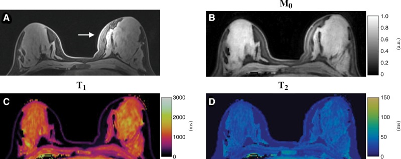
Figure 4:
Conventional and MR fingerprinting images in a 19-year-old healthy female participant. A, Standard clinical fat-saturated image. Substantial signal variation in left breast was observed (arrow), which is likely due to B1 field inhomogeneity. B, Proton density (M0), C, T1, and, D, T2 maps acquired from same section location as in A by using proposed three-dimensional MR fingerprinting method.

