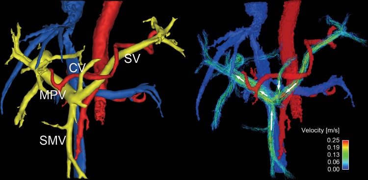
Figure 5:
Four-dimensional flow MR images in a 69-year-old woman with low-risk varices (not seen on anatomic image). Flow direction in coronary vein (CV) is hepatopetal. The fractional flow change in the main portal vein (MPV) was 0.23, which provided no evidence of high-risk varices. White arrows show the flow direction. SMV = superior mesenteric vein; SV = splenic vein.

