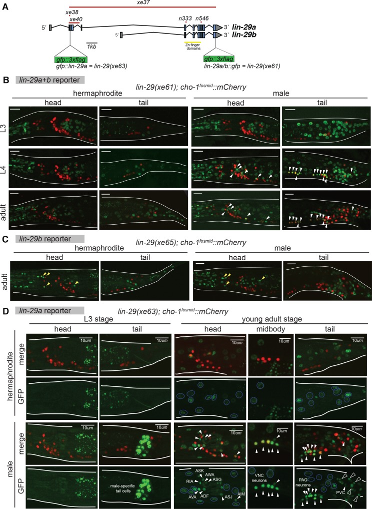
Figure 3. Temporal expression pattern of lin-29 in the nervous system of both sexes.
(A) Cartoon showing the lin-29 locus. Alternative promoter usage generates two LIN-29 protein isoforms. Exons 1 – 4 are LIN-29A specific while exons 5 – 11, that include the Zn-finger DNA binding domain, are shared by both -A and -B isoforms. The lin-29 locus was tagged using CRISPR/Cas9 genome engineering. gfp was inserted either at the C-terminal end (lin-29a/b::gfp; xe61 allele) to tag both LIN-29A and B protein isoforms or at the N-terminal end (gfp::lin-29a; xe63 allele) to tag only the LIN-29A isoform. Canonical alleles n333 and n546 as well as the newly generated alleles xe37 (null), xe38 (A-specific) and xe40 (A-specific) are indicated. (B) lin-29a/b expression pattern during larval development in both sexes. Confocal images for endogenously tagged lin-29a/b::gfp (xe61) show that GFP is expressed in the pharynx at the L3 stage and onwards in both sexes (examination of young animals showed that pharyngeal expression starts at the L1 stage). Hypodermal GFP expression all along the body started at the end of the L3 stage in both sexes. At the L4 stage, we also detected GFP expression in neurons only in the male, many of which were also labeled by a cholinergic marker, a cho-1/CHT mCherry expressing fosmid (marked by arrows). Expression in these male neurons persisted in the adult stage. No neuronal expression was observed in the head nor tail in hermaphrodite animals. Scale bar: 10 µm. (C) lin-29b expression pattern at the young adult stage in both sexes. Confocal images for gfp tagged lin-29b(xe40) (this allele is given a new name, xe65, since it contains the xe40 lesion plus the gfp insertion) show that GFP is expressed in head glia in both sexes (marked by yellow arrowheads). GFP is also observed in the pharynx and in tail cells in both sexes. Cholinergic cho-1/CHT mCherry expressing fosmid (otIs544) did not co-localize with GFP showing that LIN-29B is not expressed in neurons in the head nor tail in either sex (Note that LIN-29B is expressed in midbody neurons in both sexes not shown in this image). Scale bar: 10 µm. (D) gfp::lin-29a expression pattern during larval development in both sexes. Confocal images for endogenously tagged gfp::lin-29a (xe63) show that GFP is expressed in male-specific non-neuronal tail cells at the end of the L3 stage and onwards. No expression was observed in the head or tail of the hermaphrodite at this stage. No overlap between GFP and the cholinergic fosmid based reporter cho-1/CHT (otIs544) was observed at this stage. At the young adult stage, we observed GFP expression in neurons only in males (expression in male neurons started at the L4 stage), indicated by white arrows. No neuronal expression was observed in hermaphrodite neurons. Many GFP positive neurons in the male were also labeled by the cho-1/CHT mCherry expressing fosmid (otIs544). Cholinergic neurons expressing lin-29a included AIM, ASJ, AVA, ASK, AWA, ASG, RIA and ADF in the head, ventral nerve cord (VNC) motor neurons of the A, B, D and AS classes and the PVC interneuron in the tail. Neuronal GFP expression is indicated by arrows and neuronal identity is indicated for head and tail neurons in the GFP panels. GFP expression was observed throughout the hypodermis in both sexes, indicated with blue circles in the GFP panels. Hypodermis nuclei are larger. Cholinergic retrovesicular ganglion (RVG), ventral nerve cord (VNC) and pre-anal ganglion (PAG) neurons expressing lin-29a are shown in the head, midbody and tail respectively. Male-specific tail cells expressing lin-29a are marked with black arrows. Scale bar: 10 µm.

