Abstract
The log odds of positive lymph nodes (LODDS) and the metastatic lymph node ratio (MLR) staging systems have previously been demonstrated to exhibit advantages compared with the tumor-node-metastasis (TNM) staging system in predicting the prognosis of gastric cancer. The current study compared the prognostic significance of the newest Union for International Cancer Control Node classification with the LODDS and MLR staging systems. From September 2010 to December 2012, all medical records for patients with gastric cancer at the Third Affiliated Hospital of Soochow University were retrospectively analyzed and the clinicopathologic characteristics were reviewed. Cut-off points were selected to divide the patients with gastric cancer into different groups. Univariate and multivariate analyses were performed to identify the prognostic risk factors for gastric cancer. The Harrell's concordance index (C-index) was adopted to compare the prognostic value of the three staging systems. A total of 877 patients with gastric cancer who met the inclusion criteria were analyzed in the current study. The patients were classified according to the three MLR subgroups as follows: MLR0 (MLR=0), MLR1 (0<MLR≤0.28) and MLR2 (0.28<MLR<1). The patients were classified according to the LODDS subgroups as follows: LODDS1 (LODDS≤-0.5), LODDS2 (−0.5<LODDS≤0), LODDS3 (0<LODDS≤0.5) and LODDS4 (LODDS>0.5). Based on multivariate analysis, LODDS, MLR and pathological node (pN) stage could significantly predict survival rates of patients with gastric cancer. According to the C-index, the LODDS staging system more accurately predicted the 5-year overall survival for patients with gastric cancer compared with the other two staging systems. In summary, the current study has identified that LODDS may be superior to the MLR and pN staging systems in predicting the prognosis of patients with gastric cancer. However MLR may exhibit advantages compared with LODDS for patients who have undergone adequate lymphadenectomies.
Keywords: gastric cancer, metastatic lymph node ratio, log odds of positive lymph nodes, prognosis, staging system
Introduction
Gastric cancer is the fourth most common cancer type worldwide, with >93,000 new cases diagnosed every year, and is the second leading cause of cancer-associated cases of mortality, following lung cancer with 700,000 deaths each year (1,2). Gastric cancer is considered to be prevalent in east Asia, particularly in China (3). Currently, primary tumor resection with lymphadenectomy is the main surgical treatment for resectable gastric cancer; however, most cases of gastric cancer are diagnosed in the advanced stage as the symptoms of early stage disease are often atypical (4).
The 8th edition of the tumor-node-metastasis (TNM) staging system (5), established by the American Joint Committee on Cancer and the Union for International Cancer Control (UICC), is the most commonly used system for predicting the prognosis of gastric cancer (6). The 8th edition TNM staging system is considered to be an objective and reliable method for predicting the prognosis of patients with gastric cancer (7), however, the requirement of at least 15 retrieved lymph nodes (LNs) limits the use of this system in clinical practice (8). Furthermore, stage migration may occur if a low number of LNs are retrieved, which may underestimate the severity of the disease (9).
Previously, a number of new methods for predicting the prognosis of gastric cancer have been proposed. The metastatic lymph node ratio (MLR) is used as a supplement to the TNM staging system and is defined as the ratio of metastatic LNs to the total number of retrieved LNs (10). Several previous studies have demonstrated that MLR has advantages compared with the UICC pathological node (pN) staging system in predicting the prognosis of gastric cancer (11–14). Another prognostic parameter, the log odds of positive LNs (LODDS), is defined as the log of the ratio of positive LNs to negative LNs (15). A number of studies have indicated that the prognostic value of LODDS is superior to the MLR and pN systems (15–17). The current study evaluated the prognostic significance of the LODDS and MLR staging systems compared with the 8th UICC pN staging system.
Materials and methods
Patients
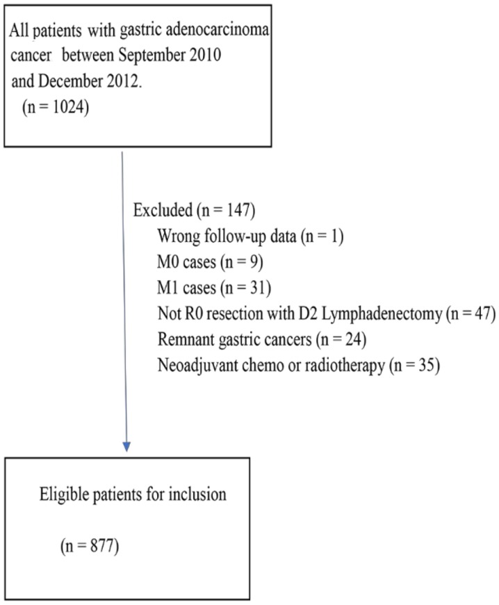
Between September 2010 and December 2012, all medical records for patients with gastric cancer treated at the Third Affiliated Hospital of Soochow University (Changzhou, China) were retrospectively analyzed. The inclusion criterion was adenocarcinoma R0 resection with D2 LN dissection. Patients with M0 and M1 statuses were excluded from the study. Patients who had received neoadjuvant chemotherapy or radiotherapy were also excluded due to the possibility of incorrect staging. Specific data are presented in Fig. 1. Clinicopathologic characteristics, including age, sex, tumor size, tumor location, tumor differentiation, tumor depth (pT stage), pN stage and TNM stage, were reviewed. Follow-up was conducted by telephone calls, e-mails and on-site visits. Informed written consent was received from all patients and the current study was approved by the Ethics Committee of The Third Affiliated Hospital of Soochow University.
Figure 1.
Inclusion criteria of the current study.
Different LN categories
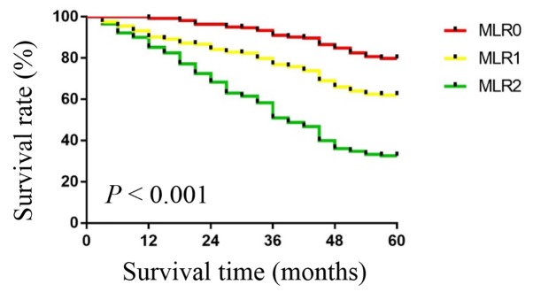
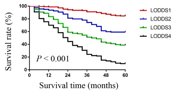
All included patients were staged using the 8th edition of the TNM staging system. The pN stage classification was performed as follows: N0, negative; N1, 1–2 positive LNs; N2, 3–6 positive LNs; N3a, 7–15 positive LNs; and N3b >15 positive LNs. Negative was used to mean no lymph node metastasis, while positive was used to mean lymph node metastasis. MLR was calculated as follows: MLR=metastatic LNs/retrieved LNs. The median MLR was selected as the cut-off value to divide the patients into three subgroups. The median MLR was 0.28, therefore the patients were classified into three subgroups as follows: MLR0 (MLR=0), MLR1 (0<MLR≤0.28) and MLR2 (0.28<MLR<1). LODDS was calculated as follows: LODDS=log (pnod + 0.5)/(nnod + 0.5), where pnod is the number of positive LNs and nnod is the number of negative LNs. The LODDS cut-off value was determined by comparing the 5-year overall survival rate with an interval of 0.5. As presented in Table I, patients were classified into four subgroups based on their LODDS value, as follows: LODDS1 (LODDS≤-0.5), LODDS2 (−0.5<LODDS≤0), LODDS3 (0<LODDS≤0.5) and LODDS4 (LODDS>0.5).
Table I.
Overall survival rates according to the value of LODDS with an interval of 0.5.
| LODDS value | No. of patients | 5-year OS rate, % | aP-value |
|---|---|---|---|
| LODDS≤-1.5 | 76 | 89.5 | 0.364 |
| −1.5<LODDS≤-1.0 | 101 | 86.1 | 0.335 |
| −1.0<LODDS≤-0.5 | 84 | 78.6 | 0.003 |
| −0.5<LODDS≤0 | 160 | 59.4 | <0.001 |
| 0<LODDS≤0.5 | 312 | 38.8 | 0.001 |
| 0.5<LODDS≤1.0 | 62 | 9.7 | <0.001 |
| 1.0<LODDS≤1.5 | 36 | 11.1 | 0.298 |
| LODDS>1.5 | 46 | 8.7 |
LODDS, log odds of positive nodes; OS, overall survival.
Compared between adjacent subgroups (e.g., a subgroup row and its following subgroup row in the table).
Statistical analysis
All analyses were performed using SPSS (version 16.0; SPSS, Inc., Chicago, IL, USA) and R software (version 3.0.0; www.r-project.org). Kaplan-Meier analysis followed by a log-rank test was used to compare the survival between subgroups. Univariate and multivariate analyses were performed using a Cox proportional hazards model. The Harrell's concordance index (C-index) was used to compare the accuracy of the prognostic predictions of different staging systems. A higher C-index indicates a better predictive accuracy. P<0.05 was considered to indicate a statistically significant difference.
Results
Patient characteristics
A total of 877 patients with gastric cancer met the aforementioned criteria and were analyzed in the current study. The clinicopathological characteristics are presented in Table II. The number of patients ≤60 and >60 years of age was 459 and 418, respectively. There were 605 male patients and 272 female patients. A total of 275 (31.4%) patients received postoperative chemotherapy. The majority of patients were in an advanced stage, with T3 and T4 patients accounting for 48.2 and 25.3%, respectively, and T1 and T2 patients accounting for 10.4 and 16.1%, respectively.
Table II.
Clinicopathological characteristics of 877 patients with gastric cancer.
| Characteristic | No. (%) |
|---|---|
| Age, years | |
| ≤60 | 459 (52.3) |
| >60 | 418 (47.7) |
| Sex | |
| Male | 605 (69.0) |
| Female | 272 (31.0) |
| Tumor location | |
| Upper | 179 (20.4) |
| Middle | 137 (15.6) |
| Lower | 550 (62.7) |
| Entire | 11 (1.3) |
| Tumor size, cm | |
| ≤5 | 568 (64.8) |
| >5 | 309 (35.2) |
| Tumor differentiation | |
| Well | 44 (5.0) |
| Moderately | 317 (36.1) |
| Poorly | 498 (56.8) |
| Undifferentiated | 18 (2.1) |
| pT stage | |
| T1 | 91 (10.4) |
| T2 | 141 (16.1) |
| T3 | 423 (48.2) |
| T4a | 211 (24.1) |
| T4b | 11 (1.3) |
| pN stage | |
| N0 | 223 (25.4) |
| N1 | 175 (20.0) |
| N2 | 265 (30.2) |
| N3a | 146 (16.6) |
| N3b | 68 (7.8) |
| TNM stage | |
| I | 126 (14.4) |
| II | 327 (37.3) |
| IIIA | 183 (20.9) |
| IIIB | 175 (20.0) |
| IIIC | 66 (7.5) |
| MLR | |
| MLR0 | 223 (25.4) |
| MLR1 | 203 (23.1) |
| MLR2 | 451 (51.4) |
| LODDS | |
| LODDS1 | 261 (29.8) |
| LODDS2 | 160 (18.2) |
| LODDS3 | 312 (35.6) |
| LODDS4 | 144 (16.4) |
| Number of LN retrieved | |
| <15 | 404 (46.1) |
| ≥15 | 473 (53.9) |
| Postoperative chemotherapy | |
| Yes | 275 (31.4) |
| No | 602 (68.6) |
pT, tumor depth; pN, pathological node; TNM, tumor-node-metastasis; MLR, metastatic lymph node ratio; LODDS, log odds of positive nodes; LN, lymph nodes.
Analysis of prognostic factors and survival
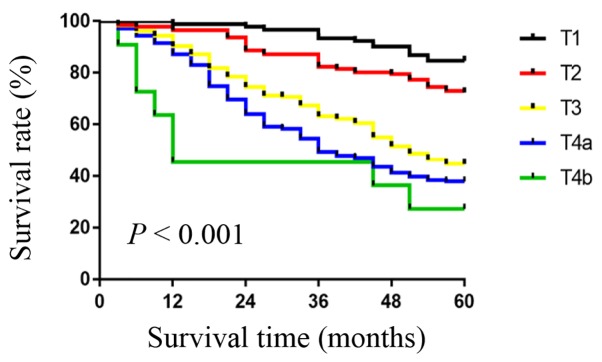
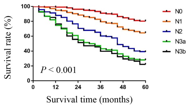
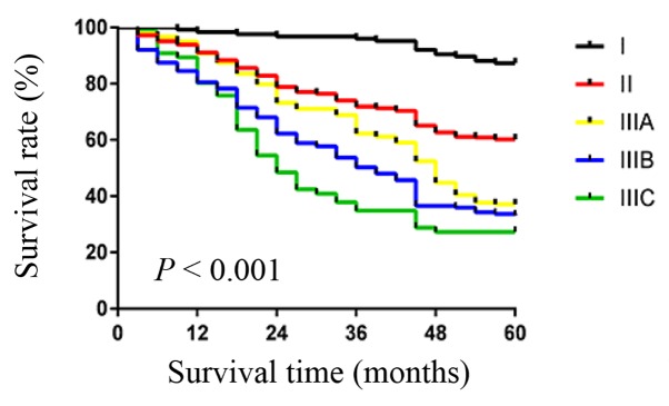
As presented in Table III, risk factors were evaluated using univariate and multivariate analyses. Prognostic factors included age, sex, tumor location, tumor size, tumor differentiation, pT stage, pN stage, TNM stage, MLR and LODDS. Overall survival rates were shown for patients in different T subgroups (Fig. 2), N subgroups (Fig. 3), TNM subgroups (Fig. 4), MLR subgroups (Fig. 5) and LODDS subgroups (Fig. 6). Based on univariate analysis, pT stage, pN stage, tumor size, tumor differentiation, MLR, LODDS and TNM were identified as significant prognostic risk factors for gastric cancer. However, based on multivariate analysis, tumor size and pT stage were not identified as significant prognostic factors.
Table III.
Univariate and multivariate analyses of prognostic factors.
| Multivariate analysis | ||||||
|---|---|---|---|---|---|---|
| Parameter | No. of patients | 5-year OS rate, % | Univariate analysis P-value | HR | 95% CI | P-value |
| Age, years | 0.677 | |||||
| ≤60 | 459 | 56.2 | ||||
| >60 | 418 | 46.2 | ||||
| Sex | 0.51 | |||||
| Male | 605 | 50.9 | ||||
| Female | 272 | 52.6 | ||||
| Tumor location | 0.374 | |||||
| Upper | 179 | 52 | ||||
| Middle | 137 | 51.8 | ||||
| Lower | 550 | 51.6 | ||||
| Entire | 11 | 27.3 | ||||
| Tumor size, cm | 0.001 | 1.983 | 0.942–3.011 | 0.094 | ||
| ≤5 | 568 | 61.3 | ||||
| >5 | 309 | 33.3 | ||||
| Tumor differentiation | 0.003 | 1.335 | 1.022–1.844 | 0.029 | ||
| Well | 44 | 72.7 | ||||
| Moderately | 317 | 59 | ||||
| Poorly | 498 | 46 | ||||
| Undifferentiated | 18 | 16.7 | ||||
| pT stage | 0.039 | 1.892 | 0.933–2.984 | 0.556 | ||
| T1 | 91 | 84.6 | ||||
| T2 | 141 | 72.3 | ||||
| T3 | 423 | 44.7 | ||||
| T4 | 222 | 37.4 | ||||
| pN stage | <0.001 | 2.012 | 1.113–2.868 | <0.001 | ||
| N0 | 223 | 80.3 | ||||
| N1 | 175 | 64.6 | ||||
| N2 | 265 | 38.9 | ||||
| N3a | 146 | 28.1 | ||||
| N3b | 68 | 22.1 | ||||
| TNM stage | <0.001 | 2.343 | 1.572–3.125 | 0.006 | ||
| II | 126 | 87.3 | ||||
| III | 327 | 60.2 | ||||
| IV | 424 | 34 | ||||
| MLR | <0.001 | 1.766 | 1.023–2.318 | <0.001 | ||
| MLR0 | 223 | 80.2 | ||||
| MLR1 | 203 | 62.1 | ||||
| MLR2 | 451 | 32.6 | ||||
| LODDS | <0.001 | 1.875 | 1.101–2.877 | <0.001 | ||
| LODDS1 | 261 | 84.7 | ||||
| LODDS2 | 160 | 59.4 | ||||
| LODDS3 | 312 | 38.8 | ||||
| LODDS4 | 144 | 9.7 | ||||
MLR, metastatic lymph node ratio; LODDS, log odds of positive nodes; OS, overall survival; HR, hazard ratio; CI, confidence interval; pT, tumor depth; pN, pathological node.
Figure 2.
Overall survival rates for patients in different T subgroups. According to mixed analysis of the five groups, the 5-year survival rate was 84.6% for T1, 72.3% for T2, 44.7% for T3, 37.9% for T4a and 27.3% for T4b. P<0.001. T, tumor depth.
Figure 3.
Overall survival rates for patients in different N subgroups. According to mixed analysis of the five groups, the 5-year survival rate was 80.3% for N0, 64.6% for N1, 38.9% for N2, 28.1% for N3a and 22.1% for N3b. P<0.001. N, lymph node.
Figure 4.
Overall survival rates for patients in different TNM subgroups. According to mixed analysis of the five groups, the 5-year survival rate was 87.3% for stage I, 60.2% for stage II, 37.2% for stage IIIA, 33.1% for stage IIIB and 27.3% for stage IIIC. P<0.001. TNM, tumor-node-metastasis.
Figure 5.
Overall survival rates for patients in different MLR subgroups. According to mixed analysis of the three groups, the 5-year survival rate was 80.2% for MLR0, 62.1% for MLR1 and 32.6% for MLR2. P<0.001. MLR, metastatic lymph node ratio.
Figure 6.
Overall survival rates for patients in different LODDS subgroups. According to mixed analysis of the four groups, the 5-year survival rate was 84.7% for LODDS1, 59.4% for LODDS2, 38.8% for LODDS3 and 9.7% for LODDS4. P<0.001. LODDS, log odds of positive nodes.
Comparison of prognostic value among the three systems
The C-index was used to compare the prognostic discrimination of the three staging systems. As demonstrated in Table IV, when all patients were included (LN ≥0), the C-index of the LODDS staging system was significantly higher compared with the C-indexes of the MLR and pN staging systems (C-index=0.795, 0.790 and 0.779, respectively; P<0.001). The patients were divided into two groups according to the number of LNs retrieved. When the number of retrieved LNs was <15, the C-index of the LODDS staging system was significantly higher compared with that of the MLR and pN staging systems (C-index=0.792, 0.781 and 0.790, respectively; P<0.001). However, when the number of retrieved LNs was ≥15, the C-index of the MLR was significantly higher compared with the LODDS and pN staging systems (C-index=0.772, 0.780 and 0.698, respectively; P=0.001).
Table IV.
Comparison of systems in predicting prognosis based on different numbers of retrieved LNs.
| No. of LNs retrieved | No. of patients | System | C-index | 95% CI | aP-value |
|---|---|---|---|---|---|
| ≥0 | 877 | LODDS | 0.795 | 0.568–1.421 | <0.001 |
| MLR | 0.790 | 0.512–1.344 | |||
| pN staging | 0.779 | 0.606–1.108 | |||
| <15 | 404 | LODDS | 0.792 | 0.433–1.246 | <0.001 |
| MLR | 0.781 | 0.446–1.298 | |||
| pN staging | 0.790 | 0.522–0.998 | |||
| ≥15 | 473 | LODDS | 0.772 | 0.502–1.450 | 0.001 |
| MLR | 0.780 | 0.476–0.966 | |||
| pN staging | 0.698 | 0.412–0.988 |
MLR, metastatic lymph node ratio; LODDS, log odds of positive nodes; LN, lymph nodes; C-index, Harrell's concordance index; CI, confidence interval; TNM, tumor-node-metastasis.
Compared with the system with the highest C-index.
Discussion
The TNM system is widely used to offer guidance for the treatment of gastric cancer (7). The 8th edition of the TNM staging system for gastric cancer was released in 2016, replacing the 2009 7th edition (8). Compared with the previous edition, the 8th edition has no changes in the definition of T and N classifications. A noteworthy difference in the 8th edition system involves separate consideration of N3a and N3b in the TNM staging system, which has been demonstrated to achieve improved prognosis prediction in patients with stage III gastric cancer (5,6). A number of previous studies have indicated that the MLR is an improved method for evaluating the prognosis of patients with gastric cancer compared with the previous TNM staging system (18,19). In addition, the LODDS staging system has been demonstrated to be superior in terms of accuracy compared with the MLR and TNM systems with regard to predicting survival (20–22). LN stage is considered to be the most important prognostic factor for patients with gastric cancer (14). In addition, clinicopathological characteristics are associated with prognosis (13).
In the current study, the LODDS, MLR and the 8th UICC pN staging systems were evaluated to compare prognostic prediction. A number of factors were responsible for making the current study novel. Firstly, the 8th UICC pN staging system was considered in the current study, which, to the best our knowledge, has not been considered in previous studies. Secondly, the 8th pN, MLR and LODDS staging systems were all compared together while the majority of previous studies have only compared the UICC pN staging system with one other system. Finally, the current study divided the patients into two groups based on the number of retrieved LNs (<15 or ≥15), which may increase the accuracy of determining the best system. The current study concluded that the LODDS staging system is superior to the MLR and TNM staging systems, particularly in patients with <15 retrieved LNs.
Based on univariate and multivariate analysis, the MLR, LODDS and pN staging systems were all significantly associated with prognosis. Similar outcomes were identified in a number of previous studies. Jian-Hui et al (16) analyzed 935 patients undergoing radical surgery treatment and demonstrated that the three systems were all independent factors for overall survival based on multivariate analysis. Furthermore, this study concluded that LODDS was the superior staging system. Tóth et al (19) revealed that the LODDS staging system was the best predictor of prognosis when <16 LNs were retrieved and the MLR should be applied in patients who underwent extended lymphadenectomies. Aurello et al (23) analyzed 177 patients and identified that the LODDS staging system, nodal ratio and pN are all prognostic factors based on multivariate analysis, and the LODDS staging system was capable of predicting survival even when <15 LNs were harvested. These studies, in addition to the current study, all supported the hypothesis that LODDS can better minimize stage migration compared with the pN system, particularly when an insufficient number of LNs are retrieved (20).
There is no agreement regarding a cut-off value for MLR in patients with gastric cancer. Zeng et al (24) divided patients into four subgroups and used X-tile to determine the optimal cut-off value. Cut-off values can also be adopted based on commonly used values in previous studies. Tóth et al (19) categorized MLR with four subgroups according to previously published cut-off values. In addition, the median MLR may be used as the cut-off value (1,2). The current study selected the median MLR as the cut-off value and divided the patients into three subgroups. It was identified that patients with high MLRs had low 5-year overall survival rates, as reported by Ke et al (25) in an analysis of 370 patients who underwent R0 surgery. This method of LODDS classification is relatively consistent. The current study stratified LODDS at an interval of 0.5 and compared the overall survival between adjacent subgroups. This method has been adopted by a number of previous studies (15,19,26,27).
The current study compared the C-index of the LODDS, MLR and 8th UICC pN staging systems. The LODDS staging system demonstrated the largest C-index when all patients were included. The same result appeared in the group with <15 LNs retrieved. In the group with ≥15 LNs retrieved, the MLR demonstrated the largest C-index. The results were generally consistent with a study conducted in 2016, in which the LODDS staging system was revealed to be an independent prognostic factor and superior to the MLR and 7th UICC pN staging systems (15). Aurello et al (23) concluded that the success of the LODDS staging system was not associated with the number of LNs examined and revealed that LODDS could predict survival when <15 LNs were retrieved, which was verified by the current study.
In conclusion, the LODDS and MLR staging systems may be adopted as alternative pN staging systems to predict the prognosis of patients with gastric cancer. Both systems were identified to be superior compared with the 8th edition of the UICC pN staging system. The LODDS staging system exhibits a higher prognostic accuracy compared with MLR when an inadequate number of LNs are retrieved, while MLR is applicable to predict prognosis in cases with adequate lymphadenectomies.
Acknowledgements
Not applicable.
Funding
The current study was supported by the Changzhou Municipal Scientific Research grant (grant no. CE20125020).
Availability of data and materials
The datasets used and/or analyzed during the current study are available from the corresponding author on reasonable request.
Authors' contributions
HC, ZT and YW wrote the manuscript and analyzed clinicopathologic data. ZY, QW, ZL and QL carried out the follow-up and collected the clinicopathologic data of patients. YW assisted HC and ZT to draft and revise the manuscript, and funded the study. All authors read and approved the final manuscript.
Ethics approval and consent to participate
The current study was approved by the Ethics Committee of The Third Affiliated Hospital of Soochow University (Changzhou, Jiangsu, China). Informed consent was obtained from all patients.
Patient consent for publication
Not applicable.
Competing interests
The authors declare that they have no competing interests.
References
- 1.Attaallah W, Uprak K, Gunal O, Yegen C. prognostic impact of the metastatic lymph node ratio on survival in gastric cancer. Indian J Surg Oncol. 2016;7:67–72. doi: 10.1007/s13193-016-0490-8. [DOI] [PMC free article] [PubMed] [Google Scholar]
- 2.Taghizadeh-Kermani A, Yahouiyan SZ, AliAkbarian M, Toussi Seilanian M. Prognostic significance of metastatic lymph node ratio in patients with gastric cancer: An evaluation in north-East of iran. Iran J Cancer Prev. 2014;7:73–79. [PMC free article] [PubMed] [Google Scholar]
- 3.Kim SG, Seo HS, Lee HH, Song KY, Park CH. Comparison of the differences in survival rates between the 7th and 8th editions of the AJCC tnm staging system for gastric adenocarcinoma: A single-institution study of 5,507 patients in Korea. J Gastric Cancer. 2017;17:212–219. doi: 10.5230/jgc.2017.17.e23. [DOI] [PMC free article] [PubMed] [Google Scholar]
- 4.Marano L, Polom K, Patriti A, Roviello G, Falco G, Stracqualursi A, De Luca R, Petrioli R, Martinotti M, Generali D, et al. Surgical management of advanced gastric cancer: An evolving issue. Eur J Surg Oncol. 2016;42:18–27. doi: 10.1016/j.ejso.2015.10.016. [DOI] [PubMed] [Google Scholar]
- 5.In H, Solsky I, Palis B, Langdon-Embry M, Ajani J, Sano T. Validation of the 8th edition of the AJCC TNM staging system for gastric cancer using the national cancer database. Ann Surg Oncol. 2017;24:3683–3691. doi: 10.1245/s10434-017-6078-x. [DOI] [PubMed] [Google Scholar]
- 6.Lu J, Zheng CH, Cao LL, Li P, Xie JW, Wang JB, Lin JX, Chen QY, Lin M, Huang CM. The effectiveness of the 8th American Joint Committee on Cancer TNM classification in the prognosis evaluation of gastric cancer patients: A comparative study between the 7th and 8th editions. Eur J Surg Oncol. 2017;43:2349–2356. doi: 10.1016/j.ejso.2017.09.001. [DOI] [PubMed] [Google Scholar]
- 7.Ji X, Bu ZD, Yan Y, Li ZY, Wu AW, Zhang LH, Zhang J, Wu XJ, Zong XL, Li SX, et al. The 8th edition of the American Joint Committee on Cancer tumor-node-metastasis staging system for gastric cancer is superior to the 7th edition: Results from a Chinese mono-institutional study of 1663 patients. Gastric Cancer. 2018;21:643–652. doi: 10.1007/s10120-017-0779-5. [DOI] [PMC free article] [PubMed] [Google Scholar]
- 8.Lu J, Zheng CH, Cao LL, Ling SW, Li P, Xie JW, Wang JB, Lin JX, Chen QY, Lin M, et al. Validation of the American Joint Commission on Cancer (8th edition) changes for patients with stage III gastric cancer: Survival analysis of a large series from a Specialized Eastern Center. Cancer Med. 2017;6:2179–2187. doi: 10.1002/cam4.1118. [DOI] [PMC free article] [PubMed] [Google Scholar]
- 9.Lee YC, Yang PJ, Zhong Y, Clancy TE, Lin MT, Wang J. Lymph node ratio-based staging system outperforms the seventh AJCC system for gastric cancer: Validation analysis with National Taiwan University Hospital Cancer Registry. Am J Clin Oncol. 2017;40:35–41. doi: 10.1097/COC.0000000000000110. [DOI] [PubMed] [Google Scholar]
- 10.Komatsu S, Ichikawa D, Miyamae M, Kosuga T, Okamoto K, Arita T, Konishi H, Morimura R, Murayama Y, Shiozaki A, et al. Positive lymph node ratio as an indicator of prognosis and local tumor clearance in N3 gastric cancer. J Gastrointest Surg. 2016;20:1565–1571. doi: 10.1007/s11605-016-3197-9. [DOI] [PubMed] [Google Scholar]
- 11.Liu C, Lu P, Lu Y, Xu H, Wang S, Chen J. Clinical implications of metastatic lymph node ratio in gastric cancer. BMC Cancer. 2007;7:200. doi: 10.1186/1471-2407-7-200. [DOI] [PMC free article] [PubMed] [Google Scholar]
- 12.Kunisaki C, Makino H, Akiyama H, Otsuka Y, Ono HA, Kosaka T, Takagawa R, Nagahori Y, Takahashi M, Kito F, Shimada H. Clinical significance of the metastatic lymph-node ratio in early gastric cancer. J Gastrointest Surg. 2008;12:542–549. doi: 10.1007/s11605-007-0239-3. [DOI] [PubMed] [Google Scholar]
- 13.Xu DZ, Geng QR, Long ZJ, Zhan YQ, Li W, Zhou ZW, Chen YB, Sun XW, Chen G, Liu Q. Positive lymph node ratio is an independent prognostic factor in gastric cancer after d2 resection regardless of the examined number of lymph nodes. Ann Surg Oncol. 2009;16:319–326. doi: 10.1245/s10434-008-0240-4. [DOI] [PubMed] [Google Scholar]
- 14.Deng J, Liang H, Wang D, Sun D, Ding X, Pan Y, Liu X. Enhancement the prediction of postoperative survival in gastric cancer by combining the negative lymph node count with ratio between positive and examined lymph nodes. Ann Surg Oncol. 2010;17:1043–1051. doi: 10.1245/s10434-009-0863-0. [DOI] [PubMed] [Google Scholar]
- 15.Lee JW, Ali B, Park CH, Song KY. Different lymph node staging systems in patients with gastric cancer from Korean: What is the best prognostic assessment tool? Medicine (Baltimore) 2016;95:e3860. doi: 10.1097/MD.0000000000003860. [DOI] [PMC free article] [PubMed] [Google Scholar]
- 16.Jian-Hui C, Shi-Rong C, Hui W, Si-le C, Jian-Bo X, Er-Tao Z, Chuang-Qi C, Yu-Long H. Prognostic value of three different lymph node staging systems in the survival of patients with gastric cancer following D2 lymphadenectomy. Tumour Biol. 2016;37:11105–11113. doi: 10.1007/s13277-015-4191-7. [DOI] [PMC free article] [PubMed] [Google Scholar]
- 17.Qiu MZ, Qiu HJ, Wang ZQ, Ren C, Wang DS, Zhang DS, Luo HY, Li YH, Xu RH. The tumor-log odds of positive lymph nodes-metastasis staging system, a promising new staging system for gastric cancer after D2 resection in China. PLoS One. 2012;7:e31736. doi: 10.1371/journal.pone.0031736. [DOI] [PMC free article] [PubMed] [Google Scholar]
- 18.Xu J, Bian YH, Jin X, Cao H. Prognostic assessment of different metastatic lymph node staging methods for gastric cancer after D2 resection. World J Gastroenterol. 2013;19:1975–1983. doi: 10.3748/wjg.v19.i12.1975. [DOI] [PMC free article] [PubMed] [Google Scholar]
- 19.Tóth D, Bíró A, Varga Z, Török M, Árkosy P. Comparison of different lymph node staging systems in prognosis of gastric cancer: A bi-institutional study from Hungary. Chin J Cancer Res. 2017;29:323–332. doi: 10.21147/j.issn.1000-9604.2017.04.05. [DOI] [PMC free article] [PubMed] [Google Scholar]
- 20.Spolverato G, Ejaz A, Kim Y, Squires MH, Poultsides G, Fields RC, Bloomston M, Weber SM, Votanopoulos K, Acher AW, et al. Prognostic performance of different lymph node staging systems after curative intent resection for gastric adenocarcinoma. Ann Surg. 2015;262:991–998. doi: 10.1097/SLA.0000000000001040. [DOI] [PubMed] [Google Scholar]
- 21.Zhou R, Zhang J, Sun H, Liao Y, Liao W. Comparison of three lymph node classifications for survival prediction in distant metastatic gastric cancer. Int J Surg. 2016;35:165–171. doi: 10.1016/j.ijsu.2016.09.096. [DOI] [PubMed] [Google Scholar]
- 22.Calero A, Escrig-Sos J, Mingol F, Arroyo A, Martinez-Ramos D, de Juan M, Salvador-Sanchis JL, Garcia-Granero E, Calpena R, Lacueva FJ. Usefulness of the log odds of positive lymph nodes to predict and discriminate prognosis in gastric carcinomas. J Gastrointest Surg. 2015;19:813–820. doi: 10.1007/s11605-014-2728-5. [DOI] [PubMed] [Google Scholar]
- 23.Aurello P, Petrucciani N, Nigri GR, La Torre M, Magistri P, Tierno S, D'Angelo F, Ramacciato G. Log odds of positive lymph nodes (LODDS): What are their role in the prognostic assessment of gastric adenocarcinoma? J Gastrointest Surg. 2014;18:1254–1260. doi: 10.1007/s11605-014-2539-8. [DOI] [PubMed] [Google Scholar]
- 24.Zeng WJ, Hu WQ, Wang LW, Yan SG, Li JD, Zhao HL, Peng CW, Yang GF, Li Y. Lymph node ratio is a better prognosticator than lymph node status for gastric cancer: A retrospective study of 138 cases. Oncol Lett. 2013;6:1693–1700. doi: 10.3892/ol.2013.1615. [DOI] [PMC free article] [PubMed] [Google Scholar]
- 25.Ke B, Song XN, Liu N, Zhang RP, Wang CL, Liang H. Prognostic value of the lymph node ratio in stage III gastric cancer patients undergoing radical resection. PLoS One. 2014;9:e96455. doi: 10.1371/journal.pone.0096455. [DOI] [PMC free article] [PubMed] [Google Scholar]
- 26.Sun Z, Xu Y, Li de M, Wang ZN, Zhu GL, Huang BJ, Li K, Xu HM. Log odds of positive lymph nodes: A novel prognostic indicator superior to the number-based and the ratio-based N category for gastric cancer patients with R0 resection. Cancer. 2010;116:2571–2580. doi: 10.1002/cncr.24989. [DOI] [PubMed] [Google Scholar]
- 27.Wang W, Xu DZ, Li YF, Guan YX, Sun XW, Chen YB, Kesari R, Huang CY, Li W, Zhan YQ, Zhou ZW. Tumor-ratio-metastasis staging system as an alternative to the 7th edition UICC TNM system in gastric cancer after D2 resection-results of a single-institution study of 1343 Chinese patients. Ann Oncol. 2011;22:2049–2056. doi: 10.1093/annonc/mdq716. [DOI] [PubMed] [Google Scholar]
Associated Data
This section collects any data citations, data availability statements, or supplementary materials included in this article.
Data Availability Statement
The datasets used and/or analyzed during the current study are available from the corresponding author on reasonable request.


