Abstract
Aquaporins (AQPs) are membrane channel proteins regulating the flux of water and other various small solutes across membranes. Significant progress has been made in understanding the roles of AQPs in plants’ physiological processes, and now their activities in various plant–microbe interactions are receiving more attention. This review summarizes the various roles of different AQPs during interactions with microbes which have positive and negative consequences on the host plants. In positive plant–microbe interactions involving rhizobia, arbuscular mycorrhizae (AM), and plant growth-promoting rhizobacteria (PGPR), AQPs play important roles in nitrogen fixation, nutrient transport, improving water status, and increasing abiotic stress tolerance. For negative interactions resulting in pathogenesis, AQPs help plants resist infections by preventing pathogen ingress by influencing stomata opening and influencing defensive signaling pathways, especially through regulating systemic acquired resistance. Interactions with bacterial or viral pathogens can be directly perturbed through direct interaction of AQPs with harpins or replicase. However, whilst these observations indicate the importance of AQPs, further work is needed to develop a fuller mechanistic understanding of their functions.
Keywords: aquaporins, plant–microbe interaction, water homeostasis, solute transport, signaling
1. Introduction
Plants are constantly exposed to a multitude of microorganisms, with which they can form interactions with negative or positive consequences. Positive interactions are exemplified by symbiotic microorganisms which are beneficial for plant growth or activate natural defences. Negative interactions involve pathogens and can lead to disease. Many studies have focused on the mechanisms of plant–microbe interactions in terms their physical, biochemical, and molecular aspects, such as water availability [1,2], nutrients [3,4,5,6], root exudates [7,8,9], or signaling [10,11,12,13] aspects. These have established that water and nutrients play fundamental roles in the establishment of plant–microbe interactions.
The maintenance of water homeostasis is critically important for plants to sustain cellular and functional homeostasis during various growth conditions. Water can flow along cell wall structures (apoplastic path) or from cell to cell, along plasmodesmata (symplastic path) and both across the cell membrane in the root or in the leaves. Plant aquaporins, as channel proteins, are located in the plasma membrane and cytosolic regions, and play key regulatory roles in plant water transport [14,15,16,17,18,19]. In addition to transporting water, aquaporins (AQPs) can control the transcellular movement of small neutral molecules, such as CO2 [20,21,22], ammonia [23], hydrogen peroxide [24,25,26], and metalloids (boric acid, silicon, antimonite, arsenite) [27,28,29], or function as gated ion channels under certain conditions [30,31,32]. Given these roles, AQP influence growth regulation, hydraulic regulation, nutrient acquisition and translocation, and carbon fixation in roots and leaves [15,33,34,35,36,37], and are also involved in plant responses to stresses [26,38,39,40,41].
The specific roles or mechanisms of plant AQPs in plant–microbe interactions have been largely unexplored. Most studies have characterized the activities and gene expression of AQPs in plants in both positive and negative plant–microbe interactions. For instance, AQPs’ genes are up-regulated by mycorrhizal colonization in the roots of Medicago truncatula [42,43]. Candidatus Liberibacter asiaticus infection repressed the expression of genes encoding AQPs, while another was induced in citrus stems [44] or increased in pepper leaves after Phytophthora capsici infection [45]. Considering the diversity of AQPs, plants, and microbes, it is necessary to consider their diversity, cellular localization, and roles in different plant–microbe interactions. These are our aims in this review.
2. Plants’ Aquaporin Diversity and Function
AQPs are channel proteins belonging to the Major Intrinsic Protein (MIP) superfamily, and play an important role in plant–water relations. More than 100 AQPs have been discovered in plants, which now comprise a large and diverse protein family [35]. These are mainly clustered into five phylogenetic subfamilies depending on the plant species, membrane localization, and amino acid sequence. These classes are plasma membrane intrinsic proteins (PIPs), tonoplast intrinsic proteins (TIPs), nodulin 26-like intrinsic proteins (NIPs), small basic intrinsic proteins (SIPs), and uncategorized X intrinsic proteins (XIPs) [46,47,48].
PIPs have mainly been identified in the plasma membranes, and exist in two further subgroups: PIP1s and PIP2s [49]. PIPs function as the transporters of water, glycerol, H2O2, and carbon dioxide. In general, all PIP2 proteins exhibit high water-channel activity, whereas PIP1 proteins are often inactive or have low activity [50,51,52]. PIP2 efficiently transports water, mainly within the cell-to-cell pathway [53]. Although PIP1 showed poor aquaporin activity, PIP1–PIP2 interaction enhances water permeability. Co-expression of ZmPIP1;2 and ZmPIP2;5 resulted in increased water-channel activity [54]. PIPs are expressed ubiquitously throughout the plant and PIP1 and PIP2 can be co- or differentially regulated depending on the conditions, such as water supply and osmotic conditions [55,56], light [57], temperature [58,59], or hormones [59,60,61,62].
TIPs are the most abundant AQPs in the tonoplast [35]. AtTIP1;1 of Arabidopsis thaliana was the first identified plant water channel [63]. Due to the abundance of these AQPs in the tonoplast, the tonoplast has much higher water permeability [64]. TIPs, apart from its transport function (glycerol, urea, and ammonia) [65,66], is indispensable for growth under environmental stress [58,66].
NIPs are found in plasma membranes or the endoplasmic reticulum. Unlike PIPs or TIPs, NIP expression is restricted to defined cell types or tissues [67]. For example, NIPs are expressed during nodules formation, and play an important role in transporting water between the bacteria and the host plant. NIPs can be divided into four paralogous clades, NIP-1 to NIP-4, with different functions [68]. Nodulin 26 (Nod26), belonging to NIP-1 proteins, is considered to be a major integral protein of the symbiosome membrane, which surrounds the nitrogen-fixing bacteria with legume:rhizobia symbiosis. Compared to PIPs and TIPs, NIPs are less able to transport water, but confer higher permeability onto small organic molecules and mineral nutrients. In particular, they mediate the transport of boron (B), silicon (Si), selenium (Se), or arsenic (As) [69,70,71,72]. In addition, NIPs can also transport glycerol, ammonia, H2O2, and other solutes between the plant and bacterial symbionts [73,74,75,76,77]. NIP can too be considered as a novel marker of mycorrhizal status during arbuscular mycorrhizae (AM) symbiosis [78].
SIPs were localized in the endoplasmic reticulum (ER) and have been shown to facilitate water transport with different solute permeability compared to other AQPs [79]. SIPs are not structurally and functionally well-characterized. Plants’ SIPs may possibly be involved in tolerance to various stresses, such as hydrogen peroxide [80], B-regulation [81], and osmotic stress [82]. Interestingly, like XIPs, SIPs also have an evolutionary link between the plants and fungi [83]. Much work is needed to clarify the specific physiological role of SIPs.
XIPs generally sit at the plasma membrane and are expressed on the entire cell surface [84]. XIPs have been characterized in protozoa, fungi, and some non-vascular and vascular plants. However, the functional roles of XIP in plants remain poorly identified. Studies have suggested that the XIP subfamily works as a multifunctional channel not highly permeable to water but which favors larger, uncharged solutes [48,84,85]. XIPs can also be transcriptionally regulated during AM symbiosis, and the potential role of XIPs remain obscure [78].
3. Aquaporins in Plant–Microbe Interactions
Plants are hosts to a multitude of microorganisms. In positive interactions, microorganisms can enhance the productivity and performance of its host plants and include rhizobia, mycorrhizae, and plant growth-promoting rhizobacteria (PGPR). They mostly contribute to promote the uptake of nutrients and water or improve plant tolerance [86,87,88,89,90,91,92], notwithstanding little negative impacts of AM interactions on crop performance [93]. Alternatively, numerous pathogenic fungi, bacteria, and viruses can decrease plant productivity in direct or indirect ways [94,95,96]. AQPs have currently established positive roles on plants in both positive and negative plant–microbe interactions. The aquaporins’ isoforms considered in this article are summarized in Table 1.
Table 1.
Plant aquaporins (AQPs) involved in plant–microbe interactions.
| Microbes | Host | AQP Isoform | References | |
|---|---|---|---|---|
| Positive plant–microbe interactions | Rhizobia | Legume | TIP1g; NIPs | [76,97] |
| Mycorrhizae: AM | Bean Tobacco Soybean Lettuce Maize Rice |
PvPIP NtAQP1; NtTIPa GmPIP2 LsPIP ZmPIP; ZmTIP; ZmNIP OsTIP |
[98] [99,100] [101] [101,102] [103] [61] |
|
| Mycorrhizae: EM | Poplar Olive White spruce |
PttPIP OePIP PgPIP |
[104,105] [106] [107] |
|
| PGPR | Barley Maize Soybean Lettuce |
HvPIP ZmPIP GmPIP LsPIP |
[108] [109] [101] [110] |
|
| Negative plant–microbe interactions | Candidatus Liberibacter Phytophthora capsici | Citrus Pepper |
CsPIP; CsTIP; CsNIP CaPIP |
[44,111] [112] |
3.1. Positive Plant–Microbe Interactions
3.1.1. Rhizobia
A well-characterized positive plant–microbe interaction is rhizobium-legume symbiosis. Legumes can establish symbioses with the nitrogen gas (N2)-fixing bacteria rhizobia, which leads to the formation of the root nodule [113]. N2 in the atmosphere is unavailable for direct use by most plants. Symbiosis can convert N2 to inorganic ammonium. N2-fixing symbiosis is significantly important for the nitrogen’s environmental-friendly input in both agricultural and natural ecosystems. In rhizobium-legume symbiosis, AQPs, especially nodulin 26-like intrinsic proteins, play an important regulatory role in nitrogen absorption and assimilation.
NIP channels likely have a potential multifunctional role in the metabolism and osmoregulation in nodules of rhizobium-legume symbiosis. The most important role of nodulin 26 in rhizobium-legume symbiosis is nitrogen fixation, including NH3 transport and NH4+ assimilation.
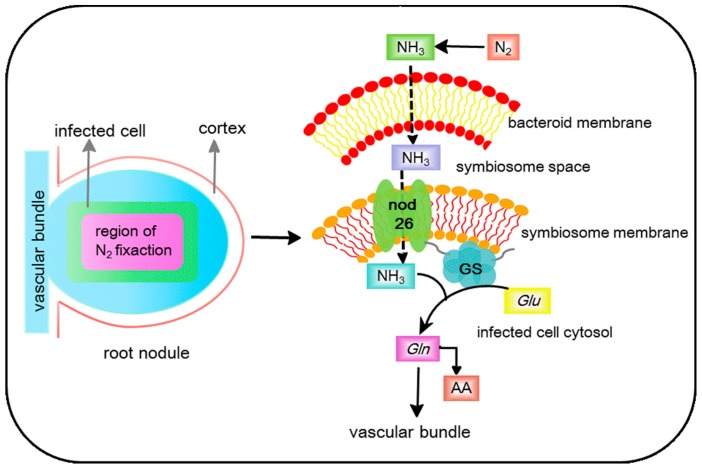
The assimilation of symbiotes fixes nitrogen to the plant cytosol, as the symbiosome membrane acts as a barrier. Nodulin 26 is an ammoniaporin [76], and there is a high concentration of this on the symbiosome membrane, which facilitates NH3/NH4+ transport in a bidirectional manner [114]. Indeed, Nodulin-26 possesses a fivefold higher preference for ammonia compared to water [76]. The direction of transport would depend on the concentration gradient of NH3 across the symbiosome membrane. Upon transport to the infected cell cytosol, ATP-dependent glutamine synthetase (GS) can promote the ammonium ion to be assimilated into organic form [115]. This process could be accelerated by the combination of soybean nodule cytosolic GS with the nodulin 26 carboxyl terminal domain [116]. The association of GS with nodulin 26 can facilitate rapid nitrogen assimilation, preventing the accumulation of free ammonia in the cytosol nodulin 26, which has a potential interplay with GS to mediate ammonia efflux and assimilation (Figure 1).
Figure 1.
Root nodules showing regions of active nitrogen fixation, and a model of nitrogen efflux and assimilation with the interaction of nodulin 26/glutamine synthetase (GS). Fixed nitrogen within the symbiosome space can be transported by the nodulin 26. GS which bind to the C-terminal domain of nodulin 26 serves as a site in the symbiosome membrane for rapid assimilation ammonia, and then gets released into the infected cell’s cytosol. GS, glutamine synthetase; Glu, glutamate; Gln, glutamine; AA, amino acid.
Additional roles for AQP in rhizobium-legume symbiosis have been suggested for TIP1. During root nodule differentiation in barrel clover (Medicago truncatula), a TIP1 homolog was shown to be transiently retargeted from the tonoplast to the symbiosome membrane [97]. Further, the re-targeting of TIP1g is important for the distribution and maturation of symbiosomes in infected cells, which can probably be achieved by increasing the availability of water [97].
3.1.2. Mycorrhizal
Mycorrhizae represent a very common symbiotic interaction between soil fungi and plant roots. The plant interface with mycorrhizal fungi may be intracellular (as with arbuscular mycorrhizae (AM)) or extracellular (as with ecto-mycorrhizae (EM)). In each case, mycorrhizal fungal mycelia improve nutrient status and water relations for their hosts, whereas the plant root provides carbon metabolites for nutrient assimilation. AQPs play an important role in both water and nutrient exchange in plant-mycorrhizal symbiosis [117]. Water conservation and absorption are two main mechanisms in plant-mycorrhizal symbiosis to cope better with stressful environmental conditions, such as drought, flooding, cold, or salinity [98,101,103,104,118]. To improve water relations, mycorrhizal symbioses can actively modify the function and gene expression of plant aquaporins to influence conditions of relative apoplastic water flow [42,119,120,121,122]. The regulation of plant AQPs seems to differ between the plant and fungal species that are involved in the symbiosis.
Mycorrhizal: AM Symbiosis
Arbuscular mycorrhizae are capable of establishing symbiotic relationships with many plants [123]. During the formation of the AM symbiosis, the plant plasma membranes undergo extensive morphological alterations that closely surround the fungal hyphae, resulting in an increase in the outer plant cell surface area [124]. As a result, symbiont can acquire nutrients (mainly phosphates) and water more efficiently than the root alone [125]. AQPs promote the morphological alterations of plants to increase symbiotic efficiency. The tobacco plasma membrane aquaporin NtAQP1 allows for CO2 passage and contributes to plant growth promotion [126]. Further, AM symbiosis indeed results in altered rates of water movement into, through, and out of the host plants [127], and also modifies root hydraulic conductivity under specific stressed conditions [98,103,128,129]. These events have been linked to AQPs. Compared to non-AM plants, AM plants significantly enhanced relative apoplastic water flow [128] and induced change in the expression of aquaporin-encoding genes, such as TIPs [119,120], PIPs [42,130], and NIPs [42]. The amount of PIP2 protein and its phosphorylation status strongly contributed to the regulation of root hydraulic properties by AM symbiosis in Phaseolus vulgaris, and enhanced root hydraulic conductivity under drought, cold, and salinity stresses in AM plants [98].
Abiotic Stresses Tolerance
It is now well-established that AQPs are involved in the regulation of host plant tolerance to stress in AM symbiosis as a process that depends on both plant and fungal interactors. Porcel et al. [99] observed that mycorrhizal wild-type plants grew faster than antisense tobacco plants which targeted mycorrhizal NtAQP1 under drought stress. This was linked to reduced symbiotic efficiency of AM fungi, mostly due to the reduced expression of PIP genes [99]. Furthermore, water conservation is an important stress tolerance mechanism, and the downregulation of AQPs protects AM plants from drought stress. PIP genes from Glycine max (GmPIP1, GmPIP2) and Lactuca sativa (LsPIP1, LsPIP2) were down-regulated following AM (Glomus mosseae) infection under drought stress and AM plants accelerated the downregulation of these genes compared to non-AM plants [101]. This aligned with the conclusions of previous experiments, where they showed that the overexpression of PIPs in transgenic tobacco or Arabidopsis had a negative effect on plants, causing fast wilting under drought stress [131,132]. Equally, under non-stressed conditions, PIP gene overexpression improved plant vigor. The decreased expression of PIP genes during drought stress in roots of AM plants can be a regulatory mechanism to decrease membrane water permeability and limit water loss from cells [101,133].
If this is the case, the AM fungal species and PIPs diversity could influence the efficacy of the above resistance mechanism. This was shown when lettuce plants were colonized with the AM fungus, Glomus intraradices where they did not exhibit such down-regulation of PIP gene expression, and the roots had higher water permeability [101]. In a comparative study involving colonization by G. intraradice or G. mosseae, the former absorbed water much more efficiently [102]. Considering the role of PIP isoforms, Aroca et al. [98] analyzed four PIP genes in roots from Phaseolus vulgaris which either were or were not colonized by AM fungi, and subjected to drought, cold, or salinity. PvPIP1;1, PvPIP1;2, and PvPIP1;3 expression showed differences in AM and non-AM plants according to the stress imposed. Different genes showed differential function and regulation by AM symbiosis under the different conditions. Notwithstanding such subtleties, the overall observation that AQPs seem to influence water permeability and nutrient flux more efficiently in AM plants with stress appears to be the overall rule.
Solute transport
The role of AQPs-mediated solute transport in AM is also important. Several of AM-regulated maize AQPs (ZmPIP1;3, ZmPIP2;2, ZmTIP1;1, ZmTIP1;2, ZmNIP1;1, ZmNIP2;1, and ZmNIP2;2) transport glycerol, ammonia, or H2O2, as well as water, which are vital to plant physiological performance under well-watered conditions [103]. Glycerol is also transported by tobacco NtTIPa [100], ZmNIP1;1, ZmTIP4;1 and ZmTIP4 of maize [103], OsTIP1;2, OsTIP3;2, and OsTIP4;1 from rice [96]. While the physiological function of glycerol in plants is unclear, glycerol is an important carbon source for pathogenic fungi [134] and may be important in establishing symbiotic relationships [135,136]. Thus, AQPs transporting glycerol from the plant to the microbe will facilitate AM symbiosis under sustained drought stress.
The regulation of nitrogen movement is also tightly related to AQPs in higher plants. The PIP, NIP, and TIP subfamilies can transport ammonia and urea to maintain their relative balance between the cytoplasm and vacuole [37,137,138]. Almost all TIP subclasses in Arabidopsis transport urea [139], as do ZmNIP2;1, ZmNIP2;4, and ZmTIP4;4 in maize [137]. This is of direct relevance to AM symbiosis, where inorganic nitrogen (ammonium and nitrate) is assimilated and transferred to the host plant [140], and urea plays a role as an intermediate solute [141,142]. Such suggestions by ZmTIP1;1 and ZmTIP1;2, which were regulated by the AM symbiosis, have been shown to transport both ammonia and urea [103].
Beyond nutrition, AQP could influence redox-mediated events. Superoxide-generating nicotinamide-adenine dinucleotide phophate (NADPH) oxidases located on the plasma membrane are major sources of apoplastic hydrogen peroxide (H2O2) production during AMF symbiosis. H2O2 accumulates in symbiont cells and around hyphal tips attempting to penetrate a host cell [143,144]. Such can reduce penetration by augmenting the plant’s innate defence response. However, the H2O2 could be removed from the interaction site by diffusion across the plasma membrane through AQP channels where it can be reduced by potent antioxidant systems [24]. For example, ZmTIP1;1 has been shown to transport H2O2 for detoxification under stress conditions [103]. The generation of reactive oxygens species is a feature which is common to many stresses and is a central event in plants’ responses to them, so the role of AQPs in the transport and relocation of H2O2 as a detoxification or a signaling mechanism needs to be studied further.
Mycorrhizal: EM Symbiosis
Another type of mycorrhizal interaction is ectomycorrhizal symbiosis. In this form of symbiosis, the fungal hyphae are extracellular to plant cells and closely surround the roots, or form a thick network known as a Hartig net between the epidermal and cortical cells [145,146]. EM fungi provide the host plant with mineral nutrients and water in exchange for carbon compounds from the host [147]. AQPs play a major role in these aspects of EM symbiosis.
As with AM fungi, AQPs regulate plant defences’ responses to facilitate EM symbiosis. Rather than crops (bean, soybean, or lettuce), the host plants of EM fungi are mostly perennial woody plants with strong seasonality, and EM symbiosis will enhance root water transport, which has been linked to increased AQP expression. This increased AQP has been shown to influence the rate of transmembrane water transport and, consequently, root hydraulic conductance, even in cell-to-cell pathways [35,106]. In poplar, PttPIP2;5 was upregulated in EM roots compared to non-mycorrhizal seedlings, and this was more important in water stress conditions [104]. Similarly, expression of PttPIP2.2 and PttPIP2.4 was higher in mycorrhized poplar plants under drought stress [105]. Tarkka et al. [148] also found an increase in the expression of one SIP and five PIP genes in Quercus robur roots inoculated with the EM fungus. Some studies showed that the upregulation of PIP genes was correlated with higher root hydraulic conductance and increased water transport from the fungus to the intercellular EM root cells in poplar [104,105]. Such higher expression of PIP genes with higher root-specific conductance could partially compensate for certain plants having smaller root sizes which could otherwise limit water transport in dwarf plants [106]. However, the role for AQPs in roots’ EM symbiosis is not a universal feature of all host plants. In contrast to the poplar, inoculation of A. muscaria on Norway spruce roots had no effect on the expression of root AQPs. Also, two PIP genes of the Picea glauca-inoculated Laccaria bicolor were downregulated with enhanced root hydraulic conductance [107].
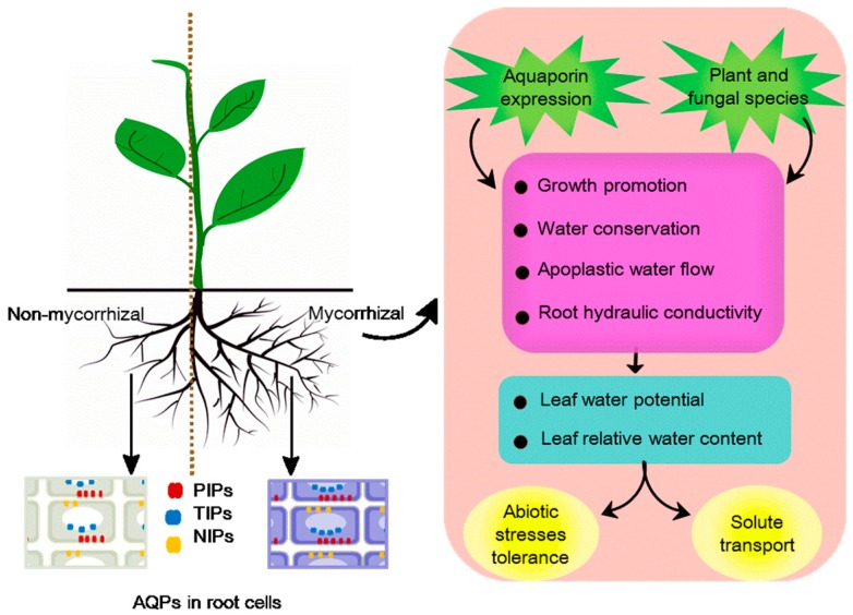
Leaving aside what could be exceptions to a general rule, the literature strongly supports the importance of AQPs in AM/EM symbioses in regulating plant–water relations (relative water content and leaf water potential) and physiology (morphology, nutrition, signaling molecules and other solutes), and these are summarized in Figure 2.
Figure 2.
Model of the role of aquaporins (AQPs) in arbuscular mycorrhizae (AM)/ecto-mycorrhizae (EM) symbioses combined with plant–water relations and physiological activity. Mycorrhizal symbioses promote growth of mycorrhizal plants, with a larger root system (the right side of the root system). The amount of AOPs in mycorrhizal plant root cells (right side, blue) is generally more than that in non-mycorrhizal plant root cells (left side, gray). Plasma membrane intrinsic proteins (PIPs) and nodulin 26-like intrinsic proteins (NIPs) are in plasma membranes, and tonoplast intrinsic proteins (TIPs) are in the tonoplast (white oval). The regulation of plant AQPs contributes to growth promotion, water conservation, apoplastic water flow, and root hydraulic conductivity to maintain the relative water content and leaf water potential. Favorable water relation promotes solute transport and increases abiotic stress tolerance. Plant and fungal species could also influence the efficacy of the above resistance mechanism.
3.1.3. Plant Growth-Promoting Rhizobacteria (PGPR)
Plant growth-promoting rhizobacteria (PGPR) are naturally occurring soil bacteria inhabiting rhizospheres, which facilitate plant growth by improving plant productivity and immunity under normal and stress conditions [149]. PGPR has largely been documented to contribute to plant tolerance against drought and salinity [108,150]. Under such conditions, plants must decrease their cell water potential to continue to take up water to reduce the impact of increased salinity or water limitation. This clearly indicates a role for AQPs, but the literature suggests that the responses of host-microbiome interactions are specific and require further characterization [151].
Inoculation with Azospirillum brasilense, the nitrogen-fixing bacterial species, has been shown to result in the upregulation of HvPIP2-1 in barley seedlings [108]. Maize plants inoculated with Bacillus megaterium has shown increased root hydraulic conductivity compared to uninoculated plants when exposed to salinity, and this was correlated with increased expression of ZmPIP1;1 and ZmPIP1;5 [109]. In a similar way, Pantoea agglomerans and B. megaterium-infected Zea mays roots showed upregulated PIP2 and ZmPIP1-1 expression regulated under salt stress conditions, which contributed to increased root hydraulic conductance in inoculated plants [152]. However, ZmPIP2;1 was down-regulated in inoculated roots, although this was up-regulated by salt addition alone. This inoculation effect was not seen post-transcriptionally where ZmPIP1;2 accumulation occurred [109], and was likely to increase the water-use efficiency of PGPR-interacting plants [153]. In lettuce plants inoculated with Pseudomonas mendocina, the maintenance of relative water content during drought stress correlated with a downregulation of PIP2. Indeed, PIP2 expression was significantly enhanced by P. mendocina only in plants grown under well-watered conditions [110].
3.2. Negative Plant–Microbe Interactions
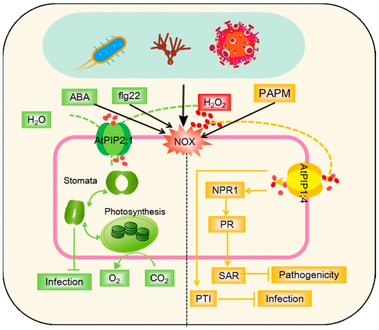
The literature strongly supports the roles of AQP in various forms of symbiotic interactions, yet the roles of AQPs in negative plant–microbe interactions are largely unknown. Plant pathogens effectively utilize plant nutrients solely for their growth and survival to the detriment of their plant hosts. AQPs expression patterns, manipulated by the pathogen, could play a role in this, as could the dehydration in the host that is a common disease symptom. The roles of AQPs in such negative interactions are depicted in Figure 3.
Figure 3.
Model of AQPs-mediated linkage of apoplastic H2O2 to stomatal closure (green) and immunity pathways (systemic acquired resistance (SAR) and PAMP-triggered immunity (PTI)) (yellow). Apoplastic H2O2 can be induced by the pathogen or pathogen-associated molecular pattern (PAMPs, flg22), generated through the NADPH oxidase (NOX) activity and rapidly translocated into the cytoplasm under regulation by AtPIP2;1 or AtPIP1;4. Abscisic acid (ABA) and flg22 (a PAMP) activate AtPIP2;1 to facilitate transport of both water and H2O2, and promote stomatal closure to restrict bacterial invasion. Translocated H2O2 cooperates with SAR or PTI to repress the pathogenicity. ABA, abscisic acid; H2O2, hydrogen peroxide; PAMP, pathogen-associated molecular patterns; flg22, a 22-amino acid eubacterial flagellin-derived peptide; NOX, NADPH oxidase; PR, pathogenesis-related genes; NPR1, non-inducer of PR genes1; SAR, systemic acquired resistance; PTI, PAMP-triggered immunity.
In considering the effects of AQP on physiological activity linked to disease, stomata are crucial to the control of plants’ water status in response to pathogens’ attack, and are potential entry points for foliar pathogens (Figure 3, green). Plants have the capacity to close their stomata after the perception of pathogen-associated molecular patterns (PAMP) to restrict bacterial invasion [154]. Rodrigues et al. [25] proposed a model whereby flg22 (a 22-amino acid eubacterial flagellin-derived peptide, a PAMP) and abscisic acid (ABA) activated PIP2;1 through phosphorylation at a conserved site, to facilitate transport of both water and H2O2 to promote stomatal closure. H2O2 production in the stomatal guard cells appears to be elicited by both ABA and PAMP, which can be transported into the cytosol by AQPs to mediate stomatal movement, acting through PIP2;1 [25,155].
Transport of apoplastic H2O2 to the cytoplasm of plant cells has been demonstrated via AtPIP1;4 following challenges to the bacterial pathogen, Pseudomonas syringae [26] (Figure 3, yellow). Once within the cell, this H2O2, and therefore AtPIP1;4, plays a key role in plant immunity pathways. Thus, in the cytoplasm, the translocated H2O2 activates the PAMP-triggered immunity (PTI) pathway to resist pathogens. It also activates the biosynthesis of salicylic acid, which acts via the signaling component, NPR1 to establish systemic acquired resistance (SAR) [26]. PIP1;4 also plays a role on effector-triggered immunity (ETI), where effectors are delivered from the pathogen into the host as part of the pathogenic process. Certain plant genotypes can recognize the effectors to initiate ETI.
In bacterial pathogens, effector delivery is done via a type of secretion mechanism which includes the harpin protein hpa1. Hpa1 interacts with aquaporin PIP1;4 of Arabidopsis [156] and OsPIP1;3 of rice. Hpa1 interactions with Arabidopsis AtPIP1;4 mediates the transport of CO2 and H2O and contributes to plant growth enhancement. In particular, this CO2 transport is linked to increases in leaf photosynthesis rates. Therefore, plant sensing of a bacterial harpin protein is connected with photosynthetic physiology to regulate plant growth [156]. For viruses, TIPs can interfere with intracellular viral replication by interacting with a cucumber mosaic virus (CMV) replication protein in the host plant’s tonoplasts [157]. Interactome data provide important clues about the involvement of aquaporins in host–pathogen interactions.
As H2O2 transport is important in responses to challenges by a pathogen, the linked changes in AQP expression are also crucial in developing our understanding of the defence network. The citrus plants major intrinsic protein CsMIPs showed differential expressions when infected with Candidatus Liberibacter asiaticus (Ca. L. asiaticus), the causal agent of huanglongbing disease [158]. Ca. L. asiaticus infection repressed expression of PIP1A, PIP1-1, PIP1-3 genes, while it induced expression of the SIP1A gene to affect the water and nutrients status [44]. The expression of AQP could be very infection stage-specific and reflects different roles in a dynamic process. This was suggested by the expression pattern of CaPIP1-1 in pepper leaves challenged with Phytophthora capsici, which were up-regulated in the stage of spore germination along with plant metabolism observed to change [112].
4. Conclusions and Perspectives
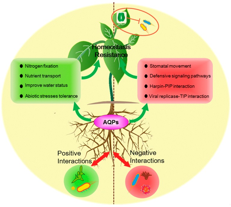
AQPs seem to play various roles in different plant–microbe interactions, as shown in Figure 4. AQPs mainly transport water and other physiological substances to maintain relative water content and leaf water potential by regulating specific AQP gene expression and activity under specific growth conditions. The above mechanism is most established for positive plant-microbe interactions. For negative plant-microbe interactions, AQPs use signaling in guard cells to adjust stomata and can interact with pathogen effectors or replicase, and can also induce resistance of plants by H2O2 signaling. In all, AQPs are responsible for maintaining water and nutrient homeostasis and increasing resistance against pathogen. Although numerous studies have elucidated the possible role of AQPs in plant–microbe interactions, detailed mechanisms of AQPs regulating plant–microbe interactions as a channel for other substances or signaling need further study. And attention should be focused on the following issues in the future:
AQP is a kind of channel, and future research should focus on identifying their capacity to transport mycelium, spores, or small secondary metabolites of pathogen.
Regulation of AQP by different plant–microbes interactions on complex processes and signaling pathways, as well as complex transcriptional, translational, and posttranscriptional factors.
The role of microbe APQs in regulation of the plant–microbe interaction.
Figure 4.
AQPs’ currently established positive roles in different pathways on plants in both positive and negative plant–microbe interactions. For positive plant–microbe interactions, AQPs mainly transport water and other physiological substances to maintain relative water content and leaf water potential by regulating specific AQP gene expression and activity under specific growth conditions. For negative plant–microbe interactions, AQPs use signaling in guard cells to adjust stomata and can interact with pathogen effectors or replicase, and can also induce resistance of plants by H2O2 signaling.
Author Contributions
R.W. and M.W. contributed to the conception and design of the layout and prepared the manuscript; K.C. and S.W. contributed table and figures; L.A.J.M. and S.G. guided proofread and polished the manuscript. R.W. and M.W. contributed equally to this work.
Funding
This work was financially supported by the National Key R&D Program (2016YFD0200300), the National Basic Research Program of China (2015CB150505), the Fundamental Research Funds for the Central Universities (KJQN201514 and KYZ201625).
Conflicts of Interest
The authors declare no conflict of interest.
References
- 1.Taketani R.G., Lançoni M.D., Kavamura V.N., Durrer A., Andreote F.D., Melo I.S. Dry season constrains bacterial phylogenetic diversity in a semi-arid rhizosphere system. Microb. Ecol. 2016;73:1–9. doi: 10.1007/s00248-016-0835-4. [DOI] [PubMed] [Google Scholar]
- 2.Aung K., Jiang Y., He S.Y. The role of water in plant–microbe interactions. Plant J. 2018;93:771–780. doi: 10.1111/tpj.13795. [DOI] [PMC free article] [PubMed] [Google Scholar]
- 3.Moreau D., Pivato B., Bru D., Busset H., Deau F., Faivre C., Matejicek A., Strbik F., Philippot L., Mougel C. Plant traits related to nitrogen uptake influence plant–microbe competition. Ecology. 2016;96:2300–2310. doi: 10.1890/14-1761.1. [DOI] [PubMed] [Google Scholar]
- 4.Panos L., Patterson G.A., Demajo W.A. Role of iron in plant–microbe interaction. Adv. Bot. Res. 2010;51:491–549. [Google Scholar]
- 5.Wang M., Gao L., Dong S., Sun Y., Shen Q., Guo S. Role of silicon on plant–pathogen interactions. Front. Plant Sci. 2017;8 doi: 10.3389/fpls.2017.00701. [DOI] [PMC free article] [PubMed] [Google Scholar]
- 6.Yadav B.K., Akhtar M.S., Panwar J. Rhizospheric Plant–Microbe Interactions: Key Factors to Soil Fertility and Plant Nutrition. Springer; New Delhi, India: 2015. pp. 127–145. [Google Scholar]
- 7.Bais H.P., Broeckling C.D., Vivanco J.M. Root Exudates Modulate Plant—Microbe Interactions in the Rhizosphere. Springer; Berlin/Heidelberg, Germany: 2007. pp. 241–252. [Google Scholar]
- 8.Biate D.L., Kumari A., Annapurna K., Kumar L.V., Ramadoss D., Reddy K.K., Naik S. Legume Root Exudates: Their Role in Symbiotic Interactions. Springer; New Delhi, India: 2015. pp. 259–271. [Google Scholar]
- 9.Haichar F.E.Z., Santaella C., Heulin T., Achouak W. Root exudates mediated interactions belowground. Soil Biol. Biochem. 2014;77:69–80. doi: 10.1016/j.soilbio.2014.06.017. [DOI] [Google Scholar]
- 10.Baker B., Zambryski P., Staskawicz B., Dinesh-Kumar S.P. Signaling in plant–microbe interactions. Science. 1997;276:726–733. doi: 10.1126/science.276.5313.726. [DOI] [PubMed] [Google Scholar]
- 11.Hückelhoven R., Kogel K.H. Reactive oxygen intermediates in plant–microbe interactions: Who is who in powdery mildew resistance? Planta. 2003;216:891–902. doi: 10.1007/s00425-003-0973-z. [DOI] [PubMed] [Google Scholar]
- 12.Kan J., Fang R., Jia Y. Interkingdom signaling in plant–microbe interactions. Sci. China Life Sci. 2017;60:1–12. doi: 10.1007/s11427-017-9092-3. [DOI] [PubMed] [Google Scholar]
- 13.Pozo M.J., Loon L.C.V., Pieterse C.M.J. Jasmonates-signals in plant–microbe interactions. J. Plant Growth Regul. 2004;23:211–222. [Google Scholar]
- 14.Maurel C., Verdoucq L., Rodrigues O. Aquaporins and plant transpiration. Plant Cell Environ. 2016;39:2580–2587. doi: 10.1111/pce.12814. [DOI] [PubMed] [Google Scholar]
- 15.Chaumont F., Tyerman S.D. Aquaporins: Highly regulated channels controlling plant water relations. Plant Physiol. 2014;164:1600–1618. doi: 10.1104/pp.113.233791. [DOI] [PMC free article] [PubMed] [Google Scholar]
- 16.Johansson I., Karlsson M., Johanson U., Larsson C., Kjellbom P. The role of aquaporins in cellular and whole plant water balance. Biochim. Biophys. Acta. 2000;1465:324–342. doi: 10.1016/S0005-2736(00)00147-4. [DOI] [PubMed] [Google Scholar]
- 17.Sun H., Wang S., Lou Y., Zhu C., Zhao H., Li Y., Li X., Gao Z. Whole-genome and expression analyses of bamboo aquaporin genes reveal their functions involved in maintaining diurnal water balance in bamboo shoots. Cells. 2018;7:195. doi: 10.3390/cells7110195. [DOI] [PMC free article] [PubMed] [Google Scholar]
- 18.Baiges I., Schaffner A.R., Mas A., Affenzeller M.J. Plant aquaporins. Physiol. Plant. 2010;115:175–182. doi: 10.1034/j.1399-3054.2002.1150201.x. [DOI] [PubMed] [Google Scholar]
- 19.Chaumont F., Tyerman S.D. Plant Aquaporins. Springer International Publishing; New York, NY, USA: 2017. [Google Scholar]
- 20.Kaldenhoff R. Mechanisms underlying CO2 diffusion in leaves. Curr. Opin. Plant Biol. 2012;15:276–281. doi: 10.1016/j.pbi.2012.01.011. [DOI] [PubMed] [Google Scholar]
- 21.Herrera M., Garvin J.L. Aquaporins as gas channels. Pflug. Arch. Eur. J. Phys. 2011;462:623–630. doi: 10.1007/s00424-011-1002-x. [DOI] [PubMed] [Google Scholar]
- 22.Kaldenhoff R., Kai L., Uehlein N. Aquaporins and membrane diffusion of CO2 in living organisms. BBA-Gen. Subj. 2014;1840:1592–1595. doi: 10.1016/j.bbagen.2013.09.037. [DOI] [PubMed] [Google Scholar]
- 23.Ludewig U., Yuan L., Wirén N.V. Tonoplast intrinsic proteins AtTIP2;1 and AtTIP2;3 facilitate NH₃ transport into the vacuole. Plant Physiol. 2005;137:671–680. doi: 10.1104/pp.104.051268. [DOI] [PMC free article] [PubMed] [Google Scholar]
- 24.Bienert G.P., Chaumont F. Aquaporin-facilitated transmembrane diffusion of hydrogen peroxide. BBA-Gen. Subj. 2014;1840:1596–1604. doi: 10.1016/j.bbagen.2013.09.017. [DOI] [PubMed] [Google Scholar]
- 25.Rodrigues O., Reshetnyak G., Grondin A., Saijo Y., Leonhardt N., Maurel C., Verdoucq L. Aquaporins facilitate hydrogen peroxide entry into guard cells to mediate ABA- and pathogen-triggered stomatal closure. Proc. Natl. Acad. Sci. USA. 2017;114:9200–9205. doi: 10.1073/pnas.1704754114. [DOI] [PMC free article] [PubMed] [Google Scholar]
- 26.Tian S., Wang X., Li P., Wang H., Ji H., Xie J., Qiu Q., Shen D., Dong H. Plant aquaporin AtPIP1;4 links apoplastic H2O2 induction to disease immunity pathways. Plant Physiol. 2016;171:1635–1650. doi: 10.1104/pp.15.01237. [DOI] [PMC free article] [PubMed] [Google Scholar]
- 27.Garneau A.P., Carpentier G.A., Marcoux A.A., Frenettecotton R., Simard C.F., Rémusborel W., Caron L., Jacobwagner M., Noël M., Powell J.J. Aquaporins mediate silicon transport in humans. PLoS ONE. 2015;10 doi: 10.1371/journal.pone.0136149. [DOI] [PMC free article] [PubMed] [Google Scholar]
- 28.Ma J.F., Yamaji N. Silicon uptake and accumulation in higher plants. Trends Plant Sci. 2006;11:392–397. doi: 10.1016/j.tplants.2006.06.007. [DOI] [PubMed] [Google Scholar]
- 29.Maurel C., Boursiac Y., Luu D.T., Santoni V., Shahzad Z., Verdoucq L. Aquaporins in plants. Physiol. Rev. 2015;95:1321–1358. doi: 10.1152/physrev.00008.2015. [DOI] [PubMed] [Google Scholar]
- 30.Byrt C.S., Zhao M., Kourghi M., Bose J., Henderson S.W., Qiu J., Gilliham M., Schultz C., Schwarz M., Ramesh S.A. Non-selective cation channel activity of aquaporin AtPIP2;1 regulated by Ca2+ and pH. Plant Cell Environ. 2017;40:802–815. doi: 10.1111/pce.12832. [DOI] [PubMed] [Google Scholar]
- 31.Yasui M., Hazama A., Kwon T.H., Nielsen S., Guggino W.B., Agre P. Rapid gating and anion permeability of an intracellular aquaporin. Nature. 1999;402:184–187. doi: 10.1038/46045. [DOI] [PubMed] [Google Scholar]
- 32.Zampighi G.A., Hall J.E., Kreman M. Purified lens junctional protein forms channels in planar lipid films. P. Natl. Acad. Sci. USA. 1985;82:8468–8472. doi: 10.1073/pnas.82.24.8468. [DOI] [PMC free article] [PubMed] [Google Scholar]
- 33.Li G., Santoni V., Maurel C. Plant aquaporins: Roles in plant physiology. BBA-Gen. Subj. 2014;1840:1574–1582. doi: 10.1016/j.bbagen.2013.11.004. [DOI] [PubMed] [Google Scholar]
- 34.Maurel C. Plant aquaporins: Novel functions and regulation properties. FEBS Lett. 2007;581:2227–2236. doi: 10.1016/j.febslet.2007.03.021. [DOI] [PubMed] [Google Scholar]
- 35.Maurel C., Verdoucq L., Luu D.T., Santoni V. Plant aquaporins: Membrane channels with multiple integrated functions. Annu. Rev. Plant. Biol. 2008;59:595–624. doi: 10.1146/annurev.arplant.59.032607.092734. [DOI] [PubMed] [Google Scholar]
- 36.Gao L., Lu Z., Ding L., Guo J., Wang M., Ling N., Guo S., Shen Q. Role of aquaporins in determining carbon and nitrogen status in higher plants. Int. J. Mol. Sci. 2018;19:35. doi: 10.3390/ijms19010035. [DOI] [PMC free article] [PubMed] [Google Scholar]
- 37.Wang M., Ding L., Gao L., Li Y., Shen Q., Guo S. The interactions of aquaporins and mineral nutrients in higher plants. Int. J. Mol. Sci. 2016;17:1229. doi: 10.3390/ijms17081229. [DOI] [PMC free article] [PubMed] [Google Scholar]
- 38.Afzal Z., Howton T.C., Sun Y., Mukhtar M.S. The roles of aquaporins in plant stress responses. Int. J. Dev. Biol. 2016;4:9. doi: 10.3390/jdb4010009. [DOI] [PMC free article] [PubMed] [Google Scholar]
- 39.Kapilan R., Vaziri M., Zwiazek J.J. Regulation of aquaporins in plants under stress. Biol. Res. 2018;51:4. doi: 10.1186/s40659-018-0152-0. [DOI] [PMC free article] [PubMed] [Google Scholar]
- 40.Srivastava A.K., Penna S., Nguyen D.V., Tran L.S. Multifaceted roles of aquaporins as molecular conduits in plant responses to abiotic stresses. Crit. Rev. Biotechnol. 2016;36:389–398. doi: 10.3109/07388551.2014.973367. [DOI] [PubMed] [Google Scholar]
- 41.Shekoofa A., Sinclair T. Aquaporin activity to improve crop drought tolerance. Cells. 2018;7:123. doi: 10.3390/cells7090123. [DOI] [PMC free article] [PubMed] [Google Scholar]
- 42.Uehlein N., Fileschi K., Eckert M., Bienert G.P., Bertl A., Kaldenhoff R. Arbuscular mycorrhizal symbiosis and plant aquaporin expression. Phytochemistry. 2007;68:122–129. doi: 10.1016/j.phytochem.2006.09.033. [DOI] [PubMed] [Google Scholar]
- 43.Watts-Williams S.J., Tyerman S.D., Cavagnaro T.R. The dual benefit of arbuscular mycorrhizal fungi under soil zinc deficiency and toxicity: Linking plant physiology and gene expression. Plant. Soil. 2017;420:1–14. doi: 10.1007/s11104-017-3409-4. [DOI] [Google Scholar]
- 44.Aritua V., Achor D., Gmitter F.G., Albrigo G., Wang N. Transcriptional and microscopic analyses of citrus stem and root responses to Candidatus Liberibacter asiaticus infection. PLoS ONE. 2013;8:e73742. doi: 10.1371/journal.pone.0073742. [DOI] [PMC free article] [PubMed] [Google Scholar]
- 45.Du Y., Gong Z., Liu G., Zhao Y. Scanning electron microscopic study of the infection process of Phytophthora capsici. Pak. J. Bot. 2013;45:1807–1811. [Google Scholar]
- 46.Bauer P.J., Scheffler B.E., Wonkeun P., Todd C.B. Identification of the family of aquaporin genes and their expression in upland cotton (Gossypium hirsutum L.) BMC Plant Biol. 2010;10:142. doi: 10.1186/1471-2229-10-142. [DOI] [PMC free article] [PubMed] [Google Scholar]
- 47.Ishibashi K., Kondo S., Hara S. The Evolutionary Aspects of Aquaporin Family. Springer; Dordrecht, The Netherlands: 2017. [Google Scholar]
- 48.Gupta A.B., Sankararamakrishnan R., Gupta A.B., Sankararamakrishnan R. Genome-wide analysis of major intrinsic proteins in the tree plant Populus trichocarpa: Characterization of XIP subfamily of aquaporins from evolutionary perspective. BMC Plant Biol. 2009;9 doi: 10.1186/1471-2229-9-134. [DOI] [PMC free article] [PubMed] [Google Scholar]
- 49.Wayne R., Tazawa M. Nature of the water channels in the internodal cells of Nitellopsis. J. Membr. Biol. 1990;116:31–39. doi: 10.1007/BF01871669. [DOI] [PubMed] [Google Scholar]
- 50.Chaumont F., Barrieu F., Jung R., Chrispeels M.J. Plasma membrane intrinsic proteins from maize cluster in two sequence subgroups with differential aquaporin activity. Plant Physiol. 2000;122:1025–1034. doi: 10.1104/pp.122.4.1025. [DOI] [PMC free article] [PubMed] [Google Scholar]
- 51.Marin-Olivier M., Chevalier T., Fobis-Loisy I., Dumas C., Gaude T. Aquaporin PIP genes are not expressed in the stigma papillae in Brassica oleracea. Plant J. 2010;24:231–240. doi: 10.1046/j.1365-313x.2000.00874.x. [DOI] [PubMed] [Google Scholar]
- 52.Suga S., Maeshima M. Water channel activity of radish plasma membrane aquaporins heterologously expressed in yeast and their modification by site-directed mutagenesis. Plant Cell Physiol. 2004;45:823–830. doi: 10.1093/pcp/pch120. [DOI] [PubMed] [Google Scholar]
- 53.Kaldenhoff R., Fischer M. Aquaporins in plants. Acta Physiol. 2006;187:169–176. doi: 10.1111/j.1748-1716.2006.01563.x. [DOI] [PubMed] [Google Scholar]
- 54.Fetter K., Van V.W., Moshelion M., Chaumont F. Interactions between plasma membrane aquaporins modulate their water channel activity. Plant Cell. 2004;16:215–228. doi: 10.1105/tpc.017194. [DOI] [PMC free article] [PubMed] [Google Scholar]
- 55.Mahdieh M., Mostajeran A., Horie T., Katsuhara M. Drought stress alters water relations and expression of PIP-type aquaporin genes in Nicotiana tabacum plants. Plant Cell Physiol. 2008;49:801–813. doi: 10.1093/pcp/pcn054. [DOI] [PubMed] [Google Scholar]
- 56.Secchi F., Lovisolo C., Uehlein N., Kaldenhoff R., Schubert A. Isolation and functional characterization of three aquaporins from olive (Olea europaea L.) Planta. 2007;225:381–392. doi: 10.1007/s00425-006-0365-2. [DOI] [PubMed] [Google Scholar]
- 57.Sato-Nara K., Demura T., Fukuda H. Expression of photosynthesis-related genes and their regulation by light during somatic embryogenesis in Daucus carota. Planta. 2004;219:23–31. doi: 10.1007/s00425-003-1201-6. [DOI] [PubMed] [Google Scholar]
- 58.Peng Y., Lin W., Cai W., Arora R. Overexpression of a Panax ginseng tonoplast aquaporin alters salt tolerance, drought tolerance and cold acclimation ability in transgenic Arabidopsis plants. Planta. 2007;226:729–740. doi: 10.1007/s00425-007-0520-4. [DOI] [PubMed] [Google Scholar]
- 59.Aroca R., Ferrante A., Vernieri P., Chrispeels M.J. Drought, abscisic acid and transpiration rate effects on the regulation of PIP aquaporin gene expression and abundance in Phaseolus vulgaris plants. Ann. Bot. 2006;98:1301–1310. doi: 10.1093/aob/mcl219. [DOI] [PMC free article] [PubMed] [Google Scholar]
- 60.Chervin C., Tira-Umphon A., Terrier N., Zouine M., Severac D., Roustan J.P. Stimulation of the grape berry expansion by ethylene and effects on related gene transcripts, over the ripening phase. Physiol. Plant. 2010;134:534–546. doi: 10.1111/j.1399-3054.2008.01158.x. [DOI] [PubMed] [Google Scholar]
- 61.Li G.W., Peng Y.H., Yu X., Zhang M.H., Cai W.M., Sun W.N., Su W.A. Transport functions and expression analysis of vacuolar membrane aquaporins in response to various stresses in rice. J. Plant Physiol. 2008;165:1879–1888. doi: 10.1016/j.jplph.2008.05.002. [DOI] [PubMed] [Google Scholar]
- 62.Ma N., Xue J., Li Y., Liu X., Dai F., Jia W., Luo Y., Gao J. Rh-PIP2;1, a rose aquaporin gene, is involved in ethylene-regulated petal expansion. Plant Physiol. 2008;148:894–907. doi: 10.1104/pp.108.120154. [DOI] [PMC free article] [PubMed] [Google Scholar]
- 63.Höfte H., Hubbard L., Reizer J., Ludevid D., Herman E.M., Chrispeels M.J. Vegetative and seed-specific forms of tonoplast intrinsic protein in the vacuolar membrane of Arabidopsis thaliana. Plant Physiol. 1992;99:561–570. doi: 10.1104/pp.99.2.561. [DOI] [PMC free article] [PubMed] [Google Scholar]
- 64.Fleurat-Lessard P., Michonneau P., Maeshima M., Drevon J.J., Serraj R. The distribution of aquaporin subtypes (PIP1, PIP2 and γ-TIP) is tissue dependent in soybean (Glycine max) Root Nodules. Ann. Bot. 2005;96:457–460. doi: 10.1093/aob/mci195. [DOI] [PMC free article] [PubMed] [Google Scholar]
- 65.Lindahl V., Gourdon P., Andersson M., Hess B. Permeability and ammonia selectivity in aquaporin TIP2;1: Linking structure to function. Sci. Rep. 2018;8 doi: 10.1038/s41598-018-21357-2. [DOI] [PMC free article] [PubMed] [Google Scholar]
- 66.Schüssler M.D., Alexandersson E., Bienert G.P., Kichey T., Laursen K.H., Johanson U., Kjellbom P., Schjoerring J.K., Jahn T.P. The effects of the loss of TIP1;1 and TIP1;2 aquaporins in Arabidopsis thaliana. Plant J. 2010;56:756–767. doi: 10.1111/j.1365-313X.2008.03632.x. [DOI] [PubMed] [Google Scholar]
- 67.Ciavatta V.T., Morillon R., Pullman G.S., Chrispeels M.J., Cairney J. An aquaglyceroporin is abundantly expressed early in the development of the suspensor and the embryo proper of loblolly pine. Plant Physiol. 2001;127:1556–1567. doi: 10.1104/pp.010793. [DOI] [PMC free article] [PubMed] [Google Scholar]
- 68.Abascal F., Irisarri I., Zardoya R. Diversity and evolution of membrane intrinsic proteins. Biochim. Biophys. Acta. 2014;1840:1468–1481. doi: 10.1016/j.bbagen.2013.12.001. [DOI] [PubMed] [Google Scholar]
- 69.Takano J., Wada M., Ludewig U., Schaaf G., Von W.N., Fujiwara T. The Arabidopsis major intrinsic protein NIP5;1 is essential for efficient boron uptake and plant development under boron limitation. Plant Cell. 2006;18:1498–1509. doi: 10.1105/tpc.106.041640. [DOI] [PMC free article] [PubMed] [Google Scholar]
- 70.Bienert G.P., Schüssler M.D., Jahn T.P. Metalloids: Essential, beneficial or toxic? Major intrinsic proteins sort it out. Trends Biochem. Sci. 2008;33:20–26. doi: 10.1016/j.tibs.2007.10.004. [DOI] [PubMed] [Google Scholar]
- 71.Xue Q.Z., Jian F.M. Involvement of silicon influx transporter OsNIP2;1 in selenite uptake in rice. Plant Physiol. 2010;153:1871–1877. doi: 10.1104/pp.110.157867. [DOI] [PMC free article] [PubMed] [Google Scholar]
- 72.Jian Feng M., Kazunori T., Naoki Y., Namiki M., Saeko K., Maki K., Masaji I., Yoshiko M., Masahiro Y. A silicon transporter in rice. Nature. 2006;440:688–691. doi: 10.1038/nature04590. [DOI] [PubMed] [Google Scholar]
- 73.Dynowski M., Mayer M., Moran O., Ludewig U. Molecular determinants of ammonia and urea conductance in plant aquaporin homologs. FEBS Lett. 2008;582:2458–2462. doi: 10.1016/j.febslet.2008.06.012. [DOI] [PubMed] [Google Scholar]
- 74.Dynowski M., Schaaf G., Loque D., Moran O., Ludewig U. Plant plasma membrane water channels conduct the signalling molecule H2O2. Biochem. J. 2008;414:53–61. doi: 10.1042/BJ20080287. [DOI] [PubMed] [Google Scholar]
- 75.Mizutani M., Watanabe S., Nakagawa T., Maeshima M. Aquaporin NIP2;1 is mainly localized to the ER membrane and shows root-specific accumulation in Arabidopsis thaliana. Plant Cell Physiol. 2006;47:1420–1426. doi: 10.1093/pcp/pcl004. [DOI] [PubMed] [Google Scholar]
- 76.Hwang J.H., Ellingson S.R., Roberts D.M. Ammonia permeability of the soybean nodulin 26 channel. FEBS Lett. 2010;584:4339–4343. doi: 10.1016/j.febslet.2010.09.033. [DOI] [PubMed] [Google Scholar]
- 77.Benjamin P., Till Arvid D., Gerd Patrick B. Metalloido-porins: Essentiality of Nodulin 26-like intrinsic proteins in metalloid transport. Plant Sci. Int. J. Exp. Plant. Biol. 2015;238:212–227. doi: 10.1016/j.plantsci.2015.06.002. [DOI] [PubMed] [Google Scholar]
- 78.Giovannetti M., Balestrini R., Volpe V., Guether M., Straub D., Costa A., Ludewig U., Bonfante P. Two putative-aquaporin genes are differentially expressed during arbuscular mycorrhizal symbiosis in Lotus japonicus. BMC Plant Biol. 2012;12:186. doi: 10.1186/1471-2229-12-186. [DOI] [PMC free article] [PubMed] [Google Scholar]
- 79.Ishikawa F., Suga S., Uemura T., Sato M.H., Maeshima M. Novel type aquaporin SIPs are mainly localized to the ER membrane and show cell-specific expression in Arabidopsis thaliana. FEBS Lett. 2005;579:5814–5820. doi: 10.1016/j.febslet.2005.09.076. [DOI] [PubMed] [Google Scholar]
- 80.Zhang D., Huang Y., Kumar M., Wan Q., Xu Z., Shao H. Heterologous expression of GmSIP1;3 from soybean in tobacco showed and growth retardation and tolerance to hydrogen peroxide. Plant Sci. 2017;263:210–218. doi: 10.1016/j.plantsci.2017.07.018. [DOI] [PubMed] [Google Scholar]
- 81.Tombuloglu H., Ozcan I., Tombuloglu G., Sakcali S., Unver T. Aquaporins in boron-tolerant barley: Identification, characterization, and expression analysis. Plant Mol. Biol. Rep. 2016;34:374–386. doi: 10.1007/s11105-015-0930-6. [DOI] [Google Scholar]
- 82.Gururani M.A., Venkatesh J., Ghosh R., Strasser R.J., Ponpandian L.N., Bae H. Chlorophyll-a fluorescence evaluation of PEG-induced osmotic stress on PSII activity in Arabidopsis plants expressing SIP1. Plant Biosyst. 2017;152:1–8. doi: 10.1080/11263504.2017.1403392. [DOI] [Google Scholar]
- 83.Verma R.K., Prabh N.D., Sankararamakrishnan R. New subfamilies of major intrinsic proteins in fungi suggest novel transport properties in fungal channels: Implications for the host-fungal interactions. BMC Evol. Biol. 2014;14 doi: 10.1186/s12862-014-0173-4. [DOI] [PMC free article] [PubMed] [Google Scholar]
- 84.Gerd Patrick B., Manuela Désirée B., Thomas Paul J., Marc B., Francois C. Solanaceae XIPs are plasma membrane aquaporins that facilitate the transport of many uncharged substrates. Plant J. 2011;66:306–317. doi: 10.1111/j.1365-313X.2011.04496.x. [DOI] [PubMed] [Google Scholar]
- 85.Lopez D., Bronner G., Brunel N., Auguin D., Bourgerie S., Brignolas F., Carpin S., Tournaireroux C., Maurel C., Fumanal B. Insights into Populus XIP aquaporins: Evolutionary expansion, protein functionality, and environmental regulation. J. Exp. Bot. 2012;63:2217–2230. doi: 10.1093/jxb/err404. [DOI] [PubMed] [Google Scholar]
- 86.Bent E. Induced systemic resistance mediated by plant growth-promoting Rhizobacteria (PGPR) and Fungi (PGPF) In: Tuzun S., Bent E., editors. Multigenic and Induced Systemic Resistance in Plants. Springer; Boston, MA, USA: 2006. pp. 225–258. [Google Scholar]
- 87.Bloemberg G.V., Lugtenberg B.J. Molecular basis of plant growth promotion and biocontrol by rhizobacteria. Curr. Opin. Plant Biol. 2001;4:343–350. doi: 10.1016/S1369-5266(00)00183-7. [DOI] [PubMed] [Google Scholar]
- 88.Cooper J.E. Early interactions between legumes and rhizobia: Disclosing complexity in a molecular dialogue. J. Appl. Microbiol. 2007;103:1355–1365. doi: 10.1111/j.1365-2672.2007.03366.x. [DOI] [PubMed] [Google Scholar]
- 89.Dodd I.C., Ruizlozano J.M. Microbial enhancement of crop resource use efficiency. Curr. Opin. Biotechnol. 2012;23:236–242. doi: 10.1016/j.copbio.2011.09.005. [DOI] [PubMed] [Google Scholar]
- 90.Kiers E.T., Duhamel M., Beesetty Y., Mensah J.A., Franken O., Verbruggen E., Fellbaum C.R., Kowalchuk G.A., Hart M.M., Bago A. Reciprocal rewards stabilize cooperation in the mycorrhizal symbiosis. Science. 2011;333:880–882. doi: 10.1126/science.1208473. [DOI] [PubMed] [Google Scholar]
- 91.Mercadoblanco J., Bakker P.A.H.M. Interactions between plants and beneficial Pseudomonas spp.: Exploiting bacterial traits for crop protection. Anton. Leeuw. Int. J. G. 2007;92:367–389. doi: 10.1007/s10482-007-9167-1. [DOI] [PubMed] [Google Scholar]
- 92.Oldroyd G.E., Murray J.D., Poole P.S., Downie J.A. The rules of engagement in the legume-rhizobial symbiosis. Annu. Rev. Genet. 2011;45:119–144. doi: 10.1146/annurev-genet-110410-132549. [DOI] [PubMed] [Google Scholar]
- 93.Jacott C., Murray J., Ridout C. Trade-Offs in arbuscular mycorrhizal symbiosis: Disease resistance, growth responses and perspectives for crop breeding. Agronomy. 2017;7:75. doi: 10.3390/agronomy7040075. [DOI] [Google Scholar]
- 94.Fisher M.C., Henk D.A., Briggs C.J., Brownstein J.S., Madoff L.C., Mccraw S.L., Gurr S.J. Emerging fungal threats to animal, plant and ecosystem health. Nature. 2012;484:186–194. doi: 10.1038/nature10947. [DOI] [PMC free article] [PubMed] [Google Scholar]
- 95.Strange R.N., Scott P.R. Plant disease: A threat to global food security. Annu. Rev. Phytopathol. 2005;43:83–116. doi: 10.1146/annurev.phyto.43.113004.133839. [DOI] [PubMed] [Google Scholar]
- 96.Boyko A., Kathiria P., Zemp F.J., Yao Y., Pogribny I., Kovalchuk I. Transgenerational changes in the genome stability and methylation in pathogen-infected plants: (Virus-induced plant genome instability) Nucleic Acids Res. 2007;35:1714–1725. doi: 10.1093/nar/gkm029. [DOI] [PMC free article] [PubMed] [Google Scholar]
- 97.Gavrin A., Kaiser B.N., Geiger D., Tyerman S.D., Wen Z., Bisseling T., Fedorova E.E. Adjustment of host cells for accommodation of symbiotic bacteria: Vacuole defunctionalization, HOPS suppression, and TIP1g retargeting in Medicago. Plant Cell. 2014;26:3809–3822. doi: 10.1105/tpc.114.128736. [DOI] [PMC free article] [PubMed] [Google Scholar]
- 98.Aroca R., Porcel R., Ruiz-Lozano J.M. How does arbuscular mycorrhizal symbiosis regulate root hydraulic properties and plasma membrane aquaporins in Phaseolus vulgaris under drought, cold or salinity stresses? New Phytol. 2010;173:808–816. doi: 10.1111/j.1469-8137.2006.01961.x. [DOI] [PubMed] [Google Scholar]
- 99.Porcel R., Gómez M., Kaldenhoff R., Ruiz-Lozano J.M. Impairment of NtAQP1 gene expression in tobacco plants does not affect root colonisation pattern by arbuscular mycorrhizal fungi but decreases their symbiotic efficiency under drought. Mycorrhiza. 2005;15:417–423. doi: 10.1007/s00572-005-0346-5. [DOI] [PubMed] [Google Scholar]
- 100.Gerbeau P., Güçlü J., Ripoche P., Maurel C. Aquaporin Nt-TIPa can account for the high permeability of tobacco cell vacuolar membrane to small neutral solutes. Plant J. 1999;18:577–587. doi: 10.1046/j.1365-313x.1999.00481.x. [DOI] [PubMed] [Google Scholar]
- 101.Porcel R., Aroca R., Azcón R., Ruiz-Lozano J.M. PIP Aquaporin Gene Expression in arbuscular mycorrhizal Glycine max and Lactuca sativa plants in relation to drought stress tolerance. Plant Mol. Biol. 2006;60:389–404. doi: 10.1007/s11103-005-4210-y. [DOI] [PubMed] [Google Scholar]
- 102.Marulanda A., Azcón R., Ruiz-Lozano J.M. Contribution of six arbuscular mycorrhizal fungal isolates to water uptake by Lactuca sativa plants under drought stress. Physiol. Plant. 2003;119:526–533. doi: 10.1046/j.1399-3054.2003.00196.x. [DOI] [Google Scholar]
- 103.Bárzana G., Aroca R., Bienert G.P., Chaumont F., Ruizlozano J.M. New insights into the regulation of aquaporins by the arbuscular mycorrhizal symbiosis in maize plants under drought stress and possible implications for plant performance. Mol. Plant Microbe Int. 2014;27:349–363. doi: 10.1094/MPMI-09-13-0268-R. [DOI] [PubMed] [Google Scholar]
- 104.Marjanović Ž., Uehlein N., Kaldenhoff R., Zwiazek J.J., Weiß M., Hampp R., Nehls U. Aquaporins in poplar: What a difference a symbiont makes! Planta. 2005;222:258–268. doi: 10.1007/s00425-005-1539-z. [DOI] [PubMed] [Google Scholar]
- 105.Marjanović Ž., Uwe N., Hampp R. Mycorrhiza formation enhances adaptive response of hybrid Poplar to drought. Ann. N. Y. Acad. Sci. 2010;1048:496–499. doi: 10.1196/annals.1342.080. [DOI] [PubMed] [Google Scholar]
- 106.Lovisolo C., Secchi F., Nardini A., Salleo S., Buffa R., Schubert A. Expression of PIP1 and PIP2 aquaporins is enhanced in olive dwarf genotypes and is related to root and leaf hydraulic conductance. Physiol. Plant. 2010;130:543–551. doi: 10.1111/j.1399-3054.2007.00902.x. [DOI] [Google Scholar]
- 107.Xu H., Kemppainen M., El Kayal W., Lee S.H., Pardo A.G., Cooke J.E.K., Zwiazek J.J. Overexpression of Laccaria bicolor aquaporin JQ585595 alters root water transport properties in ectomycorrhizal white spruce (Picea glauca) seedlings. New Phytol. 2015;205:757–770. doi: 10.1111/nph.13098. [DOI] [PubMed] [Google Scholar]
- 108.Zawoznik M.S., Ameneiros M., Benavides M.P., Vázquez S., Groppa M.D. Response to saline stress and aquaporin expression in Azospirillum-inoculated barley seedlings. Appl. Microbiol. Biot. 2011;90:1389–1397. doi: 10.1007/s00253-011-3162-1. [DOI] [PubMed] [Google Scholar]
- 109.Marulanda A., Azcón R., Chaumont F., Ruizlozano J.M., Aroca R. Regulation of plasma membrane aquaporins by inoculation with a Bacillus megaterium strain in maize (Zea mays L.) plants under unstressed and salt-stressed conditions. Planta. 2010;232:533–543. doi: 10.1007/s00425-010-1196-8. [DOI] [PubMed] [Google Scholar]
- 110.Josef K., Joséantonio H., Fuensanta C., Antonio R. Plant-growth-promoting rhizobacteria and arbuscular mycorrhizal fungi modify alleviation biochemical mechanisms in water-stressed plants. Funct. Plant. Biol. 2008;35:141–151. doi: 10.1071/FP07218. [DOI] [PubMed] [Google Scholar]
- 111.Da Y.Z., Ali Z., Chang B.W., Ling X., Jin X.Y., Zhao L.X., Xiao Q.L., Xiao L.H., Yi H.H., Khan I.A. Genome-wide sequence characterization and expression analysis of major intrinsic proteins in soybean (Glycine max L.) PLoS ONE. 2013;8 doi: 10.1371/journal.pone.0056312. [DOI] [PMC free article] [PubMed] [Google Scholar]
- 112.Yin Y.X., Wang S.B., Zhang H.X., Xiao H.J., Jin J.H., Ji J.J., Jing H., Chen R.G., Arisha M.H., Gong Z.H. Cloning and expression analysis of CaPIP1-1 gene in pepper (Capsicum annuum L.) Gene. 2015;563:87–93. doi: 10.1016/j.gene.2015.03.012. [DOI] [PubMed] [Google Scholar]
- 113.Wu Q.S., Xia R.X. Arbuscular mycorrhizal fungi influence growth, osmotic adjustment and photosynthesis of citrus under well-watered and water stress conditions. J. Plant Physiol. 2006;163:417–425. doi: 10.1016/j.jplph.2005.04.024. [DOI] [PubMed] [Google Scholar]
- 114.Niemietz C.M., Tyerman S.D. Channel-mediated permeation of ammonia gas through the peribacteroid membrane of soybean nodules. FEBS Lett. 2000;465:110–114. doi: 10.1016/S0014-5793(99)01729-9. [DOI] [PubMed] [Google Scholar]
- 115.Miflin B.J., Habash D.Z. The role of glutamine synthetase and glutamate dehydrogenase in nitrogen assimilation and possibilities for improvement in the nitrogen utilization of crops. J. Exp. Bot. 2002;53:979–987. doi: 10.1093/jexbot/53.370.979. [DOI] [PubMed] [Google Scholar]
- 116.Masalkar P., Wallace I.S., Jin H.H., Roberts D.M. Interaction of cytosolic glutamine synthetase of soybean root Nodules with the C-terminal domain of the symbiosome membrane Nodulin 26 aquaglyceroporin. J. Biol. Chem. 2010;285:23880–23888. doi: 10.1074/jbc.M110.135657. [DOI] [PMC free article] [PubMed] [Google Scholar]
- 117.Maurel C., Plassard C. Aquaporins: For more than water at the plant-fungus interface? New Phytol. 2011;190:815–817. doi: 10.1111/j.1469-8137.2011.03731.x. [DOI] [PubMed] [Google Scholar]
- 118.Calvo-Polanco M., Molina S., Zamarreño A.M., García-Mina J.M., Aroca R. The symbiosis with the arbuscular mycorrhizal fungus Rhizophagus irregularis drives root water transport in flooded tomato plants. Plant Cell Physiol. 2014;55:1017–1029. doi: 10.1093/pcp/pcu035. [DOI] [PubMed] [Google Scholar]
- 119.Krajinski F., Biela A., Schubert D., Gianinazzi-Pearson V., Kaldenhoff R., Franken P. Arbuscular mycorrhiza development regulates the mRNA abundance of Mtaqp 1 encoding a mercury-insensitive aquaporin of Medicago truncatula. Planta. 2000;211:85–90. doi: 10.1007/s004250000263. [DOI] [PubMed] [Google Scholar]
- 120.Roussel H., Bruns S., Gianinazzi-Pearson V., Hahlbrock K., Franken P. Induction of a membrane intrinsic protein-encoding mRNA in arbuscular mycorrhiza and elicitor-stimulated cell suspension cultures of parsley 1. Plant Sci. 1997;126:203–210. doi: 10.1016/S0168-9452(97)00106-4. [DOI] [Google Scholar]
- 121.Muhsin T.M., Zwiazek J.J. Ectomycorrhizas increase apoplastic water transport and root hydraulic conductivity in Ulmus americana seedlings. New Phytol. 2002;153:153–158. doi: 10.1046/j.0028-646X.2001.00297.x. [DOI] [Google Scholar]
- 122.Navarroródenas A., Bárzana G., Nicolás E., Carra A., Schubert A., Morte A. Expression analysis of aquaporins from desert truffle mycorrhizal symbiosis reveals a fine-tuned regulation under drought. Mol. Plant Microbe Int. 2013;26:1068–1078. doi: 10.1094/MPMI-07-12-0178-R. [DOI] [PubMed] [Google Scholar]
- 123.Parniske M. Arbuscular mycorrhiza: The mother of plant root endosymbioses. Nat. Rev. Microbiol. 2008;6:763–775. doi: 10.1038/nrmicro1987. [DOI] [PubMed] [Google Scholar]
- 124.Genre A., Chabaud M., Timmers T., Bonfante P., Barker D.G. Arbuscular mycorrhizal fungi elicit a novel intracellular apparatus in Medicago truncatula root epidermal cells before infection. Plant Cell. 2005;17:3489–3499. doi: 10.1105/tpc.105.035410. [DOI] [PMC free article] [PubMed] [Google Scholar]
- 125.Smith S.E., Smith F.A. Structure and function of the interfaces in biotrophic symbioses as they relate to nutrient transport. New Phytol. 2010;114:1–38. doi: 10.1111/j.1469-8137.1990.tb00370.x. [DOI] [PubMed] [Google Scholar]
- 126.Uehlein N., Lovisolo C., Siefritz F., Kaldenhoff R. The tobacco aquaporin NtAQP1 is a membrane CO2 pore with physiological functions. Nature. 2003;425:734–737. doi: 10.1038/nature02027. [DOI] [PubMed] [Google Scholar]
- 127.Augé R.M. Water relations, drought and vesicular-arbuscular mycorrhizal symbiosis. Mycorrhiza. 2001;11:3–42. doi: 10.1007/s005720100097. [DOI] [Google Scholar]
- 128.Bárzana G., Aroca R., Paz J.A., Chaumont F., Martinez-Ballesta M.C., Carvajal M., Ruiz-Lozano J.M. Arbuscular mycorrhizal symbiosis increases relative apoplastic water flow in roots of the host plant under both well-watered and drought stress conditions. Ann. Bot. 2012;109:1009–1017. doi: 10.1093/aob/mcs007. [DOI] [PMC free article] [PubMed] [Google Scholar]
- 129.Ruizlozano J.M., Alguacil M.D.M., Bárzana G., Vernieri P., Aroca R. Exogenous ABA accentuates the differences in root hydraulic properties between mycorrhizal and non mycorrhizal maize plants through regulation of PIP aquaporins. Plant Mol. Biol. 2009;70:565–579. doi: 10.1007/s11103-009-9492-z. [DOI] [PubMed] [Google Scholar]
- 130.Ouziad F., Wilde P., Schmelzer E., Hildebrandt U., Bothe H. Analysis of expression of aquaporins and Na+/H+ transporters in tomato colonized by arbuscular mycorrhizal fungi and affected by salt stress. Environ. Exp. Bot. 2006;57:177–186. doi: 10.1016/j.envexpbot.2005.05.011. [DOI] [Google Scholar]
- 131.Aharon R., Shahak Y., Wininger S., Bendov R., Kapulnik Y., Galili G. Overexpression of a plasma membrane aquaporin in transgenic Tobacco improves plant vigor under favorable growth conditions but not under drought or salt stress. Plant Cell. 2003;15:439–447. doi: 10.1105/tpc.009225. [DOI] [PMC free article] [PubMed] [Google Scholar]
- 132.Jang J., Lee S., Rhee J., Chung G., Ahn S.J. Transgenic Arabidopsis and tobacco plants overexpressing an aquaporin respond differently to various abiotic stresses. Plant Mol. Biol. 2007;64:621–632. doi: 10.1007/s11103-007-9181-8. [DOI] [PubMed] [Google Scholar]
- 133.Barrieu F., Marty-Mazars D., Thomas D., Chaumont F., Charbonnier M., Marty F. Desiccation and osmotic stress increase the abundance of mRNA of the tonoplast aquaporin BobTIP26-1 in cauliflower cells. Planta. 1999;209:77–86. doi: 10.1007/s004250050608. [DOI] [PubMed] [Google Scholar]
- 134.Wei Y., Shen W., Dauk M., Wang F., Selvaraj G., Zou J. Targeted gene disruption of glycerol-3-phosphate dehydrogenase in colletotrichum gloeosporioides reveals evidence that glycerol is a significant transferred nutrient from host plant to fungal pathogen. J. Biol. Chem. 2004;279:429–435. doi: 10.1074/jbc.M308363200. [DOI] [PubMed] [Google Scholar]
- 135.Dietz S., Bülow J.V., Beitz E., Nehls U. The aquaporin gene family of the ectomycorrhizal fungus Laccaria bicolor: Lessons for symbiotic functions. New Phytol. 2011;190:927–940. doi: 10.1111/j.1469-8137.2011.03651.x. [DOI] [PubMed] [Google Scholar]
- 136.Gustavsson S., Lebrun A.S., Nordén K., Chaumont F., Johanson U. A novel plant major intrinsic protein in Physcomitrella patens most similar to bacterial glycerol channels. Plant Physiol. 2005;139:287–295. doi: 10.1104/pp.105.063198. [DOI] [PMC free article] [PubMed] [Google Scholar]
- 137.Gu R., Chen X., Zhou Y., Yuan L. Isolation and characterization of three maize aquaporin genes, ZmNIP2;1, ZmNIP2;4 and ZmTIP4;4 involved in urea transport. BMB Rep. 2012;45:96–101. doi: 10.5483/BMBRep.2012.45.2.96. [DOI] [PubMed] [Google Scholar]
- 138.Liu L.H., Ludewig U., Gassert B., Frommer W.B., Wirén N.V. Urea transport by nitrogen-regulated tonoplast intrinsic proteins in Arabidopsis. Plant Physiol. 2003;133:1220–1228. doi: 10.1104/pp.103.027409. [DOI] [PMC free article] [PubMed] [Google Scholar]
- 139.Soto G., Alleva K., Mazzella M.A., Amodeo G., Muschietti J.P. AtTIP1;3 and AtTIP5;1, the only highly expressed Arabidopsis pollen-specific aquaporins, transport water and urea. FEBS Lett. 2008;582:4077–4082. doi: 10.1016/j.febslet.2008.11.002. [DOI] [PubMed] [Google Scholar]
- 140.Bücking H., Kafle A. Role of arbuscular mycorrhizal fungi in the nitrogen uptake of plants: Current knowledge and research gaps. Agronomy. 2015;5:587–612. doi: 10.3390/agronomy5040587. [DOI] [Google Scholar]
- 141.Govindarajulu M., Pfeffer P.E., Jin H., Abubaker J., Douds D.D., Allen J.W., Bücking H., Lammers P.J., Shacharhill Y. Nitrogen transfer in the arbuscular mycorrhizal symbiosis. Nature. 2005;435:819–823. doi: 10.1038/nature03610. [DOI] [PubMed] [Google Scholar]
- 142.Péreztienda J., Testillano P.S., Balestrini R., Fiorilli V., Azcónaguilar C., Ferrol N. GintAMT2, a new member of the ammonium transporter family in the arbuscular mycorrhizal fungus Glomus intraradices. Fungal Genet. Biol. 2011;48:1044–1055. doi: 10.1016/j.fgb.2011.08.003. [DOI] [PubMed] [Google Scholar]
- 143.Case A.J., Li S., Basu U., Tian J., Zimmerman M.C. Mitochondrial-localized NADPH oxidase 4 is a source of superoxide in angiotensin II-stimulated neurons. Am. J. Physiol.-Heart Circ. Phys. 2013;305:H19–H28. doi: 10.1152/ajpheart.00974.2012. [DOI] [PMC free article] [PubMed] [Google Scholar]
- 144.Puppo A., Pauly N., Boscari A., Mandon K., Brouquisse R. Hydrogen peroxide and nitric oxide: Key regulators of the Legume—rhizobium and mycorrhizal symbioses. Antioxid. Redox Sign. 2013;18:2202–2219. doi: 10.1089/ars.2012.5136. [DOI] [PubMed] [Google Scholar]
- 145.Barea J.M., Palenzuela J., Cornejo P., Sánchez-Castro I., Navarro-Fernández C., Lopéz-García A., Estrada B., Azcón R., Ferrol N., Azcón-Aguilar C. Ecological and functional roles of mycorrhizas in semi-arid ecosystems of Southeast Spain. J. Arid Environ. 2011;75:1292–1301. doi: 10.1016/j.jaridenv.2011.06.001. [DOI] [Google Scholar]
- 146.Bonfante P., Nuti M., Stocchi V., editors. Biotechnology of Ectomycorrhizae. Springer; New York, NY, USA: 1995. [Google Scholar]
- 147.Navarro G.A., Del P.B.Á.S., Morte A., Sánchezblanco M.J. Effects of nursery preconditioning through mycorrhizal inoculation and drought in Arbutus unedo L. plants. Mycorrhiza. 2011;21:53–64. doi: 10.1007/s00572-010-0310-x. [DOI] [PubMed] [Google Scholar]
- 148.Tarkka M.T., Herrmann S., Wubet T., Feldhahn L., Recht S., Kurth F., Mailänder S., Bönn M., Neef M., Angay O. OakContigDF159.1, a reference library for studying differential gene expression in Quercus robur during controlled biotic interactions: Use for quantitative transcriptomic profiling of oak roots in ectomycorrhizal symbiosis. New Phytol. 2013;199:529–540. doi: 10.1111/nph.12317. [DOI] [PubMed] [Google Scholar]
- 149.Martínez R., Espejo A., Sierra M., Ortiz-Bernad I., Correa D., Bedmar E., López-Jurado M., Porres J.M. Co-inoculation of Halomonas maura and Ensifer meliloti to improve alfalfa yield in saline soils. Appl. Soil Ecol. 2015;87:81–86. doi: 10.1016/j.apsoil.2014.11.013. [DOI] [Google Scholar]
- 150.Yang J., Kloepper J.W., Ryu C.M. Rhizosphere bacteria help plants tolerate abiotic stress. Trends Plant Sci. 2009;14:1–4. doi: 10.1016/j.tplants.2008.10.004. [DOI] [PubMed] [Google Scholar]
- 151.Chaumont F., Moshelion M., Daniels M.J. Regulation of plant aquaporin activity. Biol. Cell. 2012;97:749–764. doi: 10.1042/BC20040133. [DOI] [PubMed] [Google Scholar]
- 152.Gond S.K., Torres M.S., Bergen M.S., Helsel Z., White J.F. Induction of salt tolerance and up-regulation of aquaporin genes in tropical corn by rhizobacterium Pantoea agglomerans. Lett. Appl. Microbiol. 2015;60:392–399. doi: 10.1111/lam.12385. [DOI] [PubMed] [Google Scholar]
- 153.Sade N., Gebretsadik M., Seligmann R., Schwartz A., Wallach R., Moshelion M. The role of Tobacco aquaporin1 in improving water use efficiency, hydraulic conductivity, and yield production under salt stress. Plant Physiol. 2010;152:245–254. doi: 10.1104/pp.109.145854. [DOI] [PMC free article] [PubMed] [Google Scholar]
- 154.Melotto M., Underwood W., Koczan J., Nomura K., He S.Y. Plant stomata function in innate immunity against bacterial invasion. Cell. 2006;126:969–980. doi: 10.1016/j.cell.2006.06.054. [DOI] [PubMed] [Google Scholar]
- 155.Grondin A., Rodrigues O., Verdoucq L., Merlot S., Leonhardt N., Maurel C. Aquaporins contribute to ABA-triggered stomatal closure through OST1-mediated phosphorylation. Plant Cell. 2015;27:1945. doi: 10.1105/tpc.15.00421. [DOI] [PMC free article] [PubMed] [Google Scholar]
- 156.Li L., Wang H., Gago J., Cui H., Qian Z., Kodama N., Ji H., Tian S., Shen D., Chen Y. Harpin Hpa1 interacts with aquaporin PIP1;4 to promote the substrate transport and photosynthesis in Arabidopsis. Sci. Rep. 2015;5 doi: 10.1038/srep17207. [DOI] [PMC free article] [PubMed] [Google Scholar]
- 157.Jung K.M., Hwa Ran K., Kyung-Hee P. Arabidopsis tonoplast proteins TIP1 and TIP2 interact with the cucumber mosaic virus 1a replication protein. J. Gen. Virol. 2006;87:3425–3431. doi: 10.1099/vir.0.82252-0. [DOI] [PubMed] [Google Scholar]
- 158.Fan J., Chen C., Yu Q., Khalaf A., Achor D.S., Brlansky R.H., Moore G.A., Li Z.G., Gmitter F.G., Jr. Comparative transcriptional and anatomical analyses of tolerant rough lemon and susceptible sweet orange in response to ‘Candidatus Liberibacter asiaticus’ infection. Mol. Plant Microbe Int. 2012;25:1396–1407. doi: 10.1094/MPMI-06-12-0150-R. [DOI] [PubMed] [Google Scholar]


