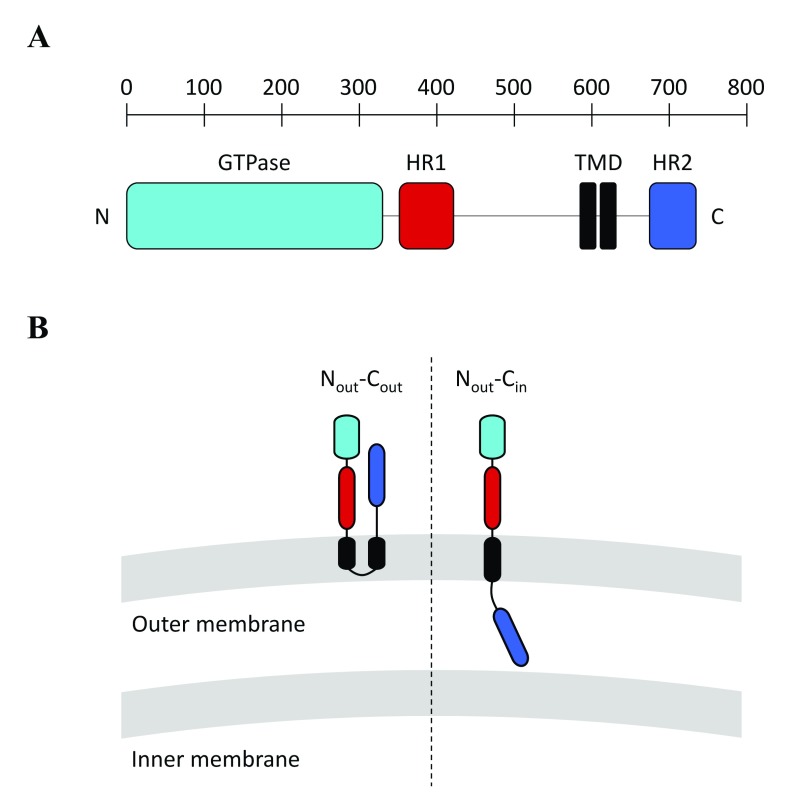
Figure 1.
( A) Molecular architecture of Mitofusin 1 (MFN1). Like MFN1, all Mitofusins include an N-terminal GTPase domain (light-blue) and two C-terminal heptad repeat domains, HR1 (red) and HR2 (dark blue), that sandwich a transmembrane region (black). The yeast Mitofusin Fzo1 includes an additional heptad repeat domain (HRN) located upstream of the GTPase domain (not depicted here). ( B) Possible topologies of Mitofusins. (Left) A transmembrane region with two transmembrane domains (TMDs) gives Mitofusins a topology in which the N- and C-terminal extremities are exposed to the cytoplasm (N out–C out topology). (Right) It was recently demonstrated that Mitofusins from vertebrates could also include a single TMD, which keeps the N-terminal GTPase and HR1 domains in the cytoplasm but places the C-terminal HR2 domain in the mitochondrial intermembrane space (N out–C in topology). Note that the BDLP1-like folding of Mitofusins observed in the X-ray structures of MFN1 ( Figure 2) is compatible with the N out–C out but not the N out–C in topology.

