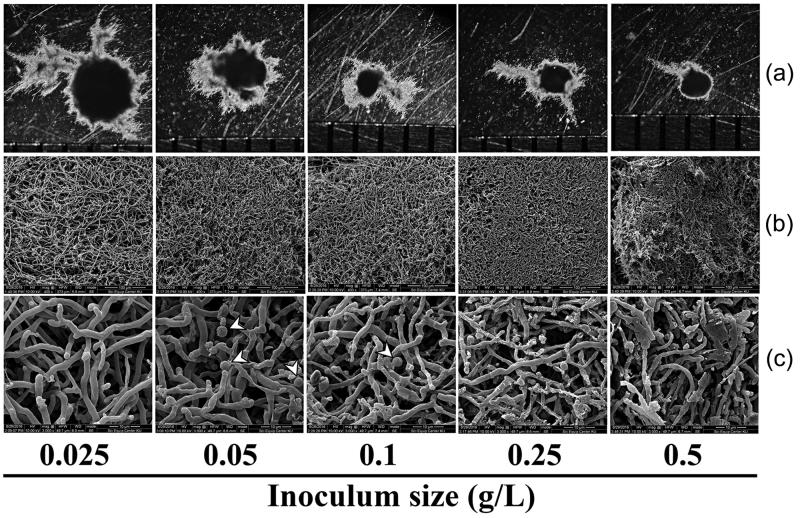
Figure 6.
Morphology of pellets of Trametes polyzona KU-RNW027 at 7-day incubation with different amounts of pellet inoculum as 0.025, 0.05, 0.1, 0.25, and 0.5 g/L. pellet morphology under stereomicroscope (a), structure of fungal pellet under SEM (400×) (b), and structure of fungal pellet under SEM (3,000×) (c). Arrows indicate terminal chlamydospore-like structures.

