Abstract
Aims/hypothesis
The blood triacylglycerol level is one of the main determinants of blood Mg2+ concentration in individuals with type 2 diabetes. Hypomagnesaemia (blood Mg2+ concentration <0.7 mmol/l) has serious consequences as it increases the risk of developing type 2 diabetes and accelerates progression of the disease. This study aimed to determine the mechanism by which triacylglycerol levels affect blood Mg2+ concentrations.
Methods
Using samples from 285 overweight individuals (BMI >27 kg/m2) who participated in the 300-Obesity study (an observational cross-sectional cohort study, as part of the Human Functional Genetics Projects), we investigated the association between serum Mg2+ with laboratory variables, including an extensive lipid profile. In a separate set of studies, hyperlipidaemia was induced in mice and in healthy humans via an oral lipid load, and blood Mg2+, triacylglycerol and NEFA concentrations were measured using colourimetric assays. In vitro, NEFAs harvested from albumin were added in increasing concentrations to several Mg2+-containing solutions to study the direct interaction between Mg2+ and NEFAs.
Results
In the cohort of overweight individuals, serum Mg2+ levels were inversely correlated with triacylglycerols incorporated in large VLDL particles (r = −0.159, p ≤ 0.01). After lipid loading, we observed a postprandial increase in plasma triacylglycerol and NEFA levels and a reciprocal reduction in blood Mg2+ concentration both in mice (Δ plasma Mg2+ −0.31 mmol/l at 4 h post oral gavage) and in healthy humans (Δ plasma Mg2+ −0.07 mmol/l at 6 h post lipid intake). Further, in vitro experiments revealed that the decrease in plasma Mg2+ may be explained by direct binding of Mg2+ to NEFAs. Moreover, Mg2+ was found to bind to albumin in a NEFA-dependent manner, evidenced by the fact that Mg2+ did not bind to fatty-acid-free albumin. The NEFA-dependent reduction in the free Mg2+ concentration was not affected by the presence of physiological concentrations of other cations.
Conclusions/interpretation
This study shows that elevated NEFA and triacylglycerol levels directly reduce blood Mg2+ levels, in part explaining the high prevalence of hypomagnesaemia in metabolic disorders. We show that blood NEFA level affects the free Mg2+ concentration, and therefore, our data challenge how the fractional excretion of Mg2+ is calculated and interpreted in the clinic.
Electronic supplementary material
The online version of this article (10.1007/s00125-018-4771-3) contains peer-reviewed but unedited supplementary material, which is available to authorised users.
Keywords: Albumin, Hypertriacylglycerolaemia, Hypomagnesaemia, Magnesium, Magnesium deficiency, Non-esterified fatty acid, Obesity, Triacylglycerols

Introduction
Hypomagnesaemia (blood Mg2+ concentration <0.7 mmol/l) is commonly observed in individuals with type 2 diabetes or the metabolic syndrome [1–3] and can result in general complaints such as fatigue, headache and weakness [4, 5]. Low oral Mg2+ intake and low blood Mg2+ levels not only increase the risk of developing type 2 diabetes but also accelerate disease progression [6–8]. A reduced blood Mg2+ is also associated with diabetes-related complications, such as cardiovascular disease and renal failure [9–12].
Blood Mg2+ levels are carefully maintained between 0.7 and 1.1 mmol/l by the interplay between intestine, bone and kidney [13]. In blood, approximately 27% of Mg2+ is bound to albumin and 8% is complexed to anions, such as phosphate, bicarbonate and citrate, leaving 65% as the free, biologically active form [14]. Although the phenomenon of albumin binding to Mg2+ has been known for decades, investigations into the buffering effect of albumin on the regulation of Mg2+ homeostasis has been largely neglected [15].
Blood fatty acid and triacylglycerol levels are largely regulated by four organs: the intestine, liver, muscle and adipose tissue. In the postprandial state, the intestine absorbs dietary lipids as fatty acids, which are re-esterified into triacylglycerols and incorporated into chylomicrons that reach metabolically active tissues via the circulation [16]. The liver can also incorporate fatty acids into triacylglycerol and secrete these as VLDL particles; this process is especially important during fasting [17]. Skeletal muscle stores fatty acids in the form of triacylglycerols but also consumes large amounts of fatty acids during exercise [18]. White adipose tissue also stores fatty acids as triacylglycerols, which can be released by lipolysis as NEFAs during a state of energy deprivation [19, 20]. In the blood, these negatively charged NEFAs are bound to carrier proteins, predominantly albumin, via non-polar interactions [21]. In physiological conditions, approximately two NEFA molecules are bound to a single albumin molecule [22]. However, in a state of hypertriacylglycerolaemia, up to seven NEFA molecules are able to bind to albumin, albeit with sequentially lower binding constants [21, 22].
In metabolic diseases, high blood triacylglycerol concentrations are associated with a lower blood Mg2+ concentration, but the directionality of this correlation remains unclear [1, 23, 24]. Severe hypomagnesaemia in animals leads to increased blood triacylglycerol levels, possibly by disrupting the function of the enzyme lecithin-cholesterol acyltransferase or by activating lipolysis in adipose tissue [25, 26]. However, whether triacylglycerols can affect Mg2+ homeostasis has not yet been investigated.
In this study, we measured serum Mg2+ concentrations and plasma lipoprotein concentrations and composition in a cohort of overweight individuals, by use of a metabolomics platform [27]. To further unravel the exact relationship between hypertriacylglycerolaemia and Mg2+ levels, we combined a population-based cross-sectional study with in vivo oral lipid loading in both humans and mice, and carried out subsequent investigations in vitro.
Methods
300-Obesity cohort
Three hundred and two individuals, aged 55–80 years, were enrolled in the 300-Obesity cohort study (an observational cross-sectional cohort study within the Human Functional Genetics Projects) at the Radboud university medical center between 2014 and 2016 [28]. The study was carried out in accordance with the Declaration of Helsinki and all participants provided informed consent. All participants had a BMI above 27 kg/m2. Individuals with a recent cardiovascular event (myocardial infarction, transient ischaemic attack, stroke <6 months ago), a history of bariatric surgery or bowel resection, inflammatory bowel disease, renal dysfunction or increased bleeding tendency, or who used oral or subcutaneous anticoagulant therapy or thrombocyte aggregation inhibitors (other than acetylsalicylic acid and carbasalate calcium) were excluded. Blood samples were taken in the morning, following an overnight fast. Blood glucose, triacylglycerols, total cholesterol and HDL-cholesterol were measured using standard laboratory procedures (Cobas C8000; Roche Diagnostics, Risch-Rotkreuz, Switzerland). The HOMA-IR was calculated using the standard formula, as published by Matthews et al [29]. High-throughput nuclear magnetic resonance (NMR) metabolomics platform (Nightingale’s Biomarker Analysis Platform, Nightingale Health, Helsinki, Finland) [27] was used for the quantification of 231 lipid and metabolite measures. The metabolites were measured in a single experiment, set-up for the quantification of different metabolite groups. In this article we focus on lipoproteins: total lipid concentrations of 14 lipoprotein subclasses, lipoprotein particles sizes, apolipoproteins and cholesterol. The NMR metabolomics platform used in this study has previously been used in various epidemiological studies [30, 31]. Details of the NMR-based metabolomics experimentation have been described previously [27]. In the present study, serum Mg2+ levels were measured in all individuals. However, 17 measurements had substantial duplo errors (large deviations [≥0.05 mmol/l from the average] between duplicate measurements) and, therefore, only 285 individuals were included in the analysis.
Oral gavage of olive oil in wild-type mice
This animal study was approved by the animal ethics board of the Radboud University Nijmegen (RU DEC 2015-0073) and by the Dutch Central Commission for Animal Experiments (CCD, AVD103002015239). Twelve male C57BL6/J mice (Charles River, Sulzfeld, Germany) were obtained at an age of 9–10 weeks. Mice were acclimatised for 2 weeks in a temperature- and light-controlled room, with six mice per cage (Eurostandard Type III, Tecniplast, Buguggiate, Italy), and were allowed free access to acidified tap water and standard pellet chow (Ssniff Spezialdiäten, Soest, Germany). After the acclimatisation period, mice received experimental chow containing 18.3% protein (wt/wt), 4.1% crude fat (wt/wt), 25.1% starch (wt/wt) and 33.6% sugar (wt/wt) (E15000-04; Ssniff Spezialdiäten). After 2 weeks on the synthetic diet, mice were fasted overnight, from 21:00 to 09:00 hours, before receiving 200 μl intragastric olive oil (extra virgin; Carbonell, Cordoba, Spain) via oral gavage. Blood was drawn via tail-bleed, using chilled sodium-heparin capillary tubes (Praxisdienst, Longuich, Germany) coated with paraoxon (Sigma, St Louis, MO, USA), before the gavage (0 h) and at 1, 2, 4, 6 and 8 h post gavage. The blood was centrifuged at 3000 g and plasma was procured.
Oral gavage of olive oil in a mouse model of diabetes
All procedures for this experiment were conducted in compliance with the UK Animals Scientific Procedures Act (1986) and University of Oxford ethical guidelines. Kir6.2-p.Val59Met mice were generated in our laboratories, as previously published [32]. All mice were 12–16 weeks old. Mice were housed with a maximum of five animals per cage (ventilated), separated by gender and with ad libitum access to food and standard pellet chow, in a 12 h light/dark cycle, at 21°C. In four male and four female C57BL6 mice, expression of a Kir6.2-p.Val59Met transgene was induced using subcutaneous injection of 400 μl tamoxifen (0.02 g/ml corn oil). This inducible mouse model recapitulates the phenotype of neonatal diabetes and these mice develop diabetes due to impaired insulin secretion [32, 33].
Three days after tamoxifen injection, mice were fasted overnight from 17:00 to 09:00 hours. Successful induction of the Kir6.2-p.Val59Met transgene was validated by measuring fasted blood glucose levels using a StatStrip Xpress glucose meter (Nova Biomedical, Waltham, MA, USA). One female mouse did not have elevated fasting glucose levels and was excluded from subsequent analyses. The remaining mice then underwent oral gavage and blood sampling using an identical experimental set up to that described above for C57BL6/J wild-type mice.
BSA and fatty-acid-free BSA solutions
BSA and fatty-acid-free BSA (FF-BSA) (both Sigma Aldrich) were separately dissolved at several concentrations in a 1 mmol/l MgCl2 solution (Merck Millipore, Darmstadt, Germany) or a physiological buffer, both set at pH 7.5 by adding NaOH. The physiological buffer contained the following (all from Merck Millipore unless stated otherwise): 27 mmol/l NaHCO3, 112 mmol/l NaCl, 5 mmol/l KCl, 1 mmol/l MgCl2, 1 mmol/l Na2HPO4 (VWR International, Radnor, PA, USA) and 2.5 mmol/l CaCl2 dissolved in Milli-Q (Radboud Institute for Molecular Life Sciences, Nijmegen, the Netherlands).
Increasing NEFA levels in BSA, FBS and MgCl2 solutions
To extract endogenous NEFAs, BSA (0.2 g/ml) dissolved in Milli-Q was mixed (1:2 vol./vol.) with ice-cold ethanol–diethyl ether (3:1 vol./vol.). The lipid phase was evaporated overnight at room temperature. To remove trace amounts of BSA, the solution was centrifuged three times with an Amicon 50 kDa filter (Merck Millipore) for 15 min at 2500 g to clog the protein in the filter. Extracted NEFAs were added to 250 μl FBS (Biowest, Nuaillé, France), 1 mmol/l MgCl2, or 0.5 mmol/l BSA in the amounts of 0, 25, 50, 100, 200, 350, 500 and 700 μl. Dilution factors were accounted for when measuring the concentrations of Mg2+ and NEFA.
Analytical measurements
Protein (Pierce, Thermo Scientific, Waltham, MA, USA), NEFA (WAKO Diagnostics, Delfzijl, the Netherlands), triacylglycerols (Roche Molecular Biochemicals, Indianapolis, IN, USA) and Mg2+ (Roche/Hitachi, Tokyo, Japan) concentrations were measured using a spectrophotometric assay according to the manufacturer’s protocols. The Mg2+ colourimetric assay was based on a Xylidyl Blue-I method and the absorbance was measured at 600 nm. NEFAs were measured at 546 nm, triacylglycerols at 500 nm and protein at 562 nm on a Bio-Rad Benchmark plus microplate spectrophotometer (Bio-Rad laboratories, Hercules, CA, USA). For inductively coupled plasma mass spectrometry (ICP-MS) analyses, serum samples were dissolved in HNO3 (>65%, Sigma) and diluted prior to being subjected to ICP-MS (X-series; Thermo Scientific).
Oral lipid load in human participants
As part of a study (ClinicalTrials.gov registration no. NCT01967459), which aimed to examine the effect of vitamin D supplementation on postprandial leucocyte activation, 24 female volunteers underwent an oral fat-loading test. The study was approved by the Institutional Review Board of the Franciscus Gasthuis & Vlietland Rotterdam and the regional independent medical ethics committee of the Maasstad Hospital Rotterdam [34]. All participants provided informed consent. Samples from the baseline oral fat load were used in this study to measure serum Mg2+, NEFA and triacylglycerol concentrations.
Inclusion criteria were an age above 18 years, a premenopausal status, BMI ≥25 kg/m2 and vitamin D deficiency. Exclusion criteria were the use of any kind of medication (except for oral contraceptives), smoking, pregnancy, participation in a clinical study less than 6 months before inclusion and the use of vitamin supplements.
Participants visited the hospital after a 10 h overnight fast and venous blood samples were obtained after an oral fat load (before Vitamin D treatment). Each participant received an oral fat load using fresh cream (Albert Heijn, Zaandam, the Netherlands) at a dose of 50 g of fat per m2 body surface calculated by the Mosteller formula [35]. During the oral fat loading, test participants were not allowed to eat or to drink (except water) and they were asked to refrain from physical activity. Venous blood sampling was repeated at 2 h intervals until 8 h after oral fat loading, and serum Mg2+ and NEFA levels and plasma triacylglycerol levels were measured (as detailed in the ‘Analytical measurements’ section above). Two individuals were excluded due to insufficient sample availability.
Statistical analyses
Results are presented as mean ± SEM, unless stated otherwise. Variables of overweight individuals were correlated univariately to serum Mg2+ levels using Pearson’s correlation analyses (SPSS for Windows v22.0.0.1, RRID:SCR_002865; IBM, New York, NY, USA). Based on the initial experiment in wild-type mice, the sample size for the experiment in Kir6.2-p.Val59Met mice was calculated using a one-way ANOVA statistic (with Dunnett’s correction for multiple comparison); to detect an effect size of 0.3 (SD 0.13) with a power of 80% and α level of 5%: a total of six mice were required per group. The sample size of the oral lipid (cream) load study in healthy individuals was assessed using a one-way ANOVA (with Dunnett’s correction for multiple comparison); to detect an effect size of 0.1 (SD 0.1) with a power of 80% and an α level of 5%: a total of 23 individuals were required. Significance of Mg2+, triacylglycerol and NEFA concentrations at 1, 2, 4, 6 and 8 h compared with those at 0 h was evaluated using a one-way ANOVA with Dunnett’s correction for multiple comparisons. Direct correlations between Mg2+ and triacylglycerol or NEFA concentrations were assessed using linear regression analyses. A p value of ≤0.05 was considered statistically significant. All statistical analyses were performed using Graphpad Prism v7 (RRID:SCR_002798; Graphpad software, La Jolla, CA, USA).
Results
Serum Mg2+ levels inversely correlate with blood triacylglycerol levels in overweight individuals
Factors affecting blood Mg2+ levels were evaluated in 285 overweight individuals (BMI >27 kg/m2) from the 300-Obesity cohort (ESM Table 1). The mean serum Mg2+ concentration in this cohort was 0.89 ± 0.09 (SD) mmol/l, with only 2% of the individuals having hypomagnesaemia (serum Mg2+ <0.7 mmol/l, see ESM Table 1 and ESM Fig. 1). Despite the fairly healthy serum Mg2+ levels in these individuals, serum Mg2+ concentrations were inversely correlated with triacylglycerol levels, predominantly those in VLDL particles (Table 1). The serum Mg2+ concentration was also inversely correlated with HOMA-IR. As HOMA-IR was strongly correlated with plasma triacylglycerol concentration (ESM Tables 2, 3), we questioned whether insulin resistance modulated the inverse correlation between serum Mg2+ and plasma triacylglycerol levels and triacylglycerols in VLDL particles. However, HOMA-IR did not influence these correlations in multivariable regression analyses (ESM Tables 4 and 5).
Table 1.
Univariate analyses for the correlation of demographics, laboratory measurements and lipoprotein particle concentration with serum Mg2+ concentration in overweight individuals from the 300-Obesity cohort
| Variable | Correlation coefficient | p value | n |
|---|---|---|---|
| Demographics | |||
| Sex (M = 0, F = 1) | −0.051 | 0.391 | 285 |
| BMI (kg/m2) | −0.063 | 0.291 | 285 |
| Age (years) | −0.099 | 0.096 | 285 |
| Waist circumference (cm) | −0.071 | 0.231 | 285 |
| SBP (mmHg) | −0.016 | 0.791 | 285 |
| DBP (mmHg) | 0.079 | 0.186 | 285 |
| Heart rate (beats/min) | −0.083 | 0.164 | 282 |
| Laboratory measures | |||
| Triacylglycerols (mmol/l) | −0.159 | 0.007 | 284 |
| Glucose (mmol/l) | −0.062 | 0.299 | 284 |
| HbA1c (mmol/mol) | −0.032 | 0.595 | 284 |
| HOMA-IR | −0.123 | 0.038 | 283 |
| Total cholesterol (mmol/l) | 0.041 | 0.495 | 284 |
| Triacylglycerols (mmol/l) | |||
| in VLDL | −0.158 | 0.008 | 284 |
| in LDL | −0.026 | 0.667 | 284 |
| in HDL | −0.052 | 0.380 | 284 |
| Cholesterol (mmol/l) | |||
| in VLDL | −0.093 | 0.118 | 284 |
| in LDL | 0.075 | 0.205 | 284 |
| HDL | 0.116 | 0.050 | 284 |
| ApoA1 (g/l) | 0.071 | 0.235 | 283 |
| ApoB (g/l) | −0.037 | 0.539 | 283 |
| Mean lipoprotein diameter (nm) | |||
| VLDL | −0.095 | 0.111 | 284 |
| LDL | −0.030 | 0.612 | 284 |
| HDL | 0.026 | 0.320 | 284 |
| Lipoprotein particle concentration (mol/l) | |||
| Chylomicrons and EL-VLDL | −0.170 | 0.004 | 284 |
| VL-VLDL | −0.174 | 0.003 | 284 |
| L-VLDL | −0.163 | 0.006 | 284 |
| M-VLDL | −0.149 | 0.012 | 284 |
| S-VLDL | −0.108 | 0.068 | 284 |
| VS-VLDL | −0.004 | 0.942 | 284 |
| IDL | 0.058 | 0.333 | 284 |
| L-LDL | 0.060 | 0.313 | 284 |
| M-LDL | 0.059 | 0.319 | 284 |
| S-LDL | 0.053 | 0.377 | 284 |
| VL-HDL | 0.020 | 0.739 | 284 |
| L-HDL | 0.089 | 0.137 | 284 |
| M-HDL | 0.163 | 0.006 | 284 |
| S-HDL | 0.179 | 0.002 | 284 |
ApoA1, apolipoprotein A1; ApoB, apolipoprotein B; DBP, diastolic blood pressure; EL-VLDL, extra-large VLDL; F, female; IDL, intermediate-density lipoprotein; L-HDL, large HDL; L-LDL, large LDL; L-VLDL, large VLDL; M, male; M-HDL, medium HDL; M-LDL, medium LDL; M-VLDL, medium VLDL; SBP, systolic blood pressure; S-HDL, small HDL; S-LDL, small LDL; S-VLDL, small VLDL; VL-HDL, very large HDL; VL-VLDL, very large VLDL; VS-VLDL, very small VLDL
To further investigate the relationship between lipoproteins and serum Mg2+ level, the composition of lipoprotein particles was investigated and the correlation of these particles with serum Mg2+ concentration was analysed (Table 1). The concentration of the larger VLDL particles showed the strongest inverse correlations with serum Mg2+ levels (Table 1). Interestingly, the concentration of smaller HDL particles was positively correlated with serum Mg2+. There was no correlation between serum Mg2+ levels and the concentration of any of the intermediate-density lipoprotein and LDL particles (Table 1). No significant correlation was observed between serum Mg2+ and the diameter of the VLDL, LDL or HDL particles (Table 1).
Increased triacylglycerol levels directly reduce plasma Mg2+ concentrations in mice
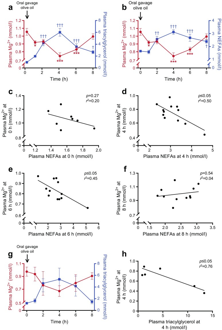
To unravel the underlying mechanism that explains how blood triacylglycerols are associated with blood Mg2+ concentrations, mice were subjected to an oral gavage of olive oil following an overnight fast. Plasma triacylglycerols and NEFA levels both peaked at 4 h post-gavage (Fig. 1a, b, Δ plasma Mg2+ −0.31 mmol/l at 4 h post-gavage). Interestingly, the plasma Mg2+ concentration showed a reciprocal decrease, reaching a nadir at 4 h post-gavage (Fig. 1a, b). At basal levels (0 h), no significant correlation was observed between plasma Mg2+ levels and NEFA concentrations (Fig. 1c, p = 0.27). However, when plasma NEFA levels increased (at 4 and 6 h), there was a clear inverse correlation between plasma Mg2+ and NEFA concentrations (Fig. 1d, e, p ≤ 0.05). When plasma NEFA levels decreased and reached a concentration approaching levels at baseline (8 h), there was no longer a significant correlation between plasma NEFA and Mg2+ concentrations (Fig. 1f, p = 0.54). Similar correlations were observed between plasma Mg2+ and triacylglycerol levels (ESM Fig. 2a–d).
Fig. 1.
Increased plasma NEFA and triacylglycerol levels directly reduce plasma Mg2+ concentration in mice. Mice were given an oral gavage of 200 μl olive oil. (a) Plasma Mg2+ (red circles) and triacylglycerol (blue squares) and (b) plasma Mg2+ (red circles,) and NEFA (blue squares) concentrations before (0 h) and at 1, 2, 4, 6 and 8 h post-gavage in wild-type mice (n = 12). (c–f) Linear regression analyses of plasma Mg2+ and NEFA concentrations at 0 h (c, n = 8), 4 h (d, n = 12), 6 h (e, n = 12) and 8 h (f, n = 11) post gavage in wild-type mice, using data from (b). Each point represents an individual mouse; several data points are missing due to insufficient sample availability. (g) Plasma Mg2+ (red circles) and triacylglycerol (blue squares) concentrations before (0 h) and at 1, 2, 4, 6 and 8 h post-gavage in hypoinsulinaemic Kir6.2-p.Val59Met mice (n = 7). (h) Linear regression analysis of plasma Mg2+ and triacylglycerol concentration at 4 h post-gavage, using data shown in (g). Each point represents an individual mouse; n = 2 mice were excluded due to insufficient sample availability. Data are mean ± SEM. *p ≤ 0.05, ***p ≤ 0.001 vs Mg2+ concentrations at 0 h; †p ≤ 0.05, ††p ≤ 0.01, †††p ≤ 0.001 vs triacylglycerol or NEFA concentrations at 0 h. Significance was evaluated using a one-way ANOVA with Dunnett’s correction for multiple comparisons
Increased blood lipids can enhance glucose-induced insulin secretion [36]. Insulin induces a compartmental shift of Mg2+, decreasing the blood concentration of Mg2+ and increasing intracellular levels [37]. Hence, to rule out insulin-dependent effects, the same oral gavage experiment was performed in inducible Kir6.2-p.Val59Met mice, which develop hypoinsulinaemia and hyperglycaemia [38]. In these mice, a similar reduction in plasma Mg2+ was observed in response to the oral gavage of olive oil as compared with wild-type mice (Fig. 1g). However, while the reduction in the plasma Mg2+ concentration was numerically similar to that observed in wild-type mice, it did not reach statistical significance in Kir6.2-p.Val59Met mice due to a high variation between the animals (which was substantially larger than in the initial experiment using wild-type mice). However, a significant inverse correlation between plasma Mg2+ and triacylglycerols was still observed in these hypoinsulinaemic mice (Fig. 1h, p ≤ 0.05).
Binding of Mg2+ to albumin is NEFA-dependent
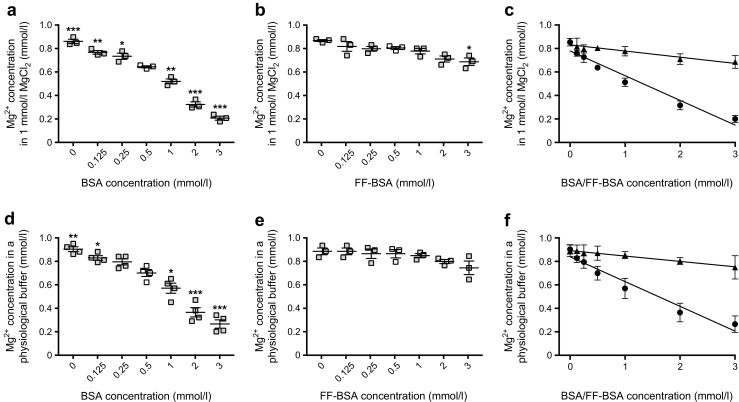
As ~30% of Mg2+ is bound to albumin, which is the predominant carrier of NEFAs, we investigated whether the binding of Mg2+ to albumin is dependent on NEFAs [14]. With increasing concentrations of BSA in MgCl2, we found that the Mg2+ concentration declined as BSA concentrations increased (Fig. 2a). Mg2+ concentrations decreased from 0.85 ± 0.02 mmol/l at 0 mmol/l BSA to 0.64 ± 0.01 mmol/l at a near-physiological concentration of BSA (0.5 mmol/l or 33.25 g/l), a 25% reduction (Fig. 2a). Interestingly, the absence of fatty acid (i.e. the use of FF-BSA instead of BSA in the MgCl2 solution) abrogated this effect, in line with the theory that binding of Mg2+ to albumin is NEFA-dependent (Fig. 2b). Linear regression analyses showed a significant inverse correlation between Mg2+ and BSA concentration (Fig. 2c); the regression constant was approximately four times stronger for BSA than for FF-BSA (Fig. 2c). To exclude the possibility that other cations present in blood compete with the binding of Mg2+ to BSA, BSA was also dissolved in a physiological buffer, which mimicked the concentration of other abundant blood electrolytes. This approach resulted in a similar decrease in the Mg2+ concentration with increasing BSA concentrations (Fig. 2d). Again, FF-BSA had no significant effect on Mg2+ levels (Fig. 2e). In the physiological buffer, BSA and FF-BSA displayed correlations similar to those observed in the MgCl2 buffer (Fig. 2f).
Fig. 2.

Binding of Mg2+ to albumin is NEFA-dependent. (a, b) The Mg2+ concentration in an increasing level of BSA (a) or FF-BSA (b) dissolved in a MgCl2 solution. The white squares indicate a BSA/FF-BSA concentration (0.5 mmol/l) that is near the physiological range; all other data points are shown in grey. (c) Linear regression analyses of the Mg2+ concentration in increasing levels of BSA (circles; y = −0.21x + 0.78, r2 = 0.96, p ≤ 0.05) or FF-BSA (triangles; y = −0.05x + 0.83, r2 = 0.90, p ≤ 0.05), using data shown in (a) and (b). (d, e) The Mg2+ concentration in an increasing level of BSA (d) or FF-BSA (e) dissolved in a physiological buffer. The white squares indicate a BSA/FF-BSA concentration (0.5 mmol/l) that is near the physiological range; all other data points are shown in grey. (f) Linear regression analyses of the Mg2+ concentration in increasing levels of BSA (circles; y = −0.21x + 0.78, r2 = 0.96, p ≤ 0.05) or FF-BSA (triangles, y = −0.05x + 0.83, r2 = 0.90, p ≤ 0.05), using data shown in (d) and (e). Data are mean ± SEM of three replicate experiments. *p ≤ 0.05, **p ≤ 0.01, ***p ≤ 0.001 vs Mg2+ concentrations in 0.5 mmol/l BSA/FF-BSA. Significance was evaluated using a one-way ANOVA with Dunnett’s correction for multiple comparisons (a, b, d, e) or using a linear regression analysis (c, f)
NEFAs directly decrease Mg2+ concentrations
We then set out to modify the binding of Mg2+ to albumin by increasing the concentration of NEFA. Elevating the concentration of NEFAs in a BSA–MgCl2 solution directly reduced the Mg2+ concentration (linear regression: y = −0.12x + 0.83, p ≤ 0.05; Fig. 3a). To resemble the in vivo setting, the experiment was repeated using FBS instead of BSA; increasing NEFA levels in FBS reduced Mg2+ concentrations to a similar extent as when BSA was used (linear regression: y = −0.10x + 1.36, p ≤ 0.05; Fig. 3b). Interestingly, the NEFA-induced reduction in Mg2+ concentration was protein-independent, as increasing NEFA levels in a MgCl2 solution also lowered Mg2+ concentration (linear regression: y = −0.08x + 1.00, p ≤ 0.05; Fig. 3c).
Fig. 3.

NEFAs directly reduce Mg2+ concentrations. Linear regression analyses of Mg2+ and NEFA concentration in (a) BSA dissolved in 1 mmol/l MgCl2 (y = −0.12x + 0.83, r2 = 0.97, p ≤ 0.05), (b) FBS (y = −0.10x + 1.36, r2 = 0.90, p ≤ 0.05) and (c) MgCl2 solution (y = −0.08x + 1.00, r2 = 0.99, p ≤ 0.05). Results from one representative experiment are shown. The experiment was repeated three additional times with similar results
Elevating triacylglycerol levels reduces serum Mg2+ concentration in healthy individuals
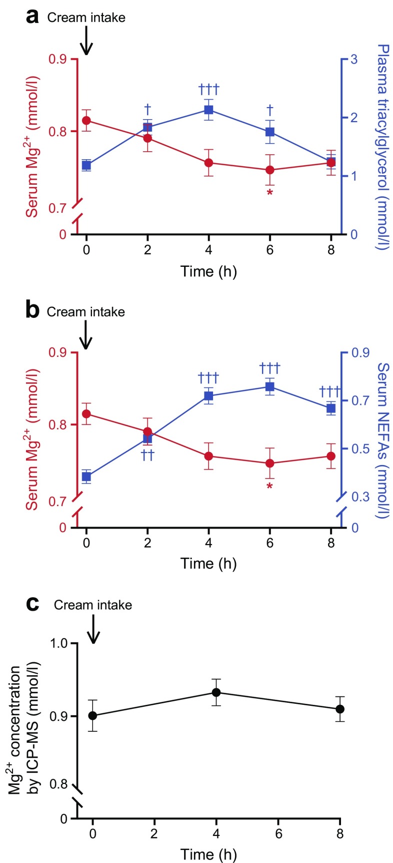
To discover whether triacylglycerols and NEFAs also affect blood Mg2+ concentration in humans, we examined samples obtained from 24 healthy female individuals, with a BMI >25 kg/m2, who received an oral fat load [34]. Serum NEFA and Mg2+ levels and plasma triacylglycerol concentrations were measured over a period of 8 h. Plasma triacylglycerol levels increased significantly, from 1.18 ± 0.10 mmol/l at 0 h to a peak of 2.13 ± 0.18 mmol/l at 4 h (Fig. 4a). Serum NEFA concentrations also significantly increased, from 0.38 ± 0.03 mmol/l at 0 h to a peak of 0.76 ± 0.04 mmol/l at 6 h (Fig. 4b). In accordance with our previous findings, serum Mg2+ levels dropped from 0.82 ± 0.01 mmol/l at 0 h to a nadir of 0.75 ± 0.02 mmol/l at 6 h (Fig. 4a, b, Δ plasma Mg2+ −0.07 mmol/l at 6 h post-intake). The total serum Mg2+ concentration, as measured by ICP-MS, was not affected by fat (cream) intake; hence, these findings suggest that only the free Mg2+ levels were affected by lipid loading (Fig. 4c).
Fig. 4.
Increased serum NEFA and plasma triacylglycerol levels directly reduce serum Mg2+ concentrations in healthy overweight or obese women. Female volunteers underwent an oral fat (cream) loading test. (a) Serum Mg2+ (red circles) and plasma triacylglycerol (blue squares) and (b) serum Mg2+ (red circles) and NEFA (blue squares) concentrations before (0 h) and at 1, 2, 4, 6 and 8 h after an oral lipid load in healthy overweight women (n = 22). (c) Total serum Mg2+ levels measured by ICP-MS before (0 h) and 4 and 8 h after oral lipid intake. Data are mean ± SEM.*p ≤ 0.05 vs Mg2+ concentrations at 0 h; †p ≤ 0.05, ††p ≤ 0.01, †††p ≤ 0.001 vs triacylglycerol or NEFA concentrations at 0 h. Significance was evaluated using a one-way ANOVA with Dunnett’s correction for multiple comparisons
Discussion
Hypomagnesaemia is a common phenomenon in type 2 diabetes. In the current study, we show that high blood NEFA and triacylglycerol concentrations directly reduce blood Mg2+ levels. We also show that high NEFA levels bind Mg2+, resulting in decreased circulating levels of free Mg2+. The conclusions of this study are based on complementary results from in vitro studies and animal and human in vivo studies. First, in a large cohort of overweight individuals, the concentration of triacylglycerols in large VLDL particles was inversely correlated with serum Mg2+ concentrations. Second, a dietary lipid load directly reduced blood Mg2+ concentration in both mice and humans, independent of insulin action. Third, in vitro, we demonstrated that this phenomenon occurs due to direct binding of NEFA molecules to Mg2+. These findings demonstrate that triacylglycerols reduce concentrations of free Mg2+ in the blood and consequently place hypertriacylglycerolaemic individuals at risk for Mg2+ deficiency.
Here, we demonstrated that increased triacylglycerol and NEFA levels reduce levels of free Mg2+(the biologically active form of Mg2+) by a direct interaction between negatively charged NEFA molecules and Mg2+ ions. This binding was shown to be highly specific for Mg2+, since the presence of physiological concentrations of other cations did not affect the interaction between Mg2+ and NEFAs. The phenomenon of reduced Mg2+ levels as a result of elevated NEFA levels has previously been observed in dogs, although no underlying mechanism was suggested [39]. In addition, previous studies have shown that Ca2+ can bind to NEFAs, and that blood Ca2+ concentrations are reduced by increasing NEFA levels in individuals [40]. However, in our in vitro experiments, the addition of physiological concentrations of Ca2+ did not affect the Mg2+–NEFA interaction, indicating a higher affinity of Mg2+ compared with Ca2+ for binding to NEFAs. This is likely due to the fact that the Mg2+ ion has a significantly higher charge density than the Ca2+ ion [41].
The findings from this study may explain why certain factors that affect circulating NEFAs are associated with changes in blood Mg2+ concentrations. For example, molecules such as β-adrenergic agonists, ethanol and adrenaline (epinephrine), which activate lipolysis and, hence, increase blood NEFA levels, are associated with reduced blood Mg2+ levels [1, 42–44]. Indeed, an intravenous infusion of the β-adrenergic agonist terbutaline causes a reduction in serum Mg2+ concentration, which is correlated to the elevated concentrations of plasma NEFAs, but not glucose [45]. Although, this does not exclude the possibility of additional potential mechanisms for Mg2+ decrease with use of these agents. In addition, prolonged fasting, which induces lipolysis and increases circulating NEFAs, also results in hypomagnesaemia [46].
Approximately 30% of blood Mg2+ is bound to albumin [14]. However, our data indicate that the binding of Mg2+ to albumin depends on the availability of NEFA. Our findings suggest that direct binding of Mg2+ to albumin is minimal, since NEFA-depleted albumin showed little binding to Mg2+. To correct for albumin binding, a factor of 0.7 is used when calculating the fractional excretion of Mg2+(FEMg): FEMg = [(uMg × sCr)/(sMg × uCr × 0.7)] × 100), where sMg and uMg are the serum and urinary Mg2+ levels, respectively, and sCr and uCr are the serum and urinary creatinine levels, respectively [47]. As the large majority of NEFAs in blood are bound to albumin, alterations in NEFA concentrations may be the determining factor in the binding of Mg2+ to albumin [22]. This paradigm change questions the current protocol for calculating FEMg. The use of the factor of ‘0.7’ is accurate in physiological conditions but will lead to inaccurate calculations in pathological conditions such as hypertriacylglycerolaemia.
Several limitations need to be considered with regard to this study. Hypertriacylglycerolaemia in mice was achieved using olive oil, while in human volunteers this was done by an oral load of cream. Olive oil contains no Mg2+, while the cream used in the human study contains 3.3 mmol/l Mg2+, leading to a potential underestimation of the reduction in serum Mg2+ in the healthy volunteers. Moreover, in the in vitro experiments, NEFAs extracted from BSA were used to increase NEFA levels in several solutions. However, the yield of this extraction procedure was not equal in each experiment performed, making it difficult to combine data from all experiments. Despite these differences in NEFA yield, the results were similar in all four replicate experiments. Finally, in overweight individuals and in individuals with type 2 diabetes, Mg2+ inversely correlates with triacylglycerols [1, 23, 24]. Our in vitro data show direct binding of Mg2+ to NEFA molecules, which, in contrast to triacylglycerol molecules, possess a negative charge. It is unlikely that Mg2+ binds to uncharged triacylglycerol molecules. However, in humans, blood triacylglycerols and NEFA levels strongly correlate, meaning that most individuals with hypertriacylglycerolaemia also have elevated NEFA levels, which would underlie the inverse correlation between Mg2+ and triacylglycerols [48–51].
This study has several strengths. Our data extend from molecule to population and have clinical implications. Moreover, we demonstrated the directionality of the inverse association between triacylglycerols and Mg2+, which could explain why hypomagnesaemia is so prominent in diseases such as type 2 diabetes. Our data do not rule out the possibility that changes in Mg2+ concentrations could also influence lipid levels.
In conclusion, we show that elevated blood NEFA and triacylglycerol levels directly reduce blood Mg2+ concentrations by the binding of Mg2+ ions to NEFA molecules. Our data explain the high prevalence of hypomagnesaemia in several metabolic diseases, characterised by elevated triacylglycerol levels [1–3]. In individuals with these diseases, those with hypertriacylglycerolaemia are at particular risk for hypomagnesaemia, and therefore, blood Mg2+ levels should be routinely measured and monitored.
Electronic supplementary material
(PDF 161 kb)
Acknowledgements
The authors thank M. Voet and A. Ruiz Llombart (Department of Physiology, Radboud university medical center, Nijmegen, the Netherlands), K. Schraa (Department of Internal Medicine, Radboud university medical center, Nijmegen, the Netherlands) and R. T. Exposito (Department of Physiology, Anatomy & Genetics, University of Oxford, Oxford, UK) for their superb technical support.
Abbreviations
- FF-BSA
Fatty-acid-free BSA
- FEMg
Fractional excretion of Mg2+
- ICP-MS
Inductively coupled plasma mass spectrometry
- NMR
Nuclear magnetic resonance
Contribution statement
SK and JHFdB conceived the project, designed the experiments, performed the experiments, analysed data and wrote the manuscript. VG performed experiments, analysed data and wrote the manuscript. CO-B and JAvD provided technical assistance, analysed data and critically read the manuscript. FMA, CJJT, RJMB and JGJH conceived the project, interpreted data and wrote the manuscript. ICLvdM, NPR, MGN and JHWR performed the population-based cross-sectional study, analysed and interpreted data, and critically read the manuscript. MAdV, BB and MCC performed the in vivo human study (oral cream study), analysed and interpreted data, and critically read the manuscript. All authors read and approved the final version of the manuscript. JGJH is the guarantor of this work.
Funding
This work was supported by funding from the Radboud Institute for Molecular Life Sciences and by grants from the Netherlands Organization for Scientific Research (NWO VICI 016.130.668 to JGJH) and by an IN-CONTROL CVON grant (CVON2012-03). JHFdB is supported by grants from NWO (Rubicon 825.14.021, Veni 016.186.012), the Dutch Kidney Foundation (Kolff 14OKG17) and the Dutch Diabetes Research Foundation (2017.81.014). JAvD is supported by a grant from the NWO (NWO VENI 91616083). The oral lipid load study in humans was financed by the Research Fund Department of Internal Medicine, Franciscus Gasthuis & Vlietland. MGN is supported by a Spinoza grant of the NWO. FMA is supported by an ERC (ERC Advanced Investigatorship), The Wellcome Trust (884655, 089795) and a Royal Society Research Wolfson Merit Award.
Data availability
The data and materials that support the findings of this study are available from the corresponding author upon reasonable request.
Duality of interest
The authors declare that there is no duality of interest associated with this manuscript.
References
- 1.Kurstjens S, de Baaij JH, Bouras H, Bindels RJ, Tack CJ, Hoenderop JG. Determinants of hypomagnesemia in patients with type 2 diabetes mellitus. Eur J Endocrinol. 2017;176(1):11–19. doi: 10.1530/EJE-16-0517. [DOI] [PubMed] [Google Scholar]
- 2.Mather HM, Nisbet JA, Burton GH, et al. Hypomagnesaemia in diabetes. Clin Chim Acta. 1979;95(2):235–242. doi: 10.1016/0009-8981(79)90364-4. [DOI] [PubMed] [Google Scholar]
- 3.Guerrero-Romero F, Bermudez-Pena C, Rodriguez-Moran M. Severe hypomagnesemia and low-grade inflammation in metabolic syndrome. Magnes Res. 2011;24(2):45–53. doi: 10.1684/mrh.2011.0281. [DOI] [PubMed] [Google Scholar]
- 4.Pham PC, Pham PA, Pham SV, Pham PT, Pham PM, Pham PT. Hypomagnesemia: a clinical perspective. Int J Nephrol Renov Dis. 2014;7:219–230. doi: 10.2147/IJNRD.S42054. [DOI] [PMC free article] [PubMed] [Google Scholar]
- 5.Grober U, Schmidt J, Kisters K. Magnesium in prevention and therapy. Nutrients. 2015;7(9):8199–8226. doi: 10.3390/nu7095388. [DOI] [PMC free article] [PubMed] [Google Scholar]
- 6.Kieboom BCT, Ligthart S, Dehghan A, et al. Serum magnesium and the risk of prediabetes: a population-based cohort study. Diabetologia. 2017;60(5):843–853. doi: 10.1007/s00125-017-4224-4. [DOI] [PMC free article] [PubMed] [Google Scholar]
- 7.Kao WH, Folsom AR, Nieto FJ, Mo JP, Watson RL, Brancati FL. Serum and dietary magnesium and the risk for type 2 diabetes mellitus: the Atherosclerosis Risk in Communities Study. Arch Intern Med. 1999;159(18):2151–2159. doi: 10.1001/archinte.159.18.2151. [DOI] [PubMed] [Google Scholar]
- 8.Fang X, Han HD, Li M, et al. Dose-response relationship between dietary magnesium intake and risk of type 2 diabetes mellitus: a systematic review and meta-regression analysis of prospective cohort studies. Nutrients. 2016;8(11):739. doi: 10.3390/nu8110739. [DOI] [PMC free article] [PubMed] [Google Scholar]
- 9.Kolte D, Vijayaraghavan K, Khera S, Sica DA, Frishman WH. Role of magnesium in cardiovascular diseases. Cardiol Rev. 2014;22(4):182–192. doi: 10.1097/CRD.0000000000000003. [DOI] [PubMed] [Google Scholar]
- 10.Sakaguchi Y, Shoji T, Hayashi T, et al. Hypomagnesemia in type 2 diabetic nephropathy: a novel predictor of end-stage renal disease. Diabetes Care. 2012;35(7):1591–1597. doi: 10.2337/dc12-0226. [DOI] [PMC free article] [PubMed] [Google Scholar]
- 11.Van Laecke S, Marechal C, Verbeke F, et al. The relation between hypomagnesaemia and vascular stiffness in renal transplant recipients. Nephrol Dial Transplant. 2011;26(7):2362–2369. doi: 10.1093/ndt/gfq728. [DOI] [PubMed] [Google Scholar]
- 12.Ter Braake AD, Shanahan CM, de Baaij JHF. Magnesium counteracts vascular calcification: passive interference or active modulation? Arterioscler Thromb Vasc Biol. 2017;37(8):1431–1445. doi: 10.1161/ATVBAHA.117.309182. [DOI] [PubMed] [Google Scholar]
- 13.de Baaij JH, Hoenderop JG, Bindels RJ. Magnesium in man: implications for health and disease. Physiol Rev. 2015;95(1):1–46. doi: 10.1152/physrev.00012.2014. [DOI] [PubMed] [Google Scholar]
- 14.Huijgen HJ, van Ingen HE, Kok WT, Sanders GT. Magnesium fractions in serum of healthy individuals and CAPD patients, measured by an ion-selective electrode and ultrafiltration. Clin Biochem. 1996;29(3):261–266. doi: 10.1016/0009-9120(96)84729-B. [DOI] [PubMed] [Google Scholar]
- 15.Kroll MH, Elin RJ. Relationships between magnesium and protein concentrations in serum. Clin Chem. 1985;31(2):244–246. [PubMed] [Google Scholar]
- 16.Hussain MM. A proposed model for the assembly of chylomicrons. Atherosclerosis. 2000;148(1):1–15. doi: 10.1016/S0021-9150(99)00397-4. [DOI] [PubMed] [Google Scholar]
- 17.Shelness GS, Sellers JA. Very-low-density lipoprotein assembly and secretion. Curr Opin Lipidol. 2001;12(2):151–157. doi: 10.1097/00041433-200104000-00008. [DOI] [PubMed] [Google Scholar]
- 18.Gorski J. Muscle triglyceride-metabolism during exercise. Can J Physiol Pharmacol. 1992;70(1):123–131. doi: 10.1139/y92-019. [DOI] [PubMed] [Google Scholar]
- 19.Herzer S, Meldner S, Grone HJ, Nordstrom V. Fasting-induced lipolysis and hypothalamic insulin signaling are regulated by neuronal glucosylceramide synthase. Diabetes. 2015;64(10):3363–3376. doi: 10.2337/db14-1726. [DOI] [PubMed] [Google Scholar]
- 20.Ahmadian M, Duncan RE, Jaworski K, Sarkadi-Nagy E, Sul HS. Triacylglycerol metabolism in adipose tissue. Futur Lipidol. 2007;2(2):229–237. doi: 10.2217/17460875.2.2.229. [DOI] [PMC free article] [PubMed] [Google Scholar]
- 21.Spector AA. Fatty acid binding to plasma albumin. J Lipid Res. 1975;16(3):165–179. [PubMed] [Google Scholar]
- 22.van der Vusse GJ. Albumin as fatty acid transporter. Drug Metab Pharmacokinet. 2009;24(4):300–307. doi: 10.2133/dmpk.24.300. [DOI] [PubMed] [Google Scholar]
- 23.Corica F, Corsonello A, Ientile R, et al. Serum ionized magnesium levels in relation to metabolic syndrome in type 2 diabetic patients. J Am Coll Nutr. 2006;25(3):210–215. doi: 10.1080/07315724.2006.10719534. [DOI] [PubMed] [Google Scholar]
- 24.Guerrero-Romero F, Rodriguez-Moran M. Low serum magnesium levels and metabolic syndrome. Acta Diabetol. 2002;39(4):209–213. doi: 10.1007/s005920200036. [DOI] [PubMed] [Google Scholar]
- 25.Gueux E, Rayssiguier Y, Piot MC, Alcindor L. Reduction of plasma lecithin--cholesterol acyltransferase activity by acute magnesium deficiency in the rat. J Nutr. 1984;114(8):1479–1483. doi: 10.1093/jn/114.8.1479. [DOI] [PubMed] [Google Scholar]
- 26.Kurstjens S, van Diepen JA, Overmars-Bos C, et al. Magnesium deficiency prevents high-fat-diet-induced obesity in mice. Diabetologia. 2018;61(9):2030–2042. doi: 10.1007/s00125-018-4680-5. [DOI] [PMC free article] [PubMed] [Google Scholar]
- 27.Soininen P, Kangas AJ, Wurtz P, et al. High-throughput serum NMR metabonomics for cost-effective holistic studies on systemic metabolism. Analyst. 2009;134(9):1781–1785. doi: 10.1039/b910205a. [DOI] [PubMed] [Google Scholar]
- 28.Netea MG, Joosten LAB, Li Y, et al. Understanding human immune function using the resources from the Human Functional Genomics Project. Nat Med. 2016;22(8):831–833. doi: 10.1038/nm.4140. [DOI] [PubMed] [Google Scholar]
- 29.Matthews DR, Hosker JR, Rudenski AS, et al. Homeostasis model assessment: insulin resistance and β-cell function from fasting plasma glucose and insulin concentrations in man. Diabetologia. 1985;28(7):412–419. doi: 10.1007/BF00280883. [DOI] [PubMed] [Google Scholar]
- 30.Wurtz P, Havulinna AS, Soininen P, et al. Metabolite profiling and cardiovascular event risk: a prospective study of 3 population-based cohorts. Circulation. 2015;131(9):774–785. doi: 10.1161/CIRCULATIONAHA.114.013116. [DOI] [PMC free article] [PubMed] [Google Scholar]
- 31.Kettunen J, Tukiainen T, Sarin AP, et al. Genome-wide association study identifies multiple loci influencing human serum metabolite levels. Nat Genet. 2012;44(3):269–276. doi: 10.1038/ng.1073. [DOI] [PMC free article] [PubMed] [Google Scholar]
- 32.Brereton MF, Iberl M, Shimomura K, et al. Reversible changes in pancreatic islet structure and function produced by elevated blood glucose. Diabetologia. 2014;57:S93–S93. doi: 10.1007/s00125-013-3077-8. [DOI] [PMC free article] [PubMed] [Google Scholar]
- 33.Girard CA, Wunderlich FT, Shimomura K, et al. Expression of an activating mutation in the gene encoding the KATP channel subunit Kir6.2 in mouse pancreatic beta cells recapitulates neonatal diabetes. J Clin Invest. 2009;119(1):80–90. doi: 10.1172/JCI35772. [DOI] [PMC free article] [PubMed] [Google Scholar]
- 34.de Vries MA, van der Meulen N, van de Geijn GM, et al. Effect of a single dose of vitamin D3 on postprandial arterial stiffness and inflammation in vitamin D-deficient women. J Clin Endocrinol Metab. 2017;102(3):992–1000. doi: 10.1210/jc.2016-3394. [DOI] [PubMed] [Google Scholar]
- 35.Mosteller RD. Simplified calculation of body-surface area. N Engl J Med. 1987;317:1098–1098. doi: 10.1056/NEJM198710223171717. [DOI] [PubMed] [Google Scholar]
- 36.Nolan CJ, Madiraju MS, Delghingaro-Augusto V, Peyot ML, Prentki M. Fatty acid signaling in the β-cell and insulin secretion. Diabetes. 2006;55(Suppl 2):S16–S23. doi: 10.2337/db06-S003. [DOI] [PubMed] [Google Scholar]
- 37.Takaya J, Higashino H, Miyazaki R, Kobayashi Y. Effects of insulin and insulin-like growth factor-1 on intracellular magnesium of platelets. Exp Mol Pathol. 1998;65(2):104–109. doi: 10.1006/exmp.1998.2232. [DOI] [PubMed] [Google Scholar]
- 38.Koster JC, Marshall BA, Ensor N, Corbett JA, Nichols CG. Targeted overactivity of beta cell K(ATP) channels induces profound neonatal diabetes. Cell. 2000;100(6):645–654. doi: 10.1016/S0092-8674(00)80701-1. [DOI] [PubMed] [Google Scholar]
- 39.Flink EB, Shane SR, Scobbo RR, Blehschmidt NG, McDowell P. Relationship of free fatty acids and magnesium in ethanol withdrawal in dogs. Metabolism. 1979;28(8):858–865. doi: 10.1016/0026-0495(79)90213-0. [DOI] [PubMed] [Google Scholar]
- 40.Zaloga GP, Willey S, Tomasic P, Chernow B. Free fatty acids alter calcium binding: a cause for misinterpretation of serum calcium values and hypocalcemia in critical illness. J Clin Endocrinol Metab. 1987;64(5):1010–1014. doi: 10.1210/jcem-64-5-1010. [DOI] [PubMed] [Google Scholar]
- 41.Jahnen-Dechent W, Ketteler M. Magnesium basics. Clin Kidney J. 2012;5(Suppl 1):i3–i14. doi: 10.1093/ndtplus/sfr163. [DOI] [PMC free article] [PubMed] [Google Scholar]
- 42.Bodenhamer J, Bergstrom R, Brown D, Gabow P, Marx JA, Lowenstein SR. Frequently nebulized beta-agonists for asthma: effects on serum electrolytes. Ann Emerg Med. 1992;21(11):1337–1342. doi: 10.1016/S0196-0644(05)81898-0. [DOI] [PubMed] [Google Scholar]
- 43.Romani AM. Magnesium homeostasis and alcohol consumption. Magnes Res. 2008;21(4):197–204. [PubMed] [Google Scholar]
- 44.Ryzen E, Servis KL, Rude RK. Effect of intravenous epinephrine on serum magnesium and free intracellular red blood cell magnesium concentrations measured by nuclear magnetic resonance. J Am Coll Nutr. 1990;9(2):114–119. doi: 10.1080/07315724.1990.10720359. [DOI] [PubMed] [Google Scholar]
- 45.Bremme K, Eneroth P, Nordstrom L, Nilsson B. Effects of infusion of the beta-adrenoceptor agonist terbutaline on serum magnesium in pregnant women. Magnesium. 1986;5(2):85–94. [PubMed] [Google Scholar]
- 46.Stewart WK, Fleming LW. Features of a successful therapeutic fast of 382 daysʼ duration. Postgrad Med J. 1973;49(569):203–209. doi: 10.1136/pgmj.49.569.203. [DOI] [PMC free article] [PubMed] [Google Scholar]
- 47.Ayuk J, Gittoes NJ. Contemporary view of the clinical relevance of magnesium homeostasis. Ann Clin Biochem. 2014;51(2):179–188. doi: 10.1177/0004563213517628. [DOI] [PubMed] [Google Scholar]
- 48.Kao LC, Cheng MH, Warburton D. Triglycerides, free fatty acids, free fatty acids/albumin molar ratio, and cholesterol levels in serum of neonates receiving long-term lipid infusions: controlled trial of continuous and intermittent regimens. J Pediatr. 1984;104(3):429–435. doi: 10.1016/S0022-3476(84)81111-7. [DOI] [PubMed] [Google Scholar]
- 49.Hubel CA, McLaughlin MK, Evans RW, Hauth BA, Sims CJ, Roberts JM. Fasting serum triglycerides, free fatty acids, and malondialdehyde are increased in preeclampsia, are positively correlated, and decrease within 48 hours post partum. Am J Obstet Gynecol. 1996;174(3):975–982. doi: 10.1016/S0002-9378(96)70336-8. [DOI] [PubMed] [Google Scholar]
- 50.Desideri-Vaillant V, Bordier L, Gidenne S, et al. Value of non-esterified fatty acids quantification in diabetes. Ann Biol Clin (Paris) 2004;62(2):177–182. [PubMed] [Google Scholar]
- 51.Baldeweg SE, Golay A, Natali A, Balkau B, Del Prato S, Coppack SW. Insulin resistance, lipid and fatty acid concentrations in 867 healthy Europeans. European Group for the Study of Insulin Resistance (EGIR) Eur J Clin Investig. 2000;30(1):45–52. doi: 10.1046/j.1365-2362.2000.00597.x. [DOI] [PubMed] [Google Scholar]
Associated Data
This section collects any data citations, data availability statements, or supplementary materials included in this article.
Supplementary Materials
(PDF 161 kb)
Data Availability Statement
The data and materials that support the findings of this study are available from the corresponding author upon reasonable request.


