Abstract
Blau syndrome (BS) is a rare autoinflammatory disorder characterized by the clinical triad of arthritis, uveitis, and dermatitis due to heterozygous gain-of-function mutations in the NOD2 gene. BS can mimic juvenile idiopathic arthritis (JIA)-associated uveitis, rheumatoid arthritis, and ocular tuberculosis. We report a family comprising a mother and her two children, all presenting with uveitis and arthritis. A NOD2 mutation was confirmed in all the three patients – the first such molecularly proven case report of familial BS from India.
Keywords: Blau syndrome, juvenile idiopathic arthritis-associated uveitis, NOD2, ocular sarcoidosis, ocular tuberculosis
We present the first case of molecularly confirmed familial BS from India in this case report
Case Report
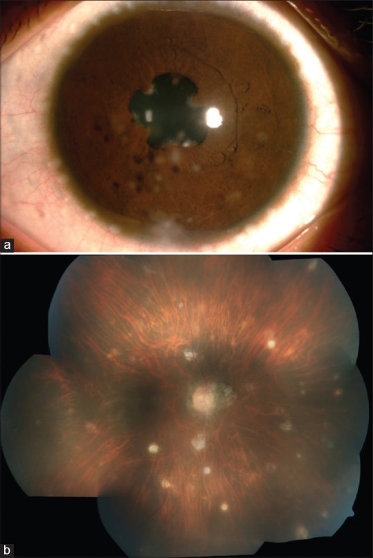
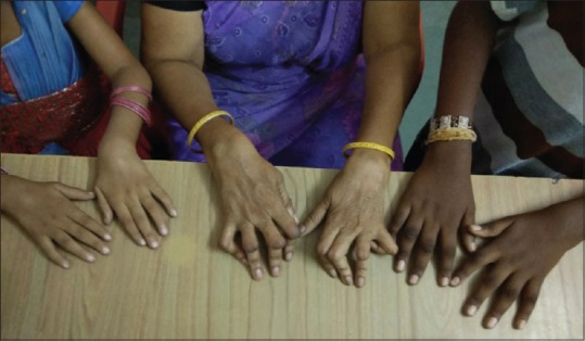
A 38-year-old mother and her two children, a boy aged 10 years and a girl aged 5 years, were referred with eye and joint symptoms present over several years. The mother was first seen by us 12 years ago with granulomatous uveitis and arthritis. She reported a history of childhood onset arthritis. Right eye vision was 3/60, N36, with active panuveitis in the right eye and no perception of light in the left eye. She had associated secondary glaucoma. She was suspected to have JIA-associated uveitis and was treated with systemic and topical steroids. She developed secondary glaucoma and was started on topical anti-glaucoma medication. At 1-year follow-up, when her inflammation was under control, she had developed complicated cataract with glaucoma and underwent phacoemulsification with intraocular lens in the bag implantation with trabeculectomy. Post-operatively she had significant inflammation with multiple recurrences and a granulomatous type of uveitis, which was treated with a further course of systemic steroids and methotrexate. She was found to have choroidal granuloma in her right eye [Fig. 1b]. She was suspected to have sarcoid-related uveitis and was started on methotrexate. Rheumatological examination revealed very deforming arthritis of the small joints of her hands with flexion contractures at the proximal interphalangeal (PIP) joints [Fig. 2]. Her systemic and ocular inflammation resolved. At follow-up her uveitis and intraocular pressure (IOP) was under control, with vision in the right eye of 6/9, N6 maintained on topical non-steroidal anti-inflammatory drugs (NSAIDs) and weekly methotrexate.
Figure 1.

(a) Slit lamp photograph of the girl child showing mutton fat keratic precipitates. (b) Montage color fundus photograph of the mother's right eye showing multiple large healed choroidal granulomas
Figure 2.

Deforming arthritis with severe contractures at proximal interphalangeal joints in mother and boggy arthritis of wrists in both children. The boy has initial stage of contracture of proximal interphalangeal joints
The first child, a boy now 10 years, presented at 5 years of age with bilateral eye pain, occasional redness, and diminished vision. His vision was 6/12, N12 in both eyes and he had bilateral intermediate uveitis. He had a history of arthritis of both wrists and small joints of hands since the age of 4 years. Both the mother and the boy were diagnosed as juvenile idiopathic arthritis with uveitis and the boy was also commenced on tapering dose schedule of topical and oral steroids along with methotrexate. He had experienced multiple recurrences and was treated with appropriate anti-inflammatory therapy during the episodes. Currently, his inflammation is under control, maintaining vision of 6/6p, N6 in both eyes with topical NSAIDs and methotrexate.
The second child, a girl now 5 years, presented at 3 years of age with bilateral granulomatous anterior uveitis with conjunctival cysts. Anterior chamber tap for mycobacterium tuberculosis polymerase chain reaction (PCR) was negative. On musculoskeletal examination she was noticed to have boggy swelling of wrists [Fig. 2], knees, and ankles bilaterally. She had dry scaly rash.
Considering the signs and symptoms in the child, her brother, and mother, an early onset sarcoidosis or BS was considered as a possible diagnosis. She was started on methotrexate and her inflammation resolved with additional course of topical steroids. She is currently on methotrexate along with topical NSAIDs and currently her inflammation is under control.
All three patients were RF, ANA, and HLAB27 negative. Chest X-ray was normal and Mantoux was negative. Angiotensin-converting enzyme (ACE) levels were also within normal limits. Genetic studies to confirm BS was done using a next-generation sequencing panel which identified all the three patients to carry a c. 1000C >T/p. Arg334Trp (R334W) heterozygous variant in the NOD2 gene. This is a previously reported recurrent mutation. All three patients are on regular follow-up with methotrexate, topical NSAIDs, and lubricants.
Discussion
BS was first described by EB Blau in 1985 as familial granulomatous disease of the skin, joints, and eyes in 11 family members over four generations.[1] When the disease occurs sporadically it is sometimes referred to as early onset sarcoidosis. BS characteristically presents within the first few years of life, with skin rash being the usual manifestation in the first year of life, followed by arthritis between 2 and 4 years and, in a majority of patients, uveitis by the age of 4 years.[2] Skin involvement is a frequent early manifestation, with a fine erythematous maculopapular rash, but which can sometimes also be scaly as was the case with our youngest patient. Arthritis in BS is typically non-destructive and boggy in nature, affecting the wrists, knees, and the PIP joints of hands.[3] Uveitis may start insidiously as posterior uveitis but later on become panuveitis, and is typically associated with significant morbidity in the absence of timely appropriate medical management.[4] Expanded systemic manifestations such as fever may be present in 52% of patients.[3] Other systemic manifestations such as fever was not seen in any of our patients. Interestingly, presentations were varied in our patients, with the mother experiencing anterior uveitis with vitritis progressing to panuveitis and choroidal granulomas, the son with intermediate uveitis, and the youngest child with granulomatous anterior uveitis alone with typical mutton fat keratic precipitates [Fig. 1a]. The disease is also characterized by the presence of non-caseating granulomas in affected tissues. Systemic and topical steroids with disease-modifying agents such as methotrexate are the mainstay of therapy in resource constraint settings. Biologics are useful particularly in severe eye disease.[5]
A confirmatory diagnosis of BS was delayed in case of the mother and the first child as the patients were from a rural area and from an economically disadvantaged background. The mother had lost vision in one eye due to complications of incompletely treated uveitis before presenting to us. It is also likely that in the absence of appropriate treatment early the disease had evolved and the typical boggy arthritis and rash were not present at the time of presentation. The patients were started on immunosuppressives after they reported to us. They both were diagnosed as JIA due to onset of disease in childhood, although the mother was suspected to have sarcoid-related uveitis after she developed choroidal granulomas and tuberculosis was ruled out. It was only when the second child presented to us in an early stage of disease with boggy arthritis, a scaly rash, and bilateral uveitis, that a diagnosis of BS was considered. Genetic testing in all the three patients confirmed the diagnosis of BS. The mother's eye disease is now quiescent. The two children are being currently treated with topical steroids, NSAIDs, and weekly methotrexate. The mother's hand joints are deformed due to previous arthritis and contractures at the PIP joints and the children's joints are improving with treatment. Though early onset sarcoidosis has been reported from India, to the best of our knowledge this is the first case series of molecularly confirmed familial BS from India.[6,7]
Conclusion
BS remains an under reported entity in India especially when ocular and joint presentations inappropriately suggest a diagnosis of JIA or tubercular uveitis among others. When treating very young children with arthritis and uveitis it is important to consider this diagnosis, particularly where there is familial recurrence, even in the absence of classical signs in all the patients. Aggressive management of uveitis and arthritis is key to successful management to avoid morbidity.
Declaration of patient consent
The authors certify that they have obtained all appropriate patient consent forms. In the form the patient(s) has/have given his/her/their consent for his/her/their images and other clinical information to be reported in the journal. The patients understand that their names and initials will not be published and due efforts will be made to conceal their identity, but anonymity cannot be guaranteed.
Financial support and sponsorship
Nil.
Conflicts of interest
There are no conflicts of interest.
References
- 1.Blau EB. Familial granulomatous arthritis, iritis, and rash. J Pediatr. 1985;107:689–93. doi: 10.1016/s0022-3476(85)80394-2. [DOI] [PubMed] [Google Scholar]
- 2.Wouters CH, Maes A, Foley KP, Bertin J, Rose CD. Blau syndrome, the prototypic auto-inflammatory granulomatous disease. Pediatr Rheumatol Online J. 2014;12:33. doi: 10.1186/1546-0096-12-33. [DOI] [PMC free article] [PubMed] [Google Scholar]
- 3.Rosé CD, Pans S, Casteels I, Anton J, Bader-Meunier B, Brissaud P, et al. Blau syndrome: Cross-sectional data from a multicentre study of clinical, radiological and functional outcomes. Rheumatology (Oxford) 2015;54:1008–16. doi: 10.1093/rheumatology/keu437. [DOI] [PubMed] [Google Scholar]
- 4.Sarens IL, Casteels I, Anton J, Bader-Meunier B, Brissaud P, Chédeville G, et al. Blau syndrome-associated uveitis: Preliminary results from an international prospective interventional case series. Am J Ophthalmol. 2018;187:158–66. doi: 10.1016/j.ajo.2017.08.017. [DOI] [PubMed] [Google Scholar]
- 5.Achille M, Ilaria P, Teresa G, Roberto C, Ilir A, Piergiorgio N, et al. Successful treatment with adalimumab for severe multifocal choroiditis and panuveitis in presumed (early-onset) ocular sarcoidosis. Int Ophthalmol. 2016;36:129–35. doi: 10.1007/s10792-015-0135-x. [DOI] [PubMed] [Google Scholar]
- 6.Jain L, Gupta N, Reddy MM, Mittal R, Barik MR, Panigrahi B, et al. A novel mutation in helical domain 2 of NOD2 in sporadic blau syndrome. Ocul Immunol Inflamm. 2018;26:292–4. doi: 10.1080/09273948.2016.1207789. [DOI] [PMC free article] [PubMed] [Google Scholar]
- 7.Agarwal K, Barua S, Adhicari P, Das S, Marak R. Early-onset sarcoidosis and juvenile idiopathic arthritis: A diagnostic dilemma. Indian J Dermatol Venereol Leprol. 2016;82:542–5. doi: 10.4103/0378-6323.183626. [DOI] [PubMed] [Google Scholar]


