Abstract
A 36-year-old female presented initially with photophobia and visual deterioration. After examination and laboratory tests, patient was diagnosed with cystinosis. Cysteamine drops 4 × 1 drops/day was given as treatment for 1 year. During follow-up, in vivo confocal microscopy (IVCM) and anterior segment optical coherence tomography (AS-OCT) was performed. Photophobia was relieved and IVCM obtained the decrease in size and density of corneal crystals 1 year after. Depth of corneal crystals did not change but crystal density score reduced with cysteamine treatment.
Keywords: Anterior segment optical coherence tomography, cysteamine, cystinosis, in vivo confocal microscopy
Cystine crystals can be deposited in all ocular tissues whereas conjunctiva and cornea are more frequently affected at cystinosis.[1,2] Topical cysteamine drops seems to be effective in reducing corneal photophobia and other symptoms.[3] Efficacy of the treatment may be assessed with anterior segment optical coherence tomography (AS-OCT) and in vivo confocal microscopy (IVCM) which seems to have more valuable data for follow-up.[4,5] In this case, we present IVCM and AS-OCT findings in a cystinosis patient with photophobia who treated with cysteamine drop for 1 year.
Case Report
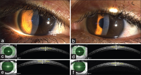
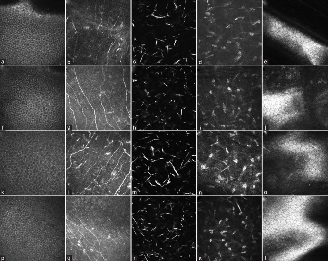
A 36-year-old woman presented to the Gazi University, Department of Ophthalmology complaining of photophobia, foreign body sensation in both eyes. On ophthalmologic examination, the visual acuity was 20/20 in both eyes. Corneal crystal deposits were observed in both eyes at the slit-lamp examination [Fig. 1]. Fundus examination was normal. Patient referred to the nephrology clinic and cystinosis was diagnosed with the result of leukocyte cystine concentration >3 nmol half-cystine per milligram of protein. Informed consent was taken and anterior segment photography, AS-OCT (Spectralis SD-OCT, Heidelberg Engineering, Heidelberg, Germany), and IVCM (Rostock Cornea Module of the Heidelberg Retina Tomography, Heidelberg Engineering GmbH, Heidelberg, Germany) of cornea was performed and central corneal thickness (CCT), depth of crystal accumulation (DC) was noted. Corneal cystine crystal score (CCCS) which was defined by Gahl et al. was 2.00 in both eyes.[4] Crystal density score (CDS) which defined by Labbe et al. was calculated retrospectively.[2,5] AS-OCT showed cystine crystals as hyperreflective dots in the anterior and mid-stroma with the depth of 415 μm (CCT: 559 μm) in the right eye and 428 μm (CCT: 556 μm) in the left eye [Fig. 1]. IVCM demonstrated that there were no cystine crystals in the epithelium and endothelium [Fig. 2]. These spindle-shaped and fusiform-shaped crystals were revealed at subbasal nerve layer, anterior, middle, and posterior stroma and Descemet's membrane. DC measured with IVCM was 338 μm (CCT: 551 μm) in the right eye and 371 μm (CCT: 554 μm) in the left eye. Bilateral CDS was 2 in the subbasal nerve layer, 3 in the anterior stroma, 3 in the middle stroma, and 1 in the posterior stroma (total 9 points). Topical treatment, cysteamine hydrochloride (cystadrops 0.55%, Orphan Europe, France), was given as 4 × 1 drops/day. Nephrology department did not initiate a systemic treatment. AS-OCT images showed no change with the depth of crystals in the stroma 1 year after treatment [Fig. 1]. CCCS was same. Density and size of cystine crystals were decreased in both eyes 1 year after treatment but there was no significant change with the depth of crystals in the IVCM of the cornea [Fig. 2]. Bilateral CDS was decreased in both subbasal nerve layer (from 2 to 1) and anterior and middle stroma (from 3 to 2). Total CDS was decreased to 6 points in both eyes. Photophobia of the patient was relieved during the treatment. We did not experience any side effect with the treatment.
Figure 1.

Corneal anterior segment photography and optical coherence tomography images of the patient. (a and b) Cystine crystals were observed in the stroma. Cystine crystals were seen as hyperreflective dots in the stroma at optical coherence tomography. The depth of crystals and central corneal thickness was measured with anterior segment photography and optical coherence tomography. (c) Right eye before treatment, central corneal thickness: 559 μm, depth of crystals: 345 μm. (d) Left eye before treatment, central corneal thickness: 556 μm, depth of crystals: 334 μm. (e) Right eye with 1 year after treatment, central corneal thickness: 558 μm, depth of crystals: 336 μm. (f) Left eye with 1 year after treatment, central corneal thickness: 553 μm, depth of crystals: 331 μm
Figure 2.
Corneal in vivo confocal microscopy images of the patient. (a–e) Demonstrate the right eye and (k–o) demonstrate the left eye before treatment. (f–j) Demonstrate the right eye and (p–t) demonstrate the left eye after treatment. Cystine crystals started to accumulate in subbasal nerve layer (b, g, l and q) and the most intense deposition was in anterior stroma (c, d, m and r). No crystal was observed in endothelium (e, j, o and t). The depth of accumulation was not significantly changed but the density and size of the crystals were decreased
Discussion
IVCM of the cornea is one of the most valuable methods to demonstrate the depth and density of cystine crystals in cellular level and very useful for following-up treatment efficacy.[5] AS-OCT seems to have less sensitivity for measuring depth of crystals.[5] In our case, density of cystine crystals was higher in anterior and mid-stroma of the cornea and there were no crystals in epithelium and endothelium. Liang et al. reported that the severity of photophobia is correlated with the density of cystine crystals.[2] Gahl et al. reported that cysteamine drop treatment may provide corneal clearance of cystine crystals and reducing photophobia.[4] In our case, we could not provide corneal clearance, but we obtained decrease in crystal density and this situation also helped reducing photophobia. We also observed that intensity of photophobia decreases and we thought that it could be related to the decrease of cystine crystals’ size and density. Cysteamine drop treatment is efficient for reducing symptoms of cystinosis as well as cystine crystals in corneal layers and IVCM is very useful tool for following-up patients.
Conclusion
Corneal cystine crystals’ size and density reduced 1 year after cysteamine treatment and IVCM was effective to demonstrate changes after treatment rather than AS-OCT.
Declaration of patient consent
The authors certify that they have obtained all appropriate patient consent forms. In the form the patient(s) has/have given his/her/their consent for his/her/their images and other clinical information to be reported in the journal. The patients understand that their names and initials will not be published and due efforts will be made to conceal their identity, but anonymity cannot be guaranteed.
Financial support and sponsorship
Nil.
Conflicts of interest
There are no conflicts of interest.
References
- 1.Gahl WA, Thoene JG, Schneider JA. Cystinosis. N Engl J Med. 2002;347:111–21. doi: 10.1056/NEJMra020552. [DOI] [PubMed] [Google Scholar]
- 2.Liang H, Baudouin C, Tahiri Joutei Hassani R, Brignole-Baudouin F, Labbe A. Photophobia and corneal crystal density in nephropathic cystinosis: An in vivo confocal microscopy and anterior-segment optical coherence tomography study. Invest Ophthalmol Vis Sci. 2015;56:3218–25. doi: 10.1167/iovs.15-16499. [DOI] [PubMed] [Google Scholar]
- 3.Alsuhaibani AH, Khan AO, Wagoner MD. Confocal microscopy of the cornea in nephropathic cystinosis. Br J Ophthalmol. 2005;89:1530–1. doi: 10.1136/bjo.2005.074468. [DOI] [PMC free article] [PubMed] [Google Scholar]
- 4.Gahl WA, Kuehl EM, Iwata F, Lindblad A, Kaiser-Kupfer MI. Corneal crystals in nephropathic cystinosis: Natural history and treatment with cysteamine eyedrops. Mol Genet Metab. 2000;71:100–20. doi: 10.1006/mgme.2000.3062. [DOI] [PubMed] [Google Scholar]
- 5.Labbé A, Niaudet P, Loirat C, Charbit M, Guest G, Baudouin C, et al. In vivo confocal microscopy and anterior segment optical coherence tomography analysis of the cornea in nephropathic cystinosis. Ophthalmology. 2009;116:870–6. doi: 10.1016/j.ophtha.2008.11.021. [DOI] [PubMed] [Google Scholar]


