Abstract
Purpose:
To study the demographic profile, clinical features, treatment outcome, and ocular morbidity of microbiologically proven Pythium keratitis in South India.
Methods:
A retrospective analysis of clinical records of microbiologically proven Pythium keratitis at a tertiary eye care referral center in South India from January 2016 to November 2017 was performed. Demographic details, predisposing risk factors, microbiological investigations, clinical course, and visual outcome were analyzed.
Results:
Seventy-one patients with microbiologically proven Pythium keratitis were identified. The mean age was 44(±18.2) years with an increase in male preponderance and 50% were farmers. Duration of delay at time of presentation to the hospital was a mean of 14(±7.2) days. The visual acuity at baseline ranged from 6/6 to no light perception (median 2.1 logMAR). A combination of 5% natamycin and 1% voriconazole was given to 42% patients, and natamycin alone was given to 39.4% patients. 1% itraconazole eye drops alone was initiated in 7 (10%) patients and 3 among this group responded. Therapeutic keratoplasty (TPK) was performed in 48 (67.6%) patients. None of the primary grafts remained clear after a period of 1 month. Twenty-six eyes (54.2%) had graft reinfection and all these eyes either developed anterior staphyloma (4) or were eviscerated (3) and 13 eyes became phthisical. The remaining 22 patients who had TPK resulted in failed graft. Among these, re-grafts were performed in 6 patients, of which 5 were doing well at the last follow-up.
Conclusion:
We report a large series of patients with Pythium keratitis. Promoting early and differential diagnosis, awareness of clinicians and specific treatment options are needed for this devastating corneal disease.
Keywords: DNA sequencing, ITS, keratitis, Pythium insidiosum, therapeutic penetrating keratoplasty
Increasing reports of Pythium keratitis in recent years has garnered much attention, with reports emerging from the Asia Pacific region.[1,2,3,4,5,6,7,8] Pythium is an oomycete that causes a devastating infection of the cornea and has been reported to have a poor outcome. It is a very difficult disease to treat with patients responding poorly to the conventional antifungal medication or to surgical procedures such as penetrating keratoplasty. Major reports of both systemic and ocular infections being caused by Pythium insidiosum have been primarily from Thailand and are found to be endemic there because of their climatic conditions.[9,10,11,12,13] Pythium species is classified in the Phylum Straminipila, Class Oomycetes, Order Pythiales, and Family Pythiaceae.[14,15] While pythiosis is often described as an emerging disease,[16] the disease was described as early as in 1884 by British veterinarians working with horses in India.[17] Systemic infections in humans with Pythium are also well documented with pythiosis being a life-threatening disease with high rates of morbidity and mortality.[18] Diagnosis and treatment still remains difficult because of the nature of this organism.[6,7]
The visual morbidity that often results from Pythium keratitis underscores the importance of studying its epidemiology. Reports have described the different modalities for recognizing this organism from the growth on culture media.[19] Another area of concern is the lack of standardized treatment protocol for these devastating organisms, and various treatment options have been recommended.[7,8,20] Keratitis due to Pythium does not scar easily and afflicted patients face a prolonged recovery often requiring multiple keratoplasty. This is an important issue of concern where the burden of corneal blindness due to microbial keratitis is already high. Recognizing, and if possible, prevention of this devastating disease is very important.
In this study, we present a large series of patients of Pythium keratitis occurring between 2016 and 2017 among patients presenting to an eye care referral center in South India. The risk factors, clinical signs and symptoms, and treatment outcomes are analyzed to see if there was any trends toward better treatment response.
Methods
This was a retrospective review of medical case records and microbiology records of all patients positive on culture for Pythium keratitis from January 2016 to November 2017. Approval was obtained from the Institutional Review Board of Aravind Eye Hospital, Madurai for this study. The demographic details, predisposing factors, clinical course, microbial results, treatment, and visual outcomes were collected. The indications for therapeutic penetrating keratoplasty were analyzed. Following recording the clinical characteristics, all the affected eyes were subjected to a microbiological evaluation. Corneal scrapings were collected under topical anesthesia using 0.5% proparacaine. Specimen included two scrapings for smear examination (one each for Grams stain and 10% potassium hydroxide wet mount) followed by a subsequent sequential scraping for culture on blood agar and potato dextrose agar. The characterized colony morphology of the Pythium species on the blood agar prompted us to the possibility of Pythium. This was further confirmed both by PCR-based DNA sequencing targeting internal transcribed spacer (ITS) region and with zoospores formation identifying the organism as P. insidiosum in all the isolates.[19,21,22] Treatment initiated according to clinical and microbiological evaluations. The eyes with positive fungal smears were treated with 5% natamycin suspension on an hourly basis during waking hours. However, if Pythium keratitis was suspected from the clinical diagnosis, the eyes were treated with natamycin alone or a combination of topical natamycin and econazole or voriconazole. Itraconazole was also used either alone or in combination with 1% azithromycin as topical drops.
Patients with poor response despite adequate and appropriate antimicrobial therapy, perforation, and ulcers threatening limbus were subjected to therapeutic keratoplasty. The excised corneal button was also cultured on blood agar and potato dextrose agar and was processed for species identification. Postoperatively, all eyes were treated with voriconazole 1% on an hourly basis for a minimum period of 3 weeks.
Statistical analysis was done using statistical software STATA V.11.0, USA. Continuous variables were expressed as mean (SD), and categorical variables were expressed as frequency (percentage).
Results
During the study period of 23 months, the prevalence of Pythium keratitis was 5.9% (71/1204) of all the cases of keratitis that were culture positive for fungus. Looking at the month wise distribution, cases had started peaking from June 2016 and continued to July 2017. Only in the month of January 2017, there was no report of similar case [Fig. 1]. We are still continuing to see relatively high numbers of cases.
Figure 1.
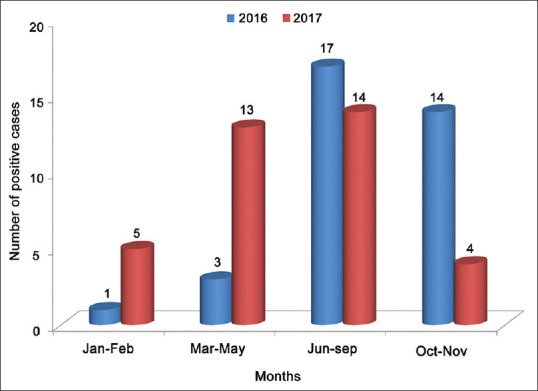
Bar diagram represents the seasonal observation of 71 culture positive Pythium keratitis cases seen at tertiary eye care center in South India from January 2016 to November 2017
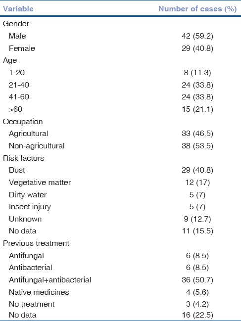
The demographic features, risk factors, and previous treatment details of the patients are summarized in Table 1. The mean age of the patients at presentation was 44 years (SD ± 18.2, range: 4–80 years), and there were 42 (59.2%) males and 29 (40.8%) female patients. Of these, 33 (46.5%) were farmers by occupation, and an equal number of them (53.5%) were either software professionals (10 [26.3%]), housewives from urban locales (13 [34.2%]), teachers (6 [15.8%]) and students (9 [23.7%]) with no exposure to vegetative matter. A predisposing factor was elicited in 51/71 (71.8%) patients [Table 1]. The time taken to presentation to hospital varied from 2 to 60 days with a mean of 14 (SD 7.2) days. The majority of patients were on some form of topical medication before presenting to us, 36/71 (50.7%) were on both anti-fungal and antibacterial drops. Native medicine such as breast milk or chicken blood was used in four patients. Symptoms of pain, redness, irritation, and photophobia were present in all patients.
Table 1.
Demographic profile of patients with Pythium keratitis
The visual acuity at the time of presentation ranged from 6/6 to no light perception (median 2.1 logMAR). Patients with BCVA less than 5/60 (50; [70.4%]), at presentation, were those who presented late to the hospital. These patients had a mean ulcer size of 38 sq. mm (SD 25.42 sq mm) [Table 2]. However, patients who had better vision at presentation, i.e., from 6/6 to 6/60, presented early to the hospital and had significantly lower mean ulcer size of 13 sq.mm.
Table 2.
Distribution of patients by visual acuity

Hyphate edges of the infiltrates were seen in most patients; slit-lamp biomicroscopy showed multiple linear tentacle-like infiltrates [Fig. 2a] in 36 of 71 (50.7%) patients, the presence of dot-like infiltrates [Fig. 2b] at the midstromal level surrounding the main infiltrate in 15 (21.1%) patients and peripheral furrowing [Fig. 2c] in 9 (12.7%) patients [Table 3]. Hypopyon was present in 18 of 71 (25.4%) patients [Fig. 2d].
Figure 2.
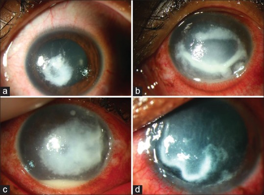
Clinical findings of patients with Pythium keratitis. Slit-lamp picture of the cornea showing (a) central, dense, grayish-white infiltrate with tentacle-like lesions. (b) Diffuse dot-like infiltrates emanating from the main infiltrate extending to the peripheral cornea. In addition peripheral furrowing is seen in this picture inferiorly from 4o clock to 7o clock hour. (c) Large dense infiltrate with dot like multiple infiltrates with peripheral furrowing from 2o clock to 4o clock hours with 1 mm hypopyon. (d) Dense grayish white infiltrate with peripheral furrowing seen from 7o clock to 9o clock hours. Tentacle like extensions and subepithelial dot like infiltrates seen extending from the main infiltrate
Table 3.
Clinical presentation of Pythium keratitis patients

Microbiology results of direct microscopy with 10% potassium hydroxide and Gram stain was positive for hyphal filaments in 77.5% of the patients [Fig. 3a and b]. All 71 Pythium isolates were cultured either from corneal scrapings (29/71), corneal buttons (27/71), or both (15/71). In 29 patients, the initial culture was negative from the corneal scraping and only culturing of the corneal button picked up the Pythium species. Flat, feathery-edged, partially submerged, colorless, or light brown colonies with filiform margins [Fig. 3c] on blood agar were grown from corneal scrapings or buttons. In our study, the mean duration for culture positivity was noted to be 3–5 days (mean + SD). PCR amplification of Pythium fungal DNA targeting ITS region yielded 495 bp product [Fig. 4].
Figure 3.
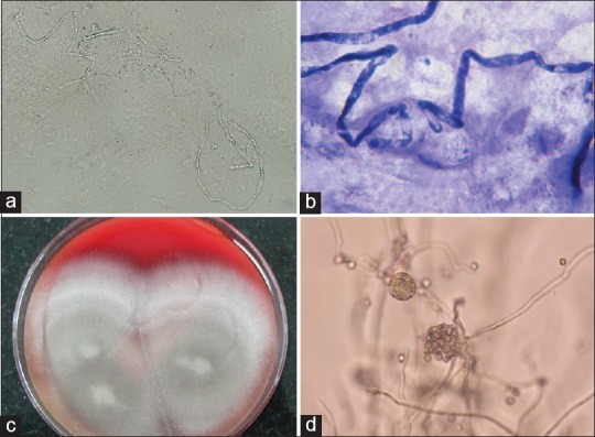
(a) 10% KOH wet mount preparation of corneal scraping showed long sparsely septate hyaline hyphae of Pythium insidiosum. The presence of numerous vesicles within the hyphae is usually observed. (b) Gram stain image showed the thick cell wall, a few septate, and mass of vesicles inside. (c) A 3 day old culture of P. insidiosum at 37°C grown on 5% sheep blood agar. (d) A vesicles with zoospores that developed after 3 h incubation before zoospore release (×10)
Fig. 4.
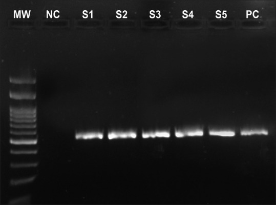
Amplification of a 495 bp specific DNA fragment of the ITS1 and ITS4 region of Pythium fungal DNA. MW: Molecular weight marker (100 bp); NC: Negative control; Lane S1-S5: Amplified Pythium fungus DNA (495bp); PC: Positive control
Medical therapy was initiated in all the patients immediately on receiving a positive report of fungus on the corneal scraping [Table 4]. The most preferred medication was a combination of natamycin and voriconazole that was given in 30 (42.3%) patients, whereas natamycin alone was given in 28 (39.4%) patients. In the group of patients who had a negative smear examination initially, a combination of natamycin and moxifloxacin was given and was modified according to the culture reports. Recently, itraconazole eye drops alone was initiated in 7 (10%) patients. To our surprise, among these 7 patients, we saw complete resolution of the ulcer in 3 patients with hourly itraconazole eye drops. The mean ulcer size for the 3 patients who resolved with itraconazole was 6.33 ± 2.5 sq. mm. These were the only 3 patients in this whole series that responded to medical treatment. The clinical profile of these 3 patients is described in Table 5; however, we could not discern any common thread in this subset of patients. They were varied age groups and professions. The only common point was that the presenting visual acuity was good in all 3 patients. One patient (no: 1) had initial progression of the ulcer and then it started responding. The duration of treatment was very prolonged in all these patients with an average of 47 days (SD).
Table 4.
Medical therapy initiated in patients with Pythium keratitis

Table 5.
Clinical profile of medically cured Pythium keratitis cases

Of the 71 patients, eleven patients did not follow-up after the initial visit. Among the rest, therapeutic keratoplasty (TPK) could not be done in 9 patients as they progressed to total corneal melt very quickly. In these 9 patients who had a corneal melt at the time of presentation, in whom a keratoplasty could not be done, eventually the eye became pthisical. TPK was performed in 48 (67.6%) patients who either had a perforated corneal ulcer at presentation itself or were clinically worsening (non-responsiveness to medical treatment or progression of clinical signs). The time interval between initial presentation to TPK varied from 0 to 59 days, with a mean of 15 days. The mean follow-up was 13 months (1–24 months).
Out of the 48 patients who had TPK, 26 (54.2%) developed a recurrence of the infection in the graft, of which 20 (77%) had limbal involvement preoperatively. The average size of ulcer in patients who had graft reinfection was 41.2 mm2 compared to 27 mm2 in those who had no re-infection postoperatively. Topical steroids were not administered postoperatively for any of the cases. Out of the patients who had graft reinfection, 13 eyes resulted in phthisis bulbi, 4 eyes into staphyloma, and three eyes had to be eviscerated. In the remaining 6 patients, the infection resolved. There was no statistically significant correlation with respect to recurrence of infection post TPK with perforation (p 0.295 Chi-square test), ulcer size (p 0.3304 Man-Whitney test), and graft size (p 0.4096 Mann-Whitney test). The remaining 22 patients who had TPK ended in failed opaque grafts. In these 22 patients, optical grafts were performed for 6 patients, out of whom 5 patients had clear graft at the last follow-up, whereas in one patient the re-graft also developed reinfection with Pythium and ultimately resulted in phthisis. In the patient who underwent TPK, anatomical success (globe salvage/no infection) was achieved in 21 (43.7%) patients and functional (vision) success was achieved in 5 patients (10.41%).
Discussion
In this study, we found that the cases of Pythium keratitis had considerably increased in the past 2 years and we are continuing to see this increase to the present in South India. The reasons for this are not yet clear. There are similar reports from this region from Agrawal et al. who reported 10 patients over 18 month's period, and Sharma et al. reported a total of 11 cases.[6,7] This disease has rightly been described as underreported, and there is a need for an increase in awareness among both the microbiologists and ophthalmologists.[23] With the occurrence of such large numbers of ocular Pythium, there is a high possibility of human systemic Pythium also occurring in the region of Southern India that deserves close observation. In other countries, systemic infection with Pythium has been reported.[9,10,11,24] Infections in plants and animals have also been reported.[14,25] Although infections in horses from India were reported as early as 1884, recent reports of infections with this organism are lacking. This large number of cases is an indication of improved awareness of this pathogen and the knowledge of its identification. It is possible that we were missing these cases earlier by falsely labeling these as unidentified fungi or cases diagnosed as fungal according to microscopy but with no growth on culture.
Unlike other studies on fungal keratitis, where it occurs predominantly in agricultural workers, this pathogen seems to infect non-agriculture workers equally as highlighted in the report by Agarwal et al. In our series also, patients with a non-agricultural background had the same predisposition to infection, with nearly 78% of the patients having a history of exposure to dust or some foreign-object falling in their eyes.
An outbreak of Pythium keratitis during the rainy season has been reported from Thailand.[2] Pythium is an aquatic organism, and many studies report an association between water and non-ocular Pythium infection,[15] but from our report, a non-aquatic environment may also be an important risk factor for the occurrence of ocular Pythium infection. However, a history of exposure to water and other aquatic environments such as agricultural fields and also soil is very imperative to raise the suspicion of Pythium keratitis.
The classic clinical features described in many series such as multiple linear tentacle-like infiltrates and the presence of dot-like infiltrates were also predominately present in our patients’ population. Although confocal was not used in this study, other studies advocate the use of confocal microscopy to get a suggestion of the possibility of Pythium keratitis. In the laboratory, the combination of the classic colony morphology along with the zoospore production as well as molecular tools for the identification of Pythium will help in the identification and confirmation.[19,26,27,28,29] Although this organism grows readily on common laboratory medium such as blood agar and potato dextrose agar, it might not grow from the early specimens, such as the first corneal scraping, and direct microscopy examination of the smear also may be negative. Although the clinical features are a strong indicator, laboratory confirmation is also very much needed to accurately classify as Pythium keratitis. As was seen in our study, in some cases, the smear was negative and only the corneal buttons removed at the time of keratoplasty grew Pythium. Hence, a combination of specimens such as corneal scrapings and buttons has to be included to get a positive culture.
Various treatment and management options have been suggested in the treatment of Pythium keratitis.[20,30,31,32] As Pythium is not a fungus, and the cell morphology is different from fungus, antifungal agents are not useful. However, the in-vitro activity of antibiotics such as azithromycin, minocycline, and tigecycline have been tested and found to be effective.[33] In the recent study by Muralidahr et al., one patient was treated with linezolid and azithromycin eye drops and the authors found the ulcer is responding.[20] One of the largest series reported so far was by Bagga et al. that had 144 patients over a 3 year period.[34] In this study, the authors studied the effect of a combination of topical linezolid and topical and oral azithromycin in the second phase of the study and found a favorable but not statistically significant response of P. insidiosum keratitis to antibacterial agents. However, the rate of TPK had been reduced in the group on antibiotics as compared to the antifungal group. This study shows promise on the efficacy of a combination of antibiotic in the treatment of Pythium keratitis. Further evaluation of this strategy in larger number of patients is recommended. In our study, in 3 patients the lesion resolved with topical itraconazole 1%. There might have been other factors contributing to the success of treatment with itraconazole such as the ulcer size, host response, and maybe even the species of Pythium. There is also the possibility that the infection may have been superficial and scraping would have resulted in debulking of infection. For any drug to be declared effective, larger studies involving more patients are needed to see the true efficacy. Penetrating keratoplasty has mixed results with some grafts getting reinfected or failing. The organism is so virulent and fast growing that there is not much time available before keratoplasty can be performed. In our series, the average time for performing keratoplasty was 15 days. Early keratoplasty may be beneficial, as recommended by some studies. In the series by Agarwal et al., cryotherapy and or absolute alcohol might prove beneficial.[7]
Conclusion
To conclude keratitis due to Pythium seems to be increasing in recent times in South India, the reasons for which are not clear, and more data and research are needed to study this vision threatening corneal ulcer. We believe that the existing anti-fungal agents are not effective against Pythium infections. However, with the recent advance in molecular technology with next generation sequencing where the whole genome of Pythium might be known, hopefully more light will be shed on the pathogenesis of this organism which will be more helpful in developing new therapeutic strategies.
Financial support and sponsorship
Nil.
Conflicts of interest
There are no conflicts of interest.
References
- 1.Virgile R, Perry HD, Pardanani B, Szabo K, Rahn EK, Stone J, et al. Human infectious corneal ulcer caused by Pythium insidiosum. Cornea. 1993;12:81–3. doi: 10.1097/00003226-199301000-00015. [DOI] [PubMed] [Google Scholar]
- 2.Thanathanee O, Enkvetchakul O, Rangsin R, Waraasawapati S, Samerpitak K, Suwan-Apichon O, et al. Outbreak of Pythium keratitis during rainy season: A case series. Cornea. 2013;32:199–204. doi: 10.1097/ICO.0b013e3182535841. [DOI] [PubMed] [Google Scholar]
- 3.Lekhanont K, Chuckpaiwong V, Chongtrakool P, Aroonroch R, Vongthongsri A. Pythium insidiosum keratitis in contact lens wear: A case report. Cornea. 2009;28:1173–7. doi: 10.1097/ICO.0b013e318199fa41. [DOI] [PubMed] [Google Scholar]
- 4.Badenoch PR, Mills RA, Chang JH, Sadlon TA, Klebe S, Coster DJ, et al. Pythium insidiosum keratitis in an Australian child. Clin Exp Ophthalmol. 2009;37:806–9. doi: 10.1111/j.1442-9071.2009.02135.x. [DOI] [PubMed] [Google Scholar]
- 5.Tanhehco TY, Stacy RC, Mendoza L, Durand ML, Jakobiec FA, Colby KA, et al. Pythium insidiosum keratitis in Israel. Eye Contact Lens. 2011;37:96–8. doi: 10.1097/ICL.0b013e3182043114. [DOI] [PubMed] [Google Scholar]
- 6.Sharma S, Balne PK, Motukupally SR, Das S, Garg P, Sahu SK, et al. Pythium insidiosum keratitis: Clinical profile and role of DNA sequencing and zoospore formation in diagnosis. Cornea. 2015;34:438–42. doi: 10.1097/ICO.0000000000000349. [DOI] [PubMed] [Google Scholar]
- 7.Agarwal S, Iyer G, Srinivasan B, Agarwal M, Panchalam Sampath Kumar S, Therese LK, et al. Clinical profile of Pythium keratitis: Perioperative measures to reduce risk of recurrence. Br J Ophthalmol. 2018;102:153–7. doi: 10.1136/bjophthalmol-2017-310604. [DOI] [PubMed] [Google Scholar]
- 8.He H, Liu H, Chen X, Wu J, He M, Zhong X, et al. Diagnosis and treatment of Pythium insidiosum corneal ulcer in a Chinese child: A case report and literature review. Am J Case Rep. 2016;17:982–8. doi: 10.12659/AJCR.901158. [DOI] [PMC free article] [PubMed] [Google Scholar]
- 9.Triscott JA, Weedon D, Cabana E. Human subcutaneous pythiosis. J Cutan Pathol. 1993;20:267–71. doi: 10.1111/j.1600-0560.1993.tb00654.x. [DOI] [PubMed] [Google Scholar]
- 10.Sudjaritruk T, Sirisanthana V. Successful treatment of a child with vascular pythiosis. BMC Infect Dis. 2011;11:33. doi: 10.1186/1471-2334-11-33. [DOI] [PMC free article] [PubMed] [Google Scholar]
- 11.Reanpang T, Orrapin S, Orrapin S, Arworn S, Kattipatanapong T, Srisuwan T, et al. Vascular pythiosis of the lower extremity in Northern Thailand: Ten years’ experience. Int J Low Extrem Wounds. 2015;14:245–50. doi: 10.1177/1534734615599652. [DOI] [PubMed] [Google Scholar]
- 12.Krajaejun T, Sathapatayavongs B, Pracharktam R, Nitiyanant P, Leelachaikul P, Wanachiwanawin W, et al. Clinical and epidemiological analyses of human pythiosis in Thailand. Clin Infect Dis. 2006;43:569–76. doi: 10.1086/506353. [DOI] [PubMed] [Google Scholar]
- 13.Imwidthaya P. Human pythiosis in Thailand. Postgrad Med J. 1994;70:558–60. doi: 10.1136/pgmj.70.826.558. [DOI] [PMC free article] [PubMed] [Google Scholar]
- 14.Mendoza L, Vilela R. The mammalian pathogenic oomycetes. Curr Fungal Infect Rep. 2013;7:198–208. [Google Scholar]
- 15.Gaastra W, Lipman LJ, De Cock AW, Exel TK, Pegge RB, Scheurwater J, et al. Pythium insidiosum: An overview. Vet Microbiol. 2010;146:1–6. doi: 10.1016/j.vetmic.2010.07.019. [DOI] [PubMed] [Google Scholar]
- 16.Laohapensang K, Rutherford RB, Supabandhu J, Vanittanakom N. Vascular pythiosis in a thalassemic patient. Vascular. 2009;17:234–8. doi: 10.2310/6670.2008.00073. [DOI] [PubMed] [Google Scholar]
- 17.Smith F. The pathology of bursattee. Vet J. 1884;19:16–7. [Google Scholar]
- 18.Permpalung N, Worasilchai N, Plongla R, Upala S, Sanguankeo A, Paitoonpong L, et al. Treatment outcomes of surgery, antifungal therapy and immunotherapy in ocular and vascular human pythiosis: A retrospective study of 18 patients. J Antimicrob Chemother. 2015;70:1885–92. doi: 10.1093/jac/dkv008. [DOI] [PubMed] [Google Scholar]
- 19.Grooters AM, Whittington A, Lopez MK, Boroughs MN, Roy AF. Evaluation of microbial culture techniques for the isolation of Pythium insidiosum from equine tissues. J Vet Diagn Invest. 2002;14:288–94. doi: 10.1177/104063870201400403. [DOI] [PubMed] [Google Scholar]
- 20.Ramappa M, Nagpal R, Sharma S, Chaurasia S. Successful medical management of presumptive Pythium insidiosum keratitis. Cornea. 2017;36:511–4. doi: 10.1097/ICO.0000000000001162. [DOI] [PubMed] [Google Scholar]
- 21.Badenoch PR, Coster DJ, Wetherall BL, Brettig HT, Rozenbilds MA, Drenth A, et al. Pythium insidiosum keratitis confirmed by DNA sequence analysis. Br J Ophthalmol. 2001;85:502–3. doi: 10.1136/bjo.85.4.496g. [DOI] [PMC free article] [PubMed] [Google Scholar]
- 22.Salipante SJ, Hoogestraat DR, SenGupta DJ, Murphey D, Panayides K, Hamilton E, et al. Molecular diagnosis of subcutaneous Pythium insidiosum infection by use of PCR screening and DNA sequencing. J Clin Microbiol. 2012;50:1480–3. doi: 10.1128/JCM.06126-11. [DOI] [PMC free article] [PubMed] [Google Scholar]
- 23.Krajaejun T, Pracharktam R, Wongwaisayawan S, Rochanawutinon M, Kunakorn M, Kunavisarut S, et al. Ocular pythiosis: Is it under-diagnosed? Am J Ophthalmol. 2004;137:370–2. doi: 10.1016/S0002-9394(03)00908-5. [DOI] [PubMed] [Google Scholar]
- 24.Franco DM, Aronson JF, Hawkins HK, Gallagher JJ, Mendoza L, McGinnis MR, et al. Systemic Pythium insidiosum in a pediatric burn patient. Burns. 2010;36:e68–71. doi: 10.1016/j.burns.2009.08.005. [DOI] [PubMed] [Google Scholar]
- 25.Pal M, Mahendra R. Pythiosis: An emerging oomycetic disease of humans and animals. Int J Livestock Res. 2014;4:1–9. [Google Scholar]
- 26.Kosrirukvongs P, Chaiprasert A, Uiprasertkul M, Chongcharoen M, Banyong R, Krajaejun T, et al. Evaluation of nested PCR technique for detection of Pythium insidiosum in pathological specimens from patients with suspected fungal keratitis. Southeast Asian J Trop Med Public Health. 2014;45:167–73. [PubMed] [Google Scholar]
- 27.Thongsri Y, Wonglakorn L, Chaiprasert A, Svobodova L, Hamal P, Pakarasang M, et al. Evaluation for the clinical diagnosis of Pythium insidiosum using a single-tube nested PCR. Mycopathologia. 2013;176:369–76. doi: 10.1007/s11046-013-9695-3. [DOI] [PMC free article] [PubMed] [Google Scholar]
- 28.Keeratijarut A, Lohnoo T, Yingyong W, Rujirawat T, Srichunrusami C, Onpeaw P, et al. Detection of the oomycete Pythium insidiosum by real-time PCR targeting the gene coding for exo-1,3-β-glucanase. J Med Microbiol. 2015;64:971–7. doi: 10.1099/jmm.0.000117. [DOI] [PubMed] [Google Scholar]
- 29.Vanittanakom N, Supabandhu J, Khamwan C, Praparattanapan J, Thirach S, Prasertwitayakij N, et al. Identification of emerging human-pathogenic Pythium insidiosum by serological and molecular assay-based methods. J Clin Microbiol. 2004;42:3970–4. doi: 10.1128/JCM.42.9.3970-3974.2004. [DOI] [PMC free article] [PubMed] [Google Scholar]
- 30.Mendoza L, Newton JC. Immunology and immunotherapy of the infections caused by Pythium insidiosum. Med Mycol. 2005;43:477–86. doi: 10.1080/13693780500279882. [DOI] [PubMed] [Google Scholar]
- 31.Wanachiwanawin W, Mendoza L, Visuthisakchai S, Mutsikapan P, Sathapatayavongs B, Chaiprasert A, et al. Efficacy of immunotherapy using antigens of Pythium insidiosum in the treatment of vascular pythiosis in humans. Vaccine. 2004;22:3613–21. doi: 10.1016/j.vaccine.2004.03.031. [DOI] [PubMed] [Google Scholar]
- 32.Cavalheiro AS, Zanette RA, Spader TB, Lovato L, Azevedo MI, Botton S, et al. In vitro activity of terbinafine associated to amphotericin B, fluvastatin, rifampicin, metronidazole and ibuprofen against Pythium insidiosum. Vet Microbiol. 2009;137:408–11. doi: 10.1016/j.vetmic.2009.01.036. [DOI] [PubMed] [Google Scholar]
- 33.Loreto ES, Tondolo JS, Pilotto MB, Alves SH, Santurio JM. New insights into the in vitro susceptibility of Pythium insidiosum. Antimicrob Agents Chemother. 2014;58:7534–7. doi: 10.1128/AAC.02680-13. [DOI] [PMC free article] [PubMed] [Google Scholar]
- 34.Bagga B, Sharma S, Madhuri Guda SJ, Nagpal R, Joseph J, Manjulatha K, et al. Leap forward in the treatment of Pythium insidiosum keratitis. Br J Ophthalmol. 2018 doi: 10.1136/bjophthalmol-2017-311360. pii: bjophthalmol-2017-311360. [DOI] [PubMed] [Google Scholar]


