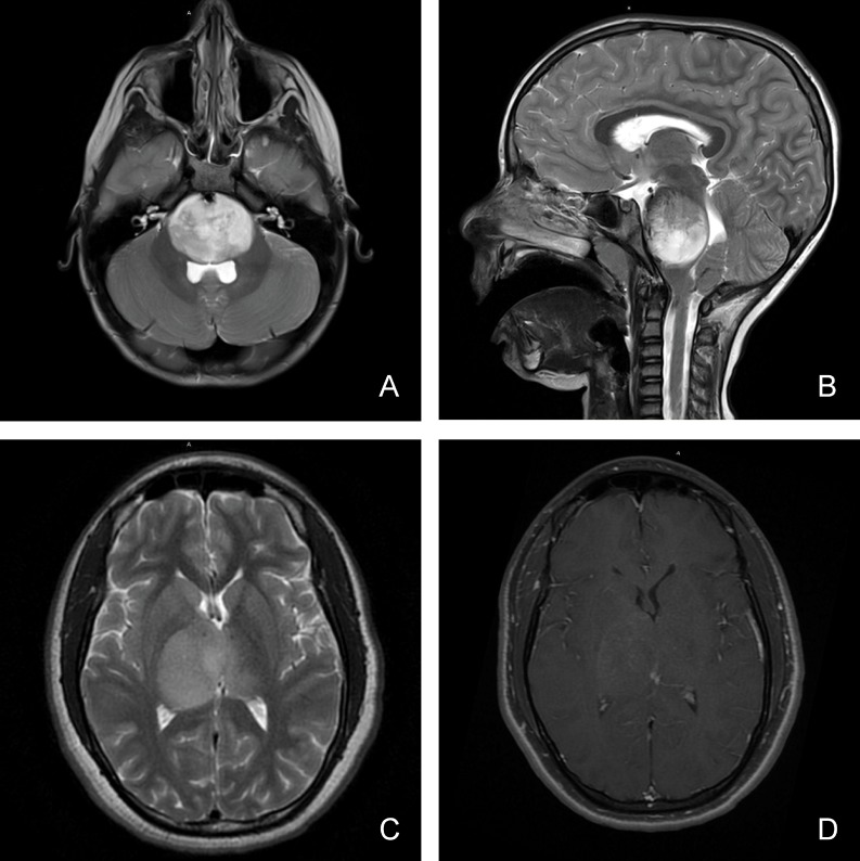
Figure 1. Magnetic resonance imaging characteristics (MRI) of pediatric diffuse midline glioma.
Pediatric diffuse midline glioma arise in the brainstem (A, B) or thalamus (C, D). T2 weighted MR imaging (A, B, C) demonstrates a homogenous, expansile, infiltrative lesion with extension within white matter tracts. Minimal to heterogenous enhancement may be observed on T1 post-gadolinium sequences (D). Local mass effect and perilesional edema may also be seen.

