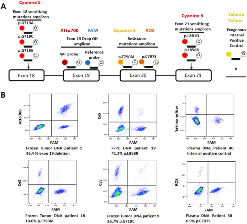
Figure 4.
(A) Design of the six-color digital PCR assay. A total of 4 primer pairs were used to amplify 4 regions on EGFR exon 18, 19, 20 and 21. Six channels of fluorescence were selected to differentiate the targets of interest. Two probes respectively labelled with FAM and Atto700 were used to detect both the EGFR wild-type sequence and exon 19 delins in a drop-off assay. Cyanine 5 labelled probes were used to detect p.L858R/p.L861Q and p.G719A/C/S mutations. Cyanine 3 and ROX labelled probes detected the p.T790M and p.C797S resistance mutations respectively. Finally, a probe with a Yakima Yellow fluorophore was added to detect an exogenous DNA sequence which serves as an internal control of PCR amplification. (B) Six-color digital PCR results in 2D dot-plots from 6 tumor and plasma samples.

