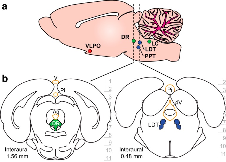
Figure 1.

Targeting the sleep–wake regulatory network. a, A sagittal brain section with structures implicated in sleep–wake regulation. The structures are distributed across multiple regions of brainstem. Color codes represent the postulated states of vigilance during which specific cell populations within each structure are predominantly active: red is non-REM (NREM) sleep; blue is REM sleep; and green is wake. Brain region names and acronyms are as follows: ventrolateral pre-optic nucleus (VLPO), laterodorsal tegmental nucleus (LDT), pedunculopontine tegmental nucleus (PPT), dorsal raphe (DR), and locus coeruleus (LC). b, Coronal brain sections that highlight SWRS populations DR and LDT underneath sensitive anatomic structures highlighted in orange. These dorsal structures should not be damaged during chronic recording studies, and so safe access to the targets requires independent and angled electrode trajectories. Labels are as follows: vein (v), pineal gland (Pi), cerebral aqueduct (Aq), and fourth ventricle (4V). Depth information is in millimeters with horizontal tics representing 1 mm. Coronal sections were adapted with permission from Paxinos and Watson (2007, their Figs. 95 and 104; copyright Elsevier).
