

Abstract
Obtaining T cells by reprogramming is one of the major goals in regenerative medicine. Here, we describe a protocol for generating functional T cells from Hoxb5-expressing pro/pre-B cells in vivo. This protocol includes the construction of Hoxb5 recombinant plasmids, retroviral packaging, isolation and viral transduction of pro/pre-B cells, cell transplantation, and phenotypic analysis of induced T cells. The procedure is reproducible and straightforward, providing an approach for generating induced T cells for translational research.
Keywords: Hoxb5, Retrovirus, Pro/pre-B cells, T cells
Abbreviations: CAR-T, Chimeric antigen receptor T-Cell Immunotherapy; TAA-TCR-T, tumor-associated antigen-TCR-T; HSC, Hematopoietic stem cells; SP, single positive; iPSC, induced pluripotent stem cells; GFP, green fluorescent protein; PB, peripheral blood; LN, Lymph node; DN, double negative; PE, Phycoerythrin; APC, Allophycocyanin; BV, Brilliant Violet; PerCP, Peridinin Chlorophyll; DAPI, 4,6-diamidino-2-phenylindole; FITC, Fluorescein isothiocyanate; 7-AAD, 7-Aminoactinomycin D
1. Introduction
Current T cell-based anti-tumor therapy, such as chimeric antigen receptor T-Cell Immunotherapy (CAR-T) and tumor-associated antigen-TCR-T (TAA-TCR-T) therapy, uses T cells collected from individual patient' peripheral blood. After in vitro activated amplification and subsequent gene editing, the engineered bulk T cells were transfused back to the patient for expected tumor clearance. The complicated manipulation process of separation, amplification, and gene editing of patients' T cells before transfusion is time- and cost-consuming. Besides, it is challenging to collect sufficient T cells from patients' peripheral blood in some cases, such as infant or T cell-deficient patients. Therefore, it is necessary to explore alternative approaches to generate functional T cells to extend T-cell based therapy.
Several attempts have been reported aiming to generate T cells. Hematopoietic stem cells (HSCs) can naturally differentiate into the entire lymphoid lineages in vivo, including T cells. However, it is unrealistic to derive abundant and functional CD4+ single positive (SP) and CD8+ SP T cells simultaneously from HSCs or iPSCs due to the failure of mimicking the thymic microenvironments in vitro. Another concept of generating de novo T cells by direct lineage-conversion in vivo takes advantage of the physiological microenvironment of T cell development, which is expected to obtain functional T cells. Pax5-deficient pro-B cells give rise to mature T cells in irradiated Rag2-deficient mice.1 Combined heterozygous loss of Ebf1 and Pax5 allows for B cell progenitors to generate T cell progenitors in irradiated Rag1-deficient recipients.2 However, the phenotypic T cells obtained above brought the risk of clonal tumors in vivo. In a recent study, we reported that the enforced expression of transcription factor Hoxb5 converted pro/pre-B cells into fully functional T cells in vivo.3
Here, we describe a step-by-step protocol of generating functional T cells in vivo from Hoxb5-expressing pro/pre-B cells. The cDNA encoding Hoxb5 was inserted into a retroviral expression cassette with a green fluorescent protein (GFP) reporting sequence. The recombinant vector was transduced into a modified Plat-E cell line containing retroviral packaging elements. Pro/pre-B cells were used as target cells due to their weak epigenetic barriers for reprogramming.4, 5, 6 The pro/pre-B cells sorted from murine bone marrow were transduced with the Hoxb5 retrovirus by two-round spin-infection and subsequently transplanted into sublethally irradiated syngeneic recipient mice via orbital veins. Four weeks after transplantation, GFP+ T cells occurred in the peripheral blood, lymph node, spleen and thymus of individual recipient mice. This protocol provides a simple approach to abundantly generate functional T cells in vivo by reprogramming.
2. Experimental design
We illustrate a multi-step PROCEDURE for obtaining functional T cells by in vivo reprogramming, in which each part (retrovirus packaging, isolation of pro/pre-B cells, transduction of pro/pre-B cells, cell transplantation, and analysis of induced T cells) should be carefully followed to reach ideal results (Fig. 1).
Fig. 1.

Schematic diagram of generating functional T lymphocytes by reprogramming in vivo. The recombinant Hoxb5 vector or GFP empty vector control was transduced into Plat-E cells to produce viruses. Pro/pre-B cells (Ter119–Mac1–CD3–CD4–CD8–B220+CD19+CD93+IgM–) were sorted from the bone marrow and then cultured in medium (15%FBS, 100 mM GlutaMAX, 10−4 M β-ME, 10 ng/ml mSCF, 10 ng/ml Flt3L and 10 ng/ml IL7) for 12–16 h before virus transduction. Pro/pre-B cells (1 × 10⁶ per ml) were transduced with the mixed viruses with equal pre-adjusted titers. The pro/pre-B cells transduced with retro-Hoxb5 virus were collected for transfection efficiency analysis and then were transplanted into sublethally irradiated syngeneic recipient mice via orbital veins. Four weeks after transplantation, cells harvested from PB, spleen, lymph node and thymus were analyzed by flow cytometry.
2.1. Retrovirus packaging
The advantages of high expression level and low toxicity of the retrovirus-expression system in mice is the reason why we choose retrovirus for ectopic expression of Hoxb5 in pro/pre-B cells. To make a high titer, Hoxb5-retrovirus is essential for efficient transduction in this experiment. We use a modified plat-E cell line containing the retroviral packaging elements.7 The accession number of the murine Hoxb5 sequence is NM_008268.2 (Genbank database). The vector used to produce retroviruses is pMYs-IRES-GFP. This vector can stably express ectopic genes in hematopoietic progenitor cells.8 We inserted the CDS fragment of Hoxb5 into this vector and used the same empty vector as negative control. Induced GFP+ T cells will be detected in the recipients with Hoxb5-retrovirus transduced pro/pre-B cells. We choose calcium phosphate transfection in this protocol due to its easy operation, relative safety for researchers, and low toxicity for cells.
2.2. Isolation of pro/pre-B cells
Why do we choose the pro/pre-B cells as the target cell for reprogramming? Firstly, several research groups have reported that B cells can be converted to macrophages,6 reprogrammed into pluripotent stem cells,4, 9, 10 and even de-differentiated into long-term repopulating HSCs.5 These reports reveal that pro/pre-B cells possess the plasticity for reprogramming. Secondly, the genomic rearrangements of Ig heavy chain variable, diverse and joining regions in individual pro/pre-B cells serve as natural “genetic barcodes” for verifying their origin in reprogrammed cell products. Thirdly, pro/pre-B cells can be abundantly isolated, possess the capacity of homing to bone marrow, and can sustain over several months for B-lymphopoiesis in vivo, which is temporally and spatially beneficial for hematopoietic reprogramming in vivo. One to two million pro/pre-B cells can be collected via sorting from individual mice (4–6 weeks old). To exclude the potential contaminants of other lineages, we added antibodies recognizing Ter119, CD11b, CD3, CD4, CD8 for lineage-depletion (lin−). Then we sorted the Lin−B220+ CD19+ CD93+ IgM− cells as the pro/pre-B target cells for reprogramming. To reduce the sorting time and enhance cell viability, we enriched the total bone marrow cells using Streptavidin-beads recognizing biotin-conjugated B220 antibody.
2.3. Transduction of pro/pre-B cells
Spin infection is preferential for viral transduction of suspended blood cells. Here, we choose two rounds spin infection to increase the infection rate. A short time of transduction duration is also essential to keep a balance between cell motility and transduction rate. Two hours' spin transduction at 32 °C is recommended. Besides, 8 μg/ml polybrene is recommended to guarantee an ideal transduction rate. The density of the pro/pre-B cells is another crucial factor in spin transduction. Three million pro/pre-B cells in 1-ml retrovirus solution is recommended in this experiment.
2.4. Transplantation and analysis of the induced T cells
The early phase-reprogramming of the Hoxb5-retrovirus transduced pro/pre-B cells requires the bone marrow niche. Meanwhile, the donors and the recipients should be syngeneic to avoid potential immune rejection. The recipients must be conditioned by irradiation prior to transplantation, to reduce the competitiveness of host cells and evacuate related niches occupied by host cells. We choose a sublethal irradiation dose of 6.5 Gy for 8–10 week old mice (C57BL/6 background). The mature T cells can be detected in the lymph node, spleen, peripheral blood and other immune tissues or organs of the individual recipients four weeks after transplantation with Hoxb5-retrovirus transduced pro/pre-B cells. Here, we choose the thymus to analyze the early development of T cells at double negative stage (DN cells) and monitor the mature induced CD4+SP T, CD8+SP T (iT) cells in the lymph node, spleen, and peripheral blood of the related recipients.
3. Material
Mice (see REAGENT SETUP)
DMEM/HIGH GLUCOSE (Hyclone, Cat: SH30243.01B)
Opti-MEM (Gibco, Cat:31985-070)
Penicillin-Streptomycin Solution (HyClone, SV30010-100 ml Cat: SV30010-100 ml)
GlutaMAX™ Supplement (Gibco™ Cat: 35050061)
β-Mercaptoethanol (Sigma–Aldrich, Cat: M3148-500 ml)
Trypan Blue (STEM CELL, Cat: 07050)
! CAUTION Trypan blue is toxic; use gloves and wear protective clothing.
3% Acetic Acid with Methylene Blue (STEM CELL, Cat:07060)
FBS (Gibco, Cat: 10100147)
BSA (EMD Millipore Calbiochem, Cat: 840224)
NTC FBS (Natocor, Cat: #SFBE)
DPBS (Gibco, Cat: 14040133)
The usage of the antibodies mentioned below should follow the manufacturer's instructions.
Purified anti-mouse CD16/32 antibody (BioLegend Clone: 93, Cat: 14-0161-86)
Anti-Human/Mouse CD45R (B220) Biotin (Invitrogen, Clone: RA3-6B2, Cat: 13-0452-82)
PE anti-mouse CD19 (Invitrogen Clone: eBio1D3 (1D3), Cat: 12-0193-82)
APC anti-mouse IgM (Invitrogen Clone: II/41, Cat: 17-5790-82)
PE-Cy7 anti-mouse CD93 (Invitrogen Clone: AA4.1, Cat: 25-5892-82)
APC-Cy7 anti-mouse CD3 (Invitrogen Clone 145-2C11, Cat: 47-0038-42)
APC/APC-Cy7 anti-mouse CD4(Invitrogen Clone: GK1.5, Cat: 17-0041-81/Biolegend Clone: RM4-5, Cat: 100525)
Alexa Fluor®700/APC-Cy7 anti-mouse CD8a(Invitrogen Clone: 53–6.7, Cat: 56-0081-80/Cat: 47-0081-82)
APC-Cy7 anti-mouse Ter119 (Invitrogen Clone: TER119, Cat: 47-5921-82)
BV510 anti-mouse CD3 (BD Biosciences Clone: 500A2 (RUO), Cat: 740113)
APC anti-mouse TCR β chain(Invitrogen Clone: H57-597, Cat: 17-5961-81)
PE-Cy5 anti-mouse TCR γ/δ(Invitrogen Clone: eBioGL3 (GL-3, GL3), Cat: 15-5711-81)
PE-Cy7 anti-mouse CD44(Invitrogen Clone: IM7, Cat: 25-0441-82)
PerCP-Cy5.5 anti-mouse CD25(Invitrogen Clone: PC61.5, Cat: 45-0251-80)
Streptavidin eFluor 450(Invitrogen Cat: 48-4317-82)
FITC anti-mouse CD11b(Invitrogen Clone: M1/70.15, Cat: MA5-17859)
Anti-mouse CD11b (Invitrogen or BD Biosciences Clone: M1/70)
! CAUTION CD11b monoclonal antibodies recognizing each of the fluorescence mentioned above were used for compensation samples.
DAPI (Beyotime Cat: C1002)
7-AAD (Invitrogen Cat: A1310)
E.Z.N.A.® Endo-Free Plasmid Maxi Kit (Omega BIO-TEK Cat: D6926)
4. Equipment
5 ml syringe with needle (Double dove, China)
Falcon® 5 ml Round Bottom Polystyrene Tube, with Snap Cap, Sterile, (BD,#352054)
Falcon® 5 ml Round Bottom Polystyrene Tube, with 25 μm Cell Strainer Snap Cap (BD,#352235)
70 μm strainer (BD, #REF 352350)
MACS LS columns (Miltenyi Biotec)
MidiMACS separator (Miltenyi Biotec)
MultiStand (Miltenyi Biotec) or the like, such as a metallic bookstand.
FACS AriaII sorting system (BD Biosciences) (see Equipment set up)
BD LSR Fortessa Analysis system.
RS-2000X-Ray Irradiator (RAD SOURCE) (see Equipment set up)
Centrifuge (Eppendorf 5810R, with swing-bucket rotor)
5. Reagents setup
Mice C57BL/6 (B6-Ly5.2) mice (4–8 weeks old) are available from Beijing Vital River Laboratory Animal Technology Co., Ltd. B6.SJL-Ptprca Pep3b (B6-Ly5.2) mice (8–10 weeks old) are available from the Jackson Laboratory (Bar Harbor, Maine).
! CAUTION All animal experiments are to be performed in accordance with relevant authorities' guidelines and regulations.
! CAUTION The donor mice should be less than 8 weeks old for isolation of abundant pro/pre-B cells.
! CAUTION Recipients mice should be 8–10 weeks old.
Plat-E medium 10% FBS in DMEM/HIGH GLUCOSE.
Staining medium 2% FBS in DPBS.
Pro/pre-B cell culture medium 15% FBS, 100 mM GlutaMAX, 10−4 M β -ME, 10 ng/ml mSCF, 10 ng/ml Flt3L and 10 ng/ml IL7.
ACK Red blood cell lysis buffer: 8.024 g of NK4Cl, 1.001 g of NKCO3 and 3.722 g of EDTA Na2H2O in 1 L H2O.
7-AAD solution: add 20ul 7-AAD into 1 ml staining medium.
DAPI solution dilute the storage DAPI solution (1 mg/ml) with staining buffer to 2.5 μg/ml.
MACS buffer 0.5% BSA/DPBS/2 mM EDTA.
6. Equipment Setup
BD AriaII Sorter used for sorting of pro/pre-B cells. Setup the sorting system according to the BD FACSAria II User's Guide.
BD LSR Fortessa Analyzer used for cell phenotype analysis. Setup according to the BD LSR Fortessa Cell Analyzer User's Guide.
RS-2000X-Ray Irradiator used for irradiation. Setup according to manual.
7. Procedure
7.1. Packaging Hoxb5 retrovirus TIMING 76–88 h
! CAUTION The cDNA encoding Hoxb5 was inserted into pMYs-IRES-EGFP (RTV-021, Cell Biolabs) to generate a recombinant vector. The purity and concentration should be qualified before transfection. The pure plasmid DNA has an A260/A280 ratio between 1.8 and 2.0. The plasmid should be endotoxin free by using endotoxin free plasmid extract kit.
1| Remove a vial of frozen Plat-E from liquid nitrogen tank and put the vial into 37 °C water bath until most cells are thawed.
? TROUBLESHOOTING.
2| Clean the vial with ethanol, and transfer the cell suspension to a 15 ml tube.
3| Add 9 ml Plat-E medium to the cells suspension dropwise. Mix the suspension by shaking the tube gently.
4| Centrifuge at 250 g for 5 min at room temperature and discard the supernatant.
5| Resuspend the cells with 10 ml Plat-E medium and transfer to a 100-mm dish. Incubate the cells in 37 °C, 5% CO2 until the cell become 80–90% confluent. Aspirate the medium and wash the cells with 2 ml DPBS.
6| Remove DPBS completely, add 1 ml of 0.25% (wt/vol) trypsin and incubate at 37 °C for 1 min.
7| Add 4 ml Plat-E medium to neutralize trypsin and suspend the cells by pipetting up and down to single-cell suspension.
8| Transfer the cell suspension to a 50 ml tube. Centrifuge at 250 g for 5 min at room temperature. Count the numbers and adjust the concentration to 9 × 10⁵ cells/ml. Transfer 10 ml suspension to 10 mm dish. Incubate the dish at 37 °C 5% CO2 until the cell become 80–90% confluent.
9| Aspirate the Plat-E medium, and add 7.5 ml fresh Plat-E medium to the dish. Incubate the cells at 37 °C 5% CO2 for 2 h.
10| Prepare a 15 ml tube to set up transfection reagent. The table below provides a typical composition of the transfection reagent for one 10 mm dish of Plat-E cell in step 8.
| Plasmid DNA | 25 μg |
| Sterile H2O | Adjust to 2500 μl |
| 2 M CaCl2 | 156.25 μl |
| 2 × HBS | 1250 ml |
1) Mix water and 25 μg pMY-Hoxb5 plasmid by pipetting up and down;
2) Add 2 M CaCl2 and mix by pipetting up and down;
3) Add 1250 μl 2 × HBS and mix by pipetting up and down over 30 times.
4) Incubate the transfection reagent at room temperature for 2 min.
Critical step: to mix the transfection reagent thoroughly is important for formation of appropriate size of DNA-Ca2+-phosphate precipitate and transfection efficiency.
? Troubleshooting.
11| Add the transfection reagent dropwise into the Plat-E dish. Incubate the cells at 37 °C, 5% CO2 for 11–14 h.
12| Aspirate the medium from Plat-E dish and add 10 ml fresh Plat-E medium. Incubate the cells at 37 °C, 5% CO2.
! CAUTION Change the medium carefully without flushing the cell.
13| Collect the virus at the time point of 48 & 72 h post transfection.
7.2. Preparation of pro/pre-B cells TIMING: 24 h
14| Euthanize the mice by cervical dislocation.
! CAUTION Animal experiments are to be performed in accordance with relevant authorities' guidelines and regulations.
15| Using small scissors and forceps to isolate the femur and tibia bones from C57BL/6 CD45.2 mice and place them in a 60 mm tissue culture dish containing 5 ml ice-cold staining medium.
16| Cut off the end tips of each bone in the dish.
17| Discard the medium from the dish using a 5 ml syringe, and flush out the bone marrow cells with staining medium.
18| Disaggregate bone marrow tissues by repeated aspirations using pipette.
19| Filter the cells through a 70 um strainer into a 50 ml tube.
20| Spin down the cells for 5 min at 500 g at 4 °C. Remove the supernatant carefully.
21| Resuspend the cells with 500 μl ACK red blood cell lysis buffer per mouse and incubate them on ice for 3 min.
22| Add 5X volume of staining buffer. Centrifuge for 5 min at 500 g and 4 °C.
23| Resuspend the cells with 200 μl staining buffer per mouse. And count the nucleated cells with 3% Acetic Acid with Methylene Blue.
24| Adjust the cell concentration to 1 × 10⁸ cells per ml with staining medium.
25| Add 0.5 μl purified anti-mouse CD16/32 antibody per 107 cells.
26| Incubate the cells on ice for 20 min.
27| Add 0.5 μl Biotin anti-Mouse B220+ antibody per 107 cells.
28| Incubate the cells on ice for 30 min.
29| Wash the cells with 5 volume of staining buffer. Centrifuge for 5 min at 500 g and 4 °C. Discard the supernatant.
30| Adjust the cell concentration to 2.5 × 10⁸/ml with MACS buffer.
31| Add 40 μl anti-biotin microbeads per 1 × 10⁸ cells, and incubate for 20 min at 4 °C in dark.
32| Wash once with 5 volume of MACS buffer. Centrifuge the cells at 500 g, 10 °C for 5 min. Resuspend the cells with MACS buffer (5 × 10⁷/ml).
33| Pass the cell suspension through a 25 μm strainer.
34| Transfer the cell suspension into an LS column prepared as described in Set up of MACS LS column.
? TROUBLESHOOTING.
35| Pipette 5 ml MACS buffer onto the LS column. Immediately flush out fraction with the magnetically labeled cells by firmly applying the plunger supplied with the column.
36| Centrifuge the cells for 5 min at 500 g and 4 °C.
37| Resuspend the cells with staining buffer, and transfer them into Falcon® 5 ml Round Bottom Polystyrene Tube.
! CAUTION Cell concentration would be around 1 × 10⁸/ml.
38| Add the related antibodies (Table 1), and incubate on ice for 20 min.
39| Wash with 5 volumes of staining buffer, and centrifuge for 5 min at 500 g and 4 °C.
40| Resuspend the cells with 7-AAD solution.
! CAUTION Keep the cells on ice and in dark until analysis.
Table 1.
Antibody information for staining pro/pre-B cells
| Anti-Human/Mouse CD45R (B220) Biotin | 1:200 |
| Anti-Mouse CD19 PE | 1:200 |
| Anti-Mouse IgM APC | 1:200 |
| Anti-Mouse CD93-PE-Cy7 | 1:200 |
| Anti-Mouse CD4 APC-Cy7 | 1:200 |
| Anti-Mouse CD3 APC-Cy7 | 1:200 |
| Anti-Mouse CD8a APC-Cy7 | 1:200 |
| Anti-Mouse Ter-119 APC-Cy7 | 1:200 |
| Anti-Mouse CD11b APC-Cy7 | 1:200 |
| Streptavidin eFluor 450 | 1:300 |
| 7-AAD | 1:50 |
7.3. Set up of MACS LS column
-
1.
Place a LS column on MidiMACS separator assembles with Multistand.
-
2.
Add 4 ml MACS buffer to pre-wet the LS column.
-
3.
Add the microbeads incubated cells and suspended in MACS buffer.
-
4.
Place a 15 ml tube to collect the B220- depleted cells.
-
5.
Wash the LS column with 4 ml enrich buffer twice.
7.4. Equipment set up of AriaII sorting system
| Fluorochrome | Excitation (nm) | Emission (nm) | Filter (nm) |
|---|---|---|---|
| FITC | 488 | 515 | 530/30 |
| PE | 488 | 576 | 585/42 |
| PE-Cy7 | 488 | 781 | 780/60 |
| APC | 633 | 660 | 660/20 |
| 7-AAD | 488 | 647 | 695/40 |
| DAPI | 375 | 461 | 450/40 |
- 41|. Sort the pro/pre-B cells with a AriaII sorter instrument.
-
1)Prepare a 15 ml tube for collecting pro/pre-B cells filled with 1 ml FBS.
-
2)Check the purity of pro/pre-B cells.
-
3)Spin down the collected pro/pre-B cells, and wash with 5 ml culture medium containing 1 × P/S (Penicillin-Streptomycin) Solution.
-
1)
? TROUBLESHOOTING.
! CAUTION Add 100 U/ml P/S to culture medium to avoid contamination.
42| Transfer the pro/pre-B cells into culture flasks with culture medium containing 100 U/ml P/S. incubate the pro/pre-B cells for 12 h.
Critical step adjust the cell concentration to 4–6 × 10⁶/ml.
7.5. Retrovirus transduction in pro/pre-B cells TIMING: 48 h
43| Collect the virus supernatant in step13 with syringe and filter the supernatant with 0.45 μm strainer. Add the same volume of Opti-MEM; add Polybrene and adjust its final concentration to 8 μg/ml.
44| Count the pro/pre-B cells. Transfer the cells to a 50 ml tube. Centrifuge at 350 g, 5 min, 10 °C. Discard the supernatant.
45| Resuspend the cells with related virus supernatant in step 42 in 6 well plate at a concentration of 106 cell/ml. Seal the 6–well plate with Parafilm.
? TROUBLESHOOTING.
Critical step adjust the cells number in each well is important for infection efficiency and cells viability.
46| Centrifuge the 6-well plate at 805 g, 35 °C for 90min.
! CAUTION Warm up the centrifuge to 35 °C before usage.
47| Unseal the 6-well plate. Incubate the 6-well plate 37 °C, 5% CO2 for 2 h.
! CAUTION After centrifuge, the 6-well plate must be unsealed and transferred to incubator within 5 min.
! CAUTION Do not shake the 6-well plates.
48| Transfer the cells in 6-well plate into 50 ml tube. Centrifuge 350 g for 5 min. Discard the supernatant.
49| Preheat the pro/pre-B culture medium. Resuspend the cells with pro/pre-B culture medium. Count the number of pro/pre-B cells. Adjust the cell concentration to 5 million/ml in each flask. Incubate the cells at 37 °C, 5% CO2 for 24 h.
! CAUTION The cell concentration should be accurate for cell viability.
? TROUBLESHOOTING.
50| Repeat step 42–48 to infect the pro/pre-B cells again.
7.6. Transplantation of Hoxb5 retrovirus transduced pro/pre-B cells TIME: 5 h
51| Irradiate the recipient mice with 6.5Gy (1.0 Gy/min) at least 4 h before transplantation.
52| After one-day culture, collect the transduced pro/pre-B cells. Centrifuge the cells at 350 g for 5 min at room temperature and discard the supernatant. Resuspend the pro/pre-B cells with staining buffer.
53| Transplant the transduced pro/pre-B cells into sub-lethally irradiated recipients via retro-orbital veins.
? TROUBLESHOOTING.
! CAUTION Transplantation should be conducted at least 4 h after irradiation.
7.7. Flow cytometry analysis of the induced T cells. TIME: 5 h
! CAUTION The immune organs of the recipients transplanted with the Hoxb5-retrovirus transduced pro/pre-B cells (Hoxb5-mice) should be analyzed 4 weeks after transplantation.
54| Anesthetize the Hoxb5-mice using 300 μl avertin by intraperitoneal injection and then collect 1 ml peripheral blood into 1.5 ml anticoagulation tube by ophthalmic vein blood sampling.
! CAUTION Place the tube on ice after sampling.
55| Dissociate the immune organs after sacrifice the recipients. The immune organs include Thymus, spleen and lymph node. Isolate the tibia of wild type mice for preparing single stain control samples for compensation setup.
! CAUTION Place the fresh immune organs in the 2% FBS·DPBS solution and keep on ice.
56| Grind the immune organs in the staining buffer using a syringe plunger. Flush out the bone marrow of the tibia with a 5 ml syringe. Pass the cell suspension through a 70 μm strainer into the 15 ml centrifuge tubes.
57| Centrifuge the cell suspension at 4 °C 500 g for 5 min and then discard the supernatant.
! CAUTION For the cells collected from thymus and lymph node, resuspend the cells using 500 μl staining buffer after discard the supernatant, and then count the cell numbers.
58| Add 1 ml ACK red blood cell lysis buffer to the peripheral blood, spleen cells and bone marrow nucleated cells to lyse the red cells. For the peripheral blood, the lysis time is 25 min, and for the spleen cells, the lysis time is 3 min.
59| Centrifuge the cells suspension at 4 °C 500 g for 5 min and then discard the supernatant.
60| Add 5 ml 2% FBS·DPBS and resuspend the cells. Centrifuge at 4 °C 500 g for 5 min and then discard the supernatant. Add 500 μl 2% FBS·DPBS and resuspend the cells and count the cells.
61| Take 5 million cells of each sample. Adjust the cell suspension volume to 200ul for staining.
62| Add 1 μl purified anti-mouse CD16/32 antibody to each sample. Incubate the samples on ice for 20 min.
63| Add the related antibody mix buffer into the related samples. Incubate on ice in dark for 20 min. The antibodies and their quantity used for staining PB, spleen and lymph node cells were listed in Table 2, and for staining the thymus cells were listed in Table 3. For compensation, CD11b antibodies conjugated each fluorescent dye were added to unstained and individual single stained samples (Table 4).
! CAUTION It's better to make the antibody mix in a volume of 20 μl for each sample.
! CAUTION The staining process should be performed on ice in dark.
64| Add 5X volume staining buffer into the staining tubes and centrifuge at 4 °C 500 g for 5 min and then discard the supernatant.
65| Add 800 μl DAPI solution to each sample and resuspend the cells. Place the stained sample on ice.
66| Flow cytometry analysis.
Table 2.
Antibodies used for staining PB, spleen, and lymph node cells
| APC-Cy7 anti-mouse Ter119 | 1μl/sample |
| PE anti-mouse CD19 | 1μl/sample |
| APC-Cy7 anti-mouse CD11b | 1μl/sample |
| BV510 anti-mouse CD3e | 1μl/sample |
| APC anti-mouse TCR β chain | 1μl/sample |
| PE-Cy5 anti-mouse TCR γ/δ | 1μl/sample |
| Alexa Fluor®700 anti-mouse CD8a | 1μl/sample |
| PE-Cy7 anti-mouse CD4 | 1μl/sample |
Table 3.
Antibodies used for staining thymus cells
| APC-Cy7 anti-mouse Ter119 | 1μl/sample |
| APC-Cy7 anti-mouse CD11b | 1μl/sample |
| PE anti-mouse CD19 | 1μl/sample |
| APC anti-mouse CD4 | 1μl/sample |
| Alexa Fluor®700 anti-mouse CD8a | 1μl/sample |
| PE-Cy7 anti-mouse CD44 | 1μl/sample |
| PerCP-Cy5.5 anti-mouse CD25 | 1μl/sample |
Table 4.
Antibodies used for single staining samples
| Unstaining control | |
|---|---|
| FITC anti-mouse CD11b | 0.5 μl |
| Percp-Cy5.5 anti-mouse CD11b | 0.5 μl |
| PE anti-mouse CD11b | 0.5 μl |
| PE-Cy5 anti-mouse CD11b | 0.5 μl |
| PE-Cy7 anti-mouse CD11b | 0.5 μl |
| APC anti-mouse CD11b | 0.5 μl |
| Alexa Fluor®700 anti-mouse CD11b | 0.5 μl |
| DAPI | 0.5 μl |
| BV510 anti-mouse CD11b | 0.5 μl |
8. Timing
Step 1–13: Packaging Hoxb5 retrovirus: 76–88 h.
Step 14–42: Preparation of pro/pre-B cells: 24 h.
Step 43–50: Retrovirus transduction in pro/pre-B cells: 48 h.
Step 51–53: Transplantation of Hoxb5 retrovirus transduced pro/pre-B cells: 5 h.
Step 54–66: Flow cytometry analysis of the induced T cells: 5 h.
9. Troubleshooting
| Step | Problem | Possible | Solution |
|---|---|---|---|
| 1,10 | Low transfection efficiency | 1. The passage of Plat-E is too high. 2. Mix the transfection reagent incompletely. 2. The pH value of the HBS (2×) is inappropriate. |
1. Recover a low passage of the Plat-E cells. 2. Mix thoroughly after adding each component of transfection reagent. 3. Adjust the pH at 6.95–7.0 of the HBS (2×). |
| 45, 46, 47, 49 | High death rate of Pro/pre-B cells | 1. The cell concentration is too high or too low. 2. IL-7 is absent or of low activity. 3. Spin infection condition is inappropriate. 4. After spin infection, the cells were not transferred on time. 5. The infection time beyond the standard time. |
1. Adjust the cell concentration of pro/pre B cells to 3–5 million/ml in the flask. 2. Prepare new medium with active IL-7. 3. Set up the centrifuge appropriately and conduct the spin infection use swing centrifugation and stable temperature (35 °C). 4. Transfer the 6-well plates into the incubator as soon as possible after spin infection. 5. The infection duration should be 2 h. |
| 34, 41 | Low yields of pro/pre-B cells | 1. The cell input is over-saturated for the LS column. 2. Problem of sorter setup. |
1. The max number of the labeled cells of the LS column is 1 × 10⁸. Adjust the proper cell number to enrich using one LS column. 2. Set up the sorting instrument properly and check the viability and purity of the sorted cells using a control sample. |
| 45 | Low transduction efficiency | 1. The titer of the retrovirus is low 2. The density of the pro/pre-B cells is too high. |
1. Adjust the titer of the retrovirus using NIH3T3 cells. 2. Adjust the cell density to one million per ml. |
| 53 | Transplantation failure | 1. No engraftment. 2. The irradiation dose is wrong. 3. The genetic backgrounds between donor and recipients are inconsistent. 4. Mouse mortality. |
1. Failure to infuse enough dose of viable cells into the retro-orbital veins. 2. Measure the weight of mouse before irradiation (6.5 Gy/20–25 g). 3. Ensure the donor cells and recipients are syngeneic C57BL/6 strain. 4. Add broad spectrum antibiotics into the drinking water to prevent infection, and maintain the mice in SPF grade facility. |
10. Anticipated Results
This protocol describes a detailed method for the generation of functional T cells from pro/pre-B cells through reprogramming in vivo. High-purity of pro/pre-B cells and high titer of Hoxb5 virus should be ensured. The purity of sorted pro/pre-B cells (Ter119–Mac1–CD3–CD4–CD8–B220+CD19+CD93+IgM–) should be close to 100% confirmed by flow cytometry analysis (Fig. 2). We usually collected 10–15 million B220+ cells per mouse after anti-biotin beads enrichment and 3–3.5 million pro/pre-B cells per mouse after sorting. The Titer of Hoxb5 virus or GFP control virus can be adjusted to 0.69 MOI using a NIH/3T3 cell line (Fig. 5). After two-round spin-infection (805 g, 90min, 35 °C), the transduction efficiency of Hoxb5 or GFP control virus should reach around 50% in pro/pre-B cells (Fig. 3). The viability of pro/pre-B cells should be 50%–60%.
Fig. 2.

Flow cytometry analysis of the purity of sorted Ter119–Mac1–CD3–CD4–CD8–B220+CD19+CD93+IgM–pro/pre-B cells. Bone marrow nucleated cells were first incubated with biotin-conjugated anti-B220 antibody and then enriched by streptavidin microbeads using AutoMACS Pro. The pro/pre-B cells were further sorted from the B220 enriched cells by defined markers. Flow cytometry analysis confirmed the purities of B220 enriched cells (top) and sorted pro/pre-B cells (bottom).
Fig. 5.
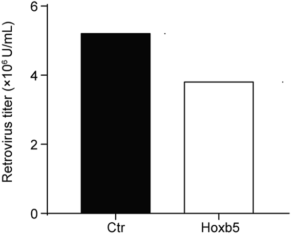
Representative titer measurement of retro-virus. The recombinant Hoxb5 vector or empty GFP control vector is transfected into a plat-E cell line with packaging elements by calcium phosphate transfection. NIH/3T3 cell line is used to measure the titer of retro-Hoxb5 virus and GFP control virus.
Fig. 3.

Flow cytometry analysis of pro/pre-B cells transduced with retrovirus expressing GFP only (Ctrl) or retro-Hoxb5. Pro/pre-B cells sorted from bone marrow cells were transduced with empty-GFP control virus or retro-Hoxb5 virus. The cDNA encoding Hoxb5 was inserted into pMYs-IRES-EGFP to generate a recombinant vector. The recombinant vector or empty vector control was transduced into Plat-E cells to produce viruses. Pro/pre-B cells were transduced with the virus by two-round spin-infection. Data are representative of four independent experiments.
Then, three million transduced GFP+ pro/pre-cells are transplanted into sublethally irradiated (6.5Gy) recipient mice (C57BL/6, CD45.2) via retro-orbital vein injection. Four weeks after transplantation, abundant mature iT cells would occur in the peripheral blood (PB, 5%–10%), spleen (10%–15%) and lymph node (30%–40%), and GFP+ iDN cells would occur in the thymus of the Hoxb5 virus transduced pro/pre-B recipient mice (Fig. 4). In general, one million Hoxb5-expressing pro/pre-B cells gave rise to approximately ten thousand induced T cell progenitors. Thus, the converting efficiency from B cell to T cell by Hoxb5 is around 1%.
Fig. 4.

Flow cytometry analysis of induced T lymphocytes in irradiated recipients injected with Hoxb5-retrovirus transduced pro/pre-B cells. GFP+Ter119–Mac1– populations obtained from the PB, spleen, and LNs (a) or GFP+Ter119–Mac1–CD19– iDN populations from the thymus (b) of recipient mice were analyzed. Three million pro/pre-B cells transduced with the retro-Hoxb5 virus were transplanted into sublethally irradiated syngeneic mice. Four weeks after transplantation, the single nucleated blood cells harvested from PB, spleen, lymph node and thymus were analyzed by flow cytometry. Representative plots were shown.
Overall, by a combination of optimized steps for viral packaging, sorting of pro/pre-B cells, retrovirus transduction, transplantation into irradiated mice, and flow cytometry analysis of regenerated T cells, this protocol enables efficient generation of functional T cells in vivo from pro/pre-B cells by retrovirally enforced expression of Hoxb5.
Conflict of interest
There is no conflict of interest.
Acknowledgements
We thank the animal center and instrument center of Guangzhou Institutes of Biomedicine and Health for the animal care, and cell sorting. This work was supported by grants from the CAS Key Research Program of Frontier Sciences (QYZDB-SSW-SMC057), the Major National Research Project of China (Grant No. 2015CB964401), the Major Scientific and Technological Project of Guangdong Province (2014B020225005), co-operation Program from Guangdong Natural Science Foundation (2014A030312012), the General Program from Guangzhou Scientific and Technological Project (201707010157), the grants from the National Natural Science Foundation of China (Grant No 31471117, 81470281, 31600948), and the grants from the Ministry of Science and Technology of China (2016YFA0100600), Guangzhou Science and Technology Program (201803040017).
Footnotes
Peer review under responsibility of Guangzhou Institutes of Biomedicine and Health, Chinese Academy of Sciences.
References
- 1.Rolink A.G., Nutt S.L., Melchers F., Busslinger M. Long-term in vivo reconstitution of T-cell development by Pax5-deficient B-cell progenitors. Nature. 1999;401:603–606. doi: 10.1038/44164. [DOI] [PubMed] [Google Scholar]
- 2.Ungerback J., Ahsberg J., Strid T., Somasundaram R., Sigvardsson M. Combined heterozygous loss of Ebf1 and Pax5 allows for T-lineage conversion of B cell progenitors. J Exp Med. 2015;212:1109–1123. doi: 10.1084/jem.20132100. [DOI] [PMC free article] [PubMed] [Google Scholar]
- 3.Zhang M., Dong Y., Hu F. Transcription factor Hoxb5 reprograms B cells into functional T lymphocytes. Nat Immunol. 2018;19:279–290. doi: 10.1038/s41590-018-0046-x. [DOI] [PMC free article] [PubMed] [Google Scholar]
- 4.Di Stefano B., Sardina J.L., van Oevelen C. C/EBPalpha poises B cells for rapid reprogramming into induced pluripotent stem cells. Nature. 2014;506:235–239. doi: 10.1038/nature12885. [DOI] [PubMed] [Google Scholar]
- 5.Riddell J., Gazit R., Garrison B.S. Reprogramming committed murine blood cells to induced hematopoietic stem cells with defined factors. Cell. 2014;157:549–564. doi: 10.1016/j.cell.2014.04.006. [DOI] [PMC free article] [PubMed] [Google Scholar]
- 6.Xie H.F., Ye M., Feng R., Graf T. Stepwise reprogramming of B cells into macrophages. Cell. 2004;117:663–676. doi: 10.1016/s0092-8674(04)00419-2. [DOI] [PubMed] [Google Scholar]
- 7.Morita S., Kojima T., Kitamura T. Plat-E: an efficient and stable system for transient packaging of retroviruses. Gene Ther. 2000;7:1063–1066. doi: 10.1038/sj.gt.3301206. [DOI] [PubMed] [Google Scholar]
- 8.Kitamura T., Koshino Y., Shibata F. Retrovirus-mediated gene transfer and expression cloning: powerful tools in functional genomics. Exp Hematol. 2003;31:1007–1014. [PubMed] [Google Scholar]
- 9.Bueno C., Sardina J.L., Di Stefano B. Reprogramming human B cells into induced pluripotent stem cells and its enhancement by C/EBP alpha. Leukemia. 2016;30:674–682. doi: 10.1038/leu.2015.294. [DOI] [PubMed] [Google Scholar]
- 10.Munoz-Lopez A., van Roon E.H., Romero-Moya D. Cellular ontogeny and hierarchy influence the reprogramming efficiency of human B cells into induced pluripotent stem cells. Stem Cells. 2016;34:581–587. doi: 10.1002/stem.2303. [DOI] [PubMed] [Google Scholar]