Lapis lazuli preserved in plaque reveals that medieval women were actively involved in producing richly illustrated manuscripts.
Abstract
During the European Middle Ages, the opening of long-distance Asian trade routes introduced exotic goods, including ultramarine, a brilliant blue pigment produced from lapis lazuli stone mined only in Afghanistan. Rare and as expensive as gold, this pigment transformed the European color palette, but little is known about its early trade or use. Here, we report the discovery of lapis lazuli pigment preserved in the dental calculus of a religious woman in Germany radiocarbon-dated to the 11th or early 12th century. The early use of this pigment by a religious woman challenges widespread assumptions about its limited availability in medieval Europe and the gendered production of illuminated texts.
INTRODUCTION
In nature, blue pigments are relatively rare, occurring in mineral seams that must be mined. Throughout the European medieval period (5th to 15th centuries AD), only a small number of natural and synthetic blue pigments were known, including ultramarine, azurite, Egyptian blue, smalt, and vivianite (table S1). Among these blues, ultramarine, made by grinding and purifying lazurite crystals from the ornamental stone lapis lazuli (1-4), was, by far, the most expensive, reserved along with gold and silver for the most luxurious manuscripts (2, 5). Unlike other blues, such as azurite and vivianite, ultramarine is both brilliant and highly stable, even at high temperatures, and when made from high-quality lapis lazuli and well purified using oil flotation, a deep blue hue can be achieved (4, 6). Mined from a single region in Afghanistan (7), lapis lazuli was a quintessential luxury trade good in the Eurasian pre–Modern period, and its waxing and waning availability in artistic centers throughout Eurasia reflects both its enormous expense and the circuitous supply lines along which it was traded over thousands of miles (8, 9).
Within the context of medieval art, the application of highly pure ultramarine in illuminated works was restricted to luxury books of high value and importance, and only scribes and painters of exceptional skill would have been entrusted with its use (5). Before the 15th century, however, scribes seldom signed their works, raising questions as to the identity of early scribes and illuminators (5, 10). Even among books in women’s monastery libraries, fewer than 15% bear female names or titles, and before the 12th century, fewer than 1% of books can be attributed to women (11). Consequently, it has long been assumed that monks, rather than nuns, were the primary producers of books throughout the Middle Ages (5). Recent historical research, however, has challenged this view, revealing that religious women were not only literate but also prolific producers and consumers of books (10-12). In Germany and Austria, religious women played a particularly active role in book production, and their work as scribes and illuminators can be traced to as early as the late eighth century (10, 12). Although surviving examples of these early works are rare and relatively modest, there is a growing body of evidence that women’s monasteries were actively producing books of the highest quality by the 12th century (10). The dual-sex monastery of Admont in Salzburg, for example, supported a community of nuns who copied many of the more than 200 surviving books from the monastery’s 12th-century book collections, and Diemut, a 12th-century female scribe at the monastery of Wessobrunn in Bavaria, was recorded to have produced more than 40 books, including a richly illuminated gospel (10). From the 13th to the 16th centuries, during which documentary evidence and record keeping in Germany is more complete, more than 4000 books attributed to over 400 women scribes have been identified, and active scriptoria have been identified at 48 women’s monasteries (11). However, identifying the early contributions of religious women to medieval book production is challenging due to the limited number of surviving books, the precarious documentation of women’s monasteries, and the tendency of scribes to leave their work unsigned (10). As a result, individual female scribes remain poorly visible in the historical record, and it is likely that most of their scribal work has gone unrecognized.
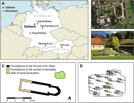
Recently, microscopic analyses have revealed that dental calculus (calcified tooth tartar) can entrap and preserve a wide range of microdebris related to craft activities (13, 14). Here, we report the identification of lazurite and phlogopite crystals, in the form of powder consistent in size and composition with lapis lazuli–derived ultramarine pigment, that were found embedded within the dental calculus of a middle-aged woman buried in association with a 9th- to 14th-century church-monastery complex at Dalheim, Germany (Fig. 1). Radiocarbon-dated to AD 997–1162, this woman represents the earliest direct evidence of ultramarine pigment usage by a religious woman in Germany. Moreover, because the monastery and the entirety of its contents were destroyed during a 14th-century fire, this finding of lapis lazuli potentially represents the sole surviving evidence of female scribal activity at the site. Our results suggest that dental calculus can be used to help identify scribes and artists in the archaeological record and to aid in the historical reconstruction of women’s monasteries and their role in book production. In addition, although the importation of this expensive foreign pigment into medieval Europe is first materially attested in the 10th century (15), its presence in an otherwise unremarkable women’s community in northern Germany powerfully testifies to the expansion of long-distance trading circuits during the 11th-century European commercial revolution.
Fig. 1. Dalheim Church of St. Peter and women’s monastery.

(A) Location of Dalheim and other monasteries discussed in the text. (B) Surviving stone architectural foundations of Dalheim’s Church of St. Peter and attached women’s monastery, shown in the circle (viewed from above and from the west). A modern building has been constructed on the site of the former cemetery. (C) Architectural plan showing the configuration of the church (black), the women’s monastery (light brown), and the location of the excavated portion of the cemetery (green). (D) Schematic view of the burial locations within the cemetery. The burial location of individual B78 is marked in green. Credit: C. Warinner.
RESULTS
Blue particle identification in dental calculus
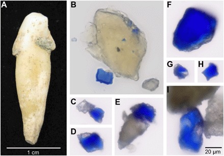
In 2014, during a separate study on the identification of plant microremains in dental calculus (16), numerous particles of blue color (Fig. 2) were observed embedded within the dental calculus of a 45- to 60-year-old woman buried in association with a medieval church-monastery complex at the site of Dalheim near Lichtenau, Germany (Fig. 1). Radiocarbon-dated to cal. AD 997–1162 (95% probability; fig. S1), this individual, B78, was otherwise unexceptional, presenting no notable skeletal pathologies or evidence of trauma or infection (16, 17). Further osteological investigation did not detect indications of hard labor, while dental analysis revealed heavy calculus deposits on the anterior teeth (fig. S2) and only mild to moderate periodontal disease accompanied by the antemortem loss of two molars, likely due to caries (16). Female biological sex was confirmed using both osteological and genetic methods (16), and the skeletal remains are now curated at the Institute of Evolutionary Medicine at the University of Zürich. Few historical records survive for the church-monastery complex, which now stands in ruins. A stone church dedicated to St. Peter was likely first constructed at the site during the ninth century and later expanded. Although the founding date of the Dalheim women’s monastery is unknown, four nearby Benedictine and Cistercian women’s monasteries were founded in AD 1127, 1140, 1142, and 1149 (18). The earliest surviving texts documenting the women’s community at Dalheim date to AD 1244, 1264, and 1278 and describe it as a house of Augustinian canonesses attached to a church dedicated to St. Peter (18). Excavated by the Westphalian Museum of Archaeology from 1988 to 1991, the monastery is believed to have housed approximately 14 religious women until its destruction by fire following a series of 14th-century battles (18, 19). An unmarked cemetery, from which B78 was excavated, is located immediately adjacent to the church.
Fig. 2. Blue particles observed embedded within archaeological dental calculus.

(A) Archaeological tooth from individual B78 showing attached dental calculus deposits before sampling. (B) View of blue particles embedded within a large piece of intact dental calculus, as well as a blue particle already freed from dental calculus. (C to I) Multiple blue particles observed following sonication of dental calculus. Note the frequent co-occurrence of associated colorless minerals. Images (B) to (I) are shown to the same scale, as indicated in (I). Credit: C. Warinner (A); M. Tromp and A. Radini (B to I).
To isolate the blue particles for further study, we first sought to demineralize the surrounding dental calculus using a dilute HCl solution (0.05 M), as is typically performed during microbotanical analysis. However, we found that this procedure led to color instability and loss (fig. S3); by comparing colors of the acid-demineralized calculus to reference pigments, we confirmed that using an acid as a decalcifying agent is detrimental to color stability and particle size in lapis lazuli, azurite, malachite, and vivianite (fig. S4; see the Supplementary Materials). We then tested an alternate approach on a second dental calculus sample from the same individual, decontaminating the calculus surface and then disrupting the calculus structure by sonication in ultrapure water. Calculus fragments and mineral particles released by this procedure were transferred to a microscope slide without mounting media or coverslip and allowed to dry under controlled conditions. Inspection under light microscopy revealed more than 100 particles of deep blue color (Fig. 2), many of which were observed in situ still encased within fragments of dental calculus (Fig. 2B). All subsequent analyses used sonication for pigment isolation.
Distribution of blue particles in dental calculus
In most cases, the blue particles appeared singly—not as clumps—having the appearance of a blue powder dispersed across many dental calculus fragments (fig. S5, A to E). Blue particles were observed across calculus pieces originating from different teeth, suggesting that the particles entered the calculus in different episodes rather than as a single localized event, and over a period of time as the dental calculus matrix calcified. Average particle size was 10.9 ± 9.5 μm (SD), which is consistent with published data for natural lapis lazuli pigment (20) and our own measurements of 10.8 ± 8.0 μm (SD) for reference Afghan lapis lazuli pigment (table S2 and data file S1).
Elemental composition of blue particles is consistent with lazurite
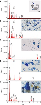
We next compared the blue particles optically to a selected reference panel of blue mineral pigments, followed by elemental analysis using scanning electron microscopy with energy-dispersive x-ray spectroscopy (SEM-EDS) (Fig. 3). With the exception of lazurite (the dominant blue mineral in lapis lazuli), all blue pigments that were available and used during the medieval period contain metal (copper, cobalt, or iron) as a major element in their composition (table S1). SEM-EDS analysis of the archaeological particles and reference blue pigments allows a clear distinction between major element compositions (Fig. 3). The archaeological blue particles lack copper, cobalt, and iron, thereby excluding pigments containing these metals as major elements, but they closely resemble the elemental composition of lazurite, the sulfur-containing tectosilicate that gives lapis lazuli its dark blue color.
Fig. 3. Elemental composition of archaeological blue particle and blue reference pigments measured by EDS.

(A) Blue particle isolated from medieval dental calculus. (B) Afghan lazurite reference pigment (RC, 410-15). (C) Azurite reference pigment (RC, 410-10). (D) Smalt reference pigment (RC, 417-14). (E) Vivianite reference pigment (RC, 410-20). The archaeological blue particles lack copper (Cu), cobalt (Co), and iron (Fe) but closely match the spectrum produced by the tectosilicate lazurite. Scale bars, 20 μm. Additional reference spectra are provided in fig. S7. Raw data are provided in data file S2. Credit: M. Tromp.
Identification of the lapis lazuli minerals lazurite and phlogopite
To confirm the identification of lapis lazuli, we analyzed the archaeological particles using micro-Raman spectroscopy. The micro-Raman spectra generated from the archaeological blue particles yielded a positive match to modern reference lapis lazuli pigment (Fig. 4) and to a previously published medieval lapis lazuli pigment identified from a medieval fresco painting (21), as well as to other lazurite spectra available in the RRUFF database (22). The spectra taken from the B78 sample show the characteristic modes for lazurite, Na3CaAl3Si3O12S, at 258, 548, 803, and 1096 cm−1, with the strongest modes being the symmetric S3− ν1-stretching mode at 548 cm−1 and its overtone at 1096 cm−1, as well as the S3− ν1-bending mode at 258 cm−1 (20). These spectral characteristics allow for an unambiguous identification of the mineral particles as lazurite.
Fig. 4. Confirmation of lazurite mineral using micro-Raman spectroscopy.

Matching micro-Raman spectra are produced by archaeological blue particles and lazurite crystals in reference pigments from both modern lapis lazuli and a blue pigment from a medieval fresco painting that was previously identified as lapis lazuli (21). Inset: Optical image of archaeological blue particle. Raw data are provided in data file S3. Credit: A. Radini, E. Tong, R. Kröger.
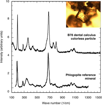
In addition, using micro-Raman spectroscopy, we identified a colorless, translucent mineral accompanying the blue lazurite crystals within the archaeological sample as phlogopite (Fig. 5). The phyllosilicate phlogopite, K(Fe, Mg)3(Si3Al)O10(F,OH)2, is an accessory mineral found to accompany the tectosilicate lazurite in lapis lazuli stone (7). Iron-rich phlogopite has some characteristic vibrational modes that are activated if Fe substitutes Mg (23). In the presence of Fe, the Si-Ob-Si mode at 681 cm−1 forms a triplet, and an additional mode appears near 550 cm−1 as observed in our sample. In addition, a systematic downshift of the Si-Ob-Si peaks has been reported with increasing iron content (23). The latter could provide an opportunity to correlate the lazurite with specific mining locations where the characteristic Fe/Mg ratios are known. Overall, our analyses show that the blue pigment found in the B78 sample is lazurite and that the colorless mineral is iron-rich phlogopite. Together, lazurite and phlogopite allow for a positive identification of the archaeological blue particles as originating from lapis lazuli.
Fig. 5. Confirmation of the phyllosilicate phlogopite mineral found adjacent to lazurite particles using micro-Raman spectroscopy.

Matching micro-Raman spectra of the colorless particle and a published reference spectrum of phlogopite (23), an accessory mineral that co-occurs with lazurite in natural lapis lazuli stone. Inset: Optical image of archaeological colorless particle. Raw data are provided in data file S3. Credit: A. Radini, E. Tong, R. Kröger.
DISCUSSION
How a middle-aged woman living a life of apparently low physical labor and buried in a cemetery associated with a woman’s religious community came to have such a rare and expensive mineral pigment in her dental calculus is not entirely certain, but we propose four possible scenarios: (i) B78 was a scribe or book painter engaged in the production of illuminated manuscripts, (ii) B78 was employed in the preparation of artist materials for herself or other scribes, (iii) B78 consumed lapis lazuli in the context of lapidary medicine, or (iv) B78 performed emotive devotional osculation of illuminated books produced by others.
Scenario 1: Book production
The most parsimonious scenario is that individual B78 was a woman engaged in the production of high-quality manuscripts. The commissioning of a talented female scribe to produce deluxe liturgical books using expensive materials has precedent in Germany at this time. For example, a pair of letters dated to between AD 1140 and 1168—nearly contemporaneous with the burial of B78—detail an exchange between Sindold, the keeper and corrector of books (armarius) of the men’s monastery at Reinhardsbrunn, and the women’s monastery of Lippoldsberg where his sisters lived, located only 70 km east of Dalheim (see the Supplementary Materials). In his letter, Sindold commissions the “skillful” production of a deluxe, illuminated matutinal (liturgical book) to be produced by sister “N” using parchment, leather, pigment, and silk that he provided for that purpose (24). That the Reinhardsbrunn armarius would outsource the production of such an important and valuable book to a women’s monastery speaks to the reputation of women as makers of books by the 12th century. While Sindold does not elaborate on the specifics of the pigments being sent, judging by the amount of parchment (the equivalent of 384 pages) and the inclusion of silk, it can be assumed that the pigments were at least commensurate in quality and expense. Among surviving books from Germany that have been tested and are known to contain lapis lazuli pigment, the earliest putatively attributed to a woman scribe is a copy of the Liber Scivias (Heidelberg University Library, Codex Salemitani X,16) by Hildegard of Bingen of the women’s monastery at Rupertsberg and produced circa AD 1200 (15); however, the unsigned paintings were colored by at least two anonymous individuals (15).
In Germany, women’s monastic communities, especially during earlier periods, were largely made up of noble or aristocratic women. Many were highly educated, and devotional reading was encouraged as an expression of piety. These women would have led lives largely free of hard labor, consistent with the absence of occupational skeletal stress observed for B78. Work was encouraged within the monastery, however, and activities related to book production were considered worthy pursuits. In adding detail to their illuminations, it is plausible to assume that artists would have occasionally licked their brushes to make a fine point, a practice that later artist manuals refer to explicitly (4). In doing so, pigments, such as lapis lazuli, may have been introduced into the oral cavity, where they could have become entrapped within dental calculus. The repeated activity of inserting the tip of the brush into the mouth could explain the distribution pattern of in situ blue particles observed across multiple calculus fragments.
Scenario 2: Pigment preparation
It is possible, although less likely, that the lapis lazuli pigment was introduced into the oral cavity of B78 through pigment production rather than painting. Ultramarine pigment production from lapis lazuli stone is described in numerous late medieval instruction manuals (4), of which the Italian 15th-century text Il Libro dell’Arte by Cennino Cennini (AD 1437) is perhaps the most detailed and best known (25). In it, Cennini describes a laborious process of grinding, progressively washing, and levigating lapis lazuli stone powder followed by oil flotation to remove impurities and concentrate the blue-bearing lazurite crystals, and he warns that the mortar in which the lapis lazuli stone is ground should be covered so that “it may not go off in dust” (see the Supplementary Materials). This airborne dust could potentially come into contact with dental calculus through accidental inhalation, either during the pigment preparation itself or afterward, such as during workshop cleaning activities. Experimental work confirms that it is possible for particles to enter the oral cavity by pigment preparation, even when only a limited amount of airborne dust is produced (see the Supplementary Materials; fig. S6). In addition, the handling of the dry pigment powder can itself create airborne dust that can settle on the face and lips, as well as enter the oral cavity.
Cennini also notes that the work of pigment production is typically performed by women, but this gendered division of labor may be a late medieval development associated with the professionalization of trade and crafts. During earlier periods, it is not specified how or who produced finished pigments from raw materials, and few recipe books are known from the 12th century AD and earlier (2, 4). Although high-quality lapis lazuli pigment first appears in European manuscripts as early as the 10th century (4, 15), the Arabic method of oil flotation described by Cennini, which is necessary to produce a brilliant blue rather than a dull bluish-gray pigment, is not attested in European artist manuals before the 15th century (4). As such, this raises questions as to whether scribes of the 11th and 12th centuries produced their own lapis lazuli pigments or received them as imported finished products from trading centers such as Alexandria, via Italian merchants. It is possible that the scribes themselves prepared their own—possibly lower quality—pigments or that scribes may have been provisioned with finished pigments produced by others, either locally or abroad. If a religious woman at Dalheim was preparing lapis lazuli pigment, then it is likely that it was for her own use or for another female scribe within her religious community.
Scenario 3: Lapidary medicine
Alternatively, B78 may have consumed powdered lapis lazuli as a form of lapidary medicine. Since antiquity, lapis lazuli stone has been ascribed magical and healing powers by many Old World cultures, who used it primarily as an amulet stone and as a component of eye ointments (3, 26). The first-century Greek medical text De Materia Medica by Dioscorides describes the medicinal libation of lapis lazuli to treat scorpion bites, ulcers, eye growths, pustules, and herniated membranes (27), and an inventory of a Jewish apothecary in Cairo dating to the 13th and 14th centuries refers to the use of lapis lazuli as both an antivenom and an eye treatment (28). Medical lapis lazuli was particularly important in the medieval Islamic world, where it is amply attested in numerous medical recipe books (3, 29). By contrast, lapis lazuli first appears in European medical texts only in the 11th century (29) in such early works as the late 11th-century Liber de lapidibus by Marbod of Rennes (30), the 12th-century Physica by Hildegard of Bingen (31), and the 12th-century Circa Instans (32). Although these works describe the medical use of lapis lazuli (see the Supplementary Materials), there is little evidence that the Mediterranean and Islamic method of ingesting lapis lazuli pigment was widespread or even practiced in 11th- and 12th-century Germany. Consequently, although the ingestion of medical lapis lazuli by B78 cannot be ruled out, it appears unlikely given the paucity of evidence for this practice.
Scenario 4: Devotional osculation
Last, women’s devotional reading has a long history, with documentation dating back to at least the sixth century in Merovingian Gaul (10). Beginning in the 14th and 15th centuries, devotional reading became more emotive under the influence of the Netherlands reformer Geert Grote of Deventer, and ritual osculation (kissing) of painted figures in illuminated prayer books became more common. In response, illuminators began adding decorative “osculation tablets” to these books to deflect kissing away from the painted figures, which suffered damage—including paint removal—through repeated kissing (33). Although it is possible that the lapis lazuli in the calculus of B78 entered the oral cavity through the kissing of painted images, the radiocarbon date of the skeleton makes it highly unlikely because such forms of emotive devotion are not attested until nearly three centuries later. In addition, osculation would have likely lifted clumps of pigment together with associated mounting media, which we did not observe in the B78 dental calculus.
Together, the presence of lapis lazuli in the dental calculus of B78 is best explained by its accidental incorporation during painting and/or pigment preparation. The discovery of lapis lazuli in the dental calculus of an 11th-century religious woman is without precedent in the European medieval archaeological record and marks the earliest direct evidence for the use of this rare and expensive pigment by a religious woman in Germany. Within medieval Europe, women’s monasteries in Germany are noteworthy for their active role in book production, but limited historical information is available for female scribes and the books they produced between the Anglo-Saxon missionary period beginning in the 8th century and the great monastic expansion of the 12th and 13th centuries (10-12). In Germany, only five women’s scriptoria are known to have been active in the 8th through the 11th centuries (11), and it is likely that most women’s book production during this period was produced more informally by individual female scribes who left few records of their work.
As was the case for many early women’s religious communities, Dalheim has left very few traces in the historical record. No books survive from the monastery, either from its libraries or in any other surviving works. Nearly invisible in the historical record, the women of Dalheim are known to us today nearly exclusively through the archaeological record and a handful of brief textual references (18, 19). The case of Dalheim raises questions as to how many other early women’s communities in Germany, including communities engaged in book production, have been similarly erased from history. Archaeological research, in combination with emerging techniques for the recovery of microremains from dental calculus and analytical methods such as SEM-EDS and micro-Raman, offers great promise for illuminating the lives of the modest and pious women who quietly produced the books of medieval Europe.
MATERIALS AND METHODS
Experimental design
Dental calculus was collected from the dentition of Dalheim individual B78, as previously described (16). Because individual calculus pieces were pooled from multiple teeth before analysis, it is not possible to determine which specific teeth harbored dental calculus containing blue particles. However, nearly all of B78’s dental calculus was concentrated on the anterior teeth (fig. S2), with very little calculus present on the molars (16), and thus, the blue particles likely originated near the lips. Comparative reference pigments were obtained from Rublev Colours by Natural Pigments LLC (RC) and Kremer Pigments Inc. (KP): lapis lazuli, Afghanistan (RC, 410-15); lapis lazuli, Chile (KP, 10550); ultramarine ash (RC, 410-14); azurite (RC, 410-10); malachite (RC, 420-20); Egyptian blue (KP, 10060); smalt (RC, 417-14); royal smalt (RC, 417-13); and vivianite (RC, 410-20). Initial decalcification was performed on 25.2 mg of dental calculus using 0.05 M HCl at 4°C, but this was found to result in color loss of the blue particles (fig. S3), as well as reference pigments (fig. S4). Further tests on reference pigments also indicated minor color alterations in 0.1 M EDTA but not in saliva or ultrapure water (fig. S4). An alternative method for the isolation of blue particles from dental calculus using sonication in ultrapure water was tested and found to result in minimal particle alteration. All further optical, elemental, and spectrographic analyses were performed on particles obtained by this method.
Optical microscopy
Nine milligrams of dental calculus was selected for sonication in ultrapure water. Before sonication, the sample was first washed in ultrapure water to remove loose soil and other surface debris. The calculus was then further decontaminated as follows. First, the sample of calculus was placed under a stereomicroscope at a magnification of up to 50× and inspected for contaminants. A fine sterile acupuncture needle was used in conjunction with a 0.6 M solution of HCl to clean the external facets of the calculus. Once completely free of any visible surface contaminants, the calculus was washed again in ultrapure water and then placed in a sterile microcentrifuge tube containing ultrapure water to remove any residual HCl. The sample was then subjected to controlled sonication for approximately 6 min until the calculus was reduced into a fine powder. The calculus powder suspension was transferred to two microscope slides and evaporated under controlled conditions. These steps were performed in a laboratory at the University of York dedicated to the extraction and mounting of microfossils from ancient dental calculus; no other material type or reference pigments were prepared in this laboratory.
Optical microscopy was initially performed at the Laboratory of Microarchaeology at the University of York, where the particles were counted and measured under transmitted and polarized light using a Zeiss Axio Scope.A1 binocular compound microscope equipped with motorized stage. Mean particle size and SD were determined by measuring 100 blue particles using AxioVision image software (data file S1). Slides were then analyzed at the microscope facilities of the Department of Archaeology at the Max Planck Institute for the Science of Human History (MPI-SHH) using an Olympus BX53M equipped with transmitted light, cross-polarized light, differential interference contrast filters, and an Olympus 20.7 Mpx camera. After locating and photographing the blue particles, a high-resolution map was made for one of the slides to aid in the targeting blue particles for SEM-EDS analysis (fig. S5F).
Scanning electron microscopy with energy-dispersive x-ray spectroscopy
SEM-EDS analysis was performed on the blue particles using a JEOL InTouchScope JSM-IT100LA coupled with a JEOL Dry Extra EDS detector in the microscopy facilities of the MPI-SHH Department of Archaeology. Spectra were collected from the archaeological blue particles and the reference pigments at a working distance of 10 mm for optimal data collection (data file S2). EDS analysis was performed qualitatively (that is, nonquantitatively) to compare the spectra produced from each reference pigment and the archaeological particles (Fig. 3and fig. S7). The archaeological particles were each targeted individually, while the reference pigments were each mounted on separate aluminum stubs, and the EDS spectra represent an average of the entire visible area at a magnification of 400×. Spectra were collected for a minimum of 15 min or until the most abundant element reached approximately 18,000 counts. All particles were examined with a 10-kV beam in low vacuum mode to avoid gold coating the samples. The sample navigation system and five-axis motor-driven stage enabled us to locate the archaeological blue particles that had previously been imaged both individually and in the high-resolution map created using the light microscope.
Micro-Raman spectroscopy
Micro-Raman spectroscopy was performed in the Department of Physics at the University of York using a Horiba Xplora System with a 532-nm semiconductor diode excitation laser providing a lateral spot extension of approximately 1 μm (data file S3). A 2400-T grating was used to obtain the highest spectral resolution of 1 cm−1 with an acquisition time of typically 1 s and 10 accumulations to minimize potential laser-induced damage to the sample and to reduce any interaction with the calculus matrix. After initial tests, it was decided that the relevant spectral range comprising the vibrational modes of interest was between 100 and 1600 cm−1. The characteristic blue color of the lazurite mineral particles in the context of otherwise translucent and colorless mineral particles enabled a rapid identification of their position at low magnification (10× objective lens) using transmission illumination with subsequent high-magnification imaging and spectral analysis (50× large working distance objective lens). The spectra were taken as single points on the largest particles that provided the best surface for the laser beam and that exhibited the largest distance from the calculus matrix. Micro-Raman spectroscopy was performed on the same slides of mounted archaeological blue particles as used for optical and SEM-EDS analysis.
Supplementary Material
Acknowledgments
We thank S. Sutherland, M. Green, C. Duffin, A. F. More, C. Cyrus, and E. Glaze for helpful advice and M. O’Reilly for graphical assistance. Funding: This work was supported by the Max Planck Society, the Leverhulme Trust (through a Leverhulme prize to C.S.), the Mäxi Foundation Zurich (to F.R.), and the National Science Foundation (BCS-1516633 to C.W.). Author contributions: C.W., A.R., and M.J.C. conceived the study. F.R., R.K., and C.W. provided materials and resources. A.R., M.T., E.T., R.K., and J.V.D. performed the experiments. C.W., A.R., M.T., E.T., and R.K. analyzed the data. A.B., M.M., M.J.C., and C.S. assisted with interpreting the results. C.W., A.R., A.B., and M.T. wrote the paper, with input from the remaining coauthors. Competing interests: The authors declare that they have no competing interests. Data and materials availability: All optical, elemental, spectrographic, and other data needed to evaluate the conclusions in the paper are present in the paper and/or the Supplementary Materials. Additional data related to this paper may be requested from the authors.
SUPPLEMENTARY MATERIALS
Supplementary material for this article is available at http://advances.sciencemag.org/cgi/content/full/5/1/eaau7126/DC1
Supplementary Text
Fig. S1. Radiocarbon date for Dalheim individual B78 bone collagen.
Fig. S2. Distribution of dental calculus deposits on the dentition of individual B78.
Fig. S3. Comparison of blue particle appearance following HCl decalcification versus sonication in ultrapure water.
Fig. S4. Comparison of the effects of 0.05 M HCl, 0.1 M EDTA, and saliva on reference pigments.
Fig. S5. In situ distribution of blue particles in the dental calculus of B78 following sonication in ultrapure water.
Fig. S6. Blue particles recovered from lips and saliva during lapis lazuli grinding experiment.
Fig. S7. Elemental composition of additional reference pigments.
Table S1. Blue mineral pigments known in medieval Europe.
Table S2. Mean size of archaeological blue particles and reference pigments.
Table S3. Characterization of airborne dust on lips and in saliva during lapis lazuli grinding.
Data file S1. Archaeological blue particle size and count.
Data file S2. Elemental composition data generated by SEM-EDS for archaeological blue particles and reference pigments.
Data file S3. Raman spectral data for archaeological particles and reference lapis lazuli.
REFERENCES AND NOTES
- 1.M. M. P. Merrifield, Medieval and Renaissance Treatises on the Arts of Painting: Original Texts with English Translations (Courier Corporation, 1999). [Google Scholar]
- 2.M. Clarke, The Art of All Colours: Mediaeval Recipe Books for Painters and Illuminators (Archetype Publications Ltd., 2011). [Google Scholar]
- 3.Frison G., Brun G., Lapis lazuli, lazurite, ultramarine ‘blue’, and the colour term ‘azure’up to the 13th century. J. Int. Col. Assoc. 16, 41–55 (2016). [Google Scholar]
- 4.R. Fuchs, D. Oltrogge, in Europäische Technik im Mittelalter 800 bis 1200—Tradition und Innovation: Ein Handbuch, U. Lindgren, Ed. (Gebr. Mann Verlag, 2001), pp. 435–450. [Google Scholar]
- 5.J. J. G. Alexander, Medieval Illuminators and Their Methods of Work (Yale Univ. Press, 1992). [Google Scholar]
- 6.Coccato A., Moens L., Vandenabeele P., On the stability of mediaeval inorganic pigments: A literature review of the effect of climate, material selection, biological activity, analysis and conservation treatments. Heritage Sci. 5, 12 (2017). [Google Scholar]
- 7.Wyart J., Bariand P., Filippi J., Lapis lazuli from Sar-e-Sang, Badakhshan, Afghanistan. Gems Gemmol. 17, 184–190 (1981). [Google Scholar]
- 8.J. Kirby, S. Nash, J. Cannon, Trade in Artists' Materials (Archetype Books, 2010). [Google Scholar]
- 9.D. V. Thompson, The Materials and Techniques of Medieval Painting (Courier Corporation, 1956), vol. 327. [Google Scholar]
- 10.A. I. Beach, Women as Scribes: Book Production and Monastic Reform in Twelfth-Century Bavaria (Cambridge Univ. Press, 2004). [Google Scholar]
- 11.C. J. Cyrus, The Scribes for Women's Convents in Late Medieval Germany (University of Toronto Press, 2009). [Google Scholar]
- 12.F. Lifshitz, Religious Women in Early Carolingian Francia: A Study of Manuscript Transmission and Monastic Culture (Oxford Univ. Press, 2014). [Google Scholar]
- 13.A. Radini, “Particles of Everyday Life: Past Diet and Living Conditions as Evidenced by Micro-debris Entrapped in Human Dental Calculus, A Case Study from Medieval Leicester and Surrounding,” PhD thesis, University of York (2016). [Google Scholar]
- 14.Warinner C., Speller C., Collins M. J., A new era in palaeomicrobiology: Prospects for ancient dental calculus as a long-term record of the human oral microbiome. Phil. Trans. R. Soc. B 370, 20130376 (2015). [DOI] [PMC free article] [PubMed] [Google Scholar]
- 15.R. Fuchs, D. Oltrogge, in Blau: Farbe der Ferne, H. Gercke, Ed. (Verlag Das Wunderhorn, 1990). [Google Scholar]
- 16.Warinner C., Rodrigues J. F. M., Vyas R., Trachsel C., Shved N., Grossmann J., Radini A., Hancock Y., Tito R. Y., Fiddyment S., Speller C., Hendy J., Charlton S., Luder H. U., Salazar-García D. C., Eppler E., Seiler R., Hansen L. H., Castruita J. A. S., Barkow-Oesterreicher S., Teoh K. Y., Kelstrup C. D., Olsen J. V., Nanni P., Kawai T., Willerslev E., von Mering C., Lewis C. M. Jr., Collins M. J., Gilbert M. T. P., Rühli F., Cappellini E., Pathogens and host immunity in the ancient human oral cavity. Nat. Genet. 46, 336–344 (2014). [DOI] [PMC free article] [PubMed] [Google Scholar]
- 17.Hofmann M. I., Böni T., Alt K. W., Woitek U., Rühli F. J., Paleopathologies of the vertebral column in medieval skeletons. Anthropologischer Anzeiger 66, 1–17 (2008). [PubMed] [Google Scholar]
- 18.Niemeyer W., Gründung und Erinnerung in der Geschichte des Klosters Dalheim. Mitteilungen der Deutschen Gesellschaft für Archäologie des Mittelalters und der Neuzeit 27, 215–226 (2014). [Google Scholar]
- 19.R. Pieper, Kloster Dalheim: Eine kurze Geschichte (Landschaftsverband Westfalen-Lippe, 2000). [Google Scholar]
- 20.Osticioli I., Mendes N. F. C., Nevin A., Gil F. P. S. C., Becucci M., Castellucci E., Analysis of natural and artificial ultramarine blue pigments using laser induced breakdown and pulsed Raman spectroscopy, statistical analysis and light microscopy. Spectr. Acta Part A Mol. Biomol. Spectr. 73, 525–531 (2009). [DOI] [PubMed] [Google Scholar]
- 21.Ballirano P., Maras A., Mineralogical characterization of the blue pigment of Michelangelo’s fresco “The Last Judgment”. American Mineralogist 91, 997–1005 (2006). [Google Scholar]
- 22.B. Lafuente, R. T. Downs, H. Yang, N. Stone, in Highlights in Mineralogical Crystallography, T. Armbruster, R. M. Danisi, Eds. (W. De Gruyter, 2015), pp. 1–30. [Google Scholar]
- 23.Wang A., Freeman J. J., Jolliff B. L., Understanding the Raman spectral features of phyllosilicates. J. Raman Spectr. 46, 829–845 (2015). [Google Scholar]
- 24.F. Peeck, Die Reinhardsbrunner Briefsammlung (MGH Epistolae Selectae, 1952). [Google Scholar]
- 25.C. D. A. Cennini, The Craftsman's Handbook: The Italian “Il Libro dell' Arte”, translated by D. V. Thompson Jr. (Dover,1954).
- 26.Duffin C. J., The pharmaceutical use of Lapis Lazuli in the Ancient East. Pharmaceutical Historian 44, 84–87 (2014). [PubMed] [Google Scholar]
- 27.T. A. Osbaldeston, R. Wood, The Herbal of Dioscorides the Greek (Ibidis Press, 2000). [Google Scholar]
- 28.Lev E., Drugs held and sold by pharmacists of the Jewish community of medieval (11–14th centuries) Cairo according to lists of materia medica found at the Taylor–Schechter Genizah collection, Cambridge. J. Ethnopharmacol. 110, 275–293 (2007). [DOI] [PubMed] [Google Scholar]
- 29.Glaze F. E., Salerno’s Lombard Prince: Johannes ‘Abbas de Curte’ as Medical Practitioner. Early Science and Medicine 23, 177–216 (2018). [Google Scholar]
- 30.Riddle J. M., Marbode of Rennes' (1035-1123) De lapidibus considered as a medical treatise with text, commentary and CW King's translation, together with text and translation of Marbode's minor works on stones. Sudhoffs Archiv 20, 1–144 (1977). [PubMed] [Google Scholar]
- 31.P. Throop, Hildegard von Bingen's Physica (Healing Arts, 1998). [Google Scholar]
- 32.K. Goehl, Das ‘Circa Instans’. Die erste große Drogenkunde des Abendlandes (Deutscher Wissenschafts-Verlag, Schriften zur Medizingeschichte, Baden-Baden, 2015), vol. Band 17.
- 33.K. M. Rudy, Kissing Images, Unfurling Rolls, Measuring Wounds, Sewing Badges and Carrying Talismans: Considering Some Harley Manuscripts Through the Physical Rituals They Reveal (British Library, 2011). [Google Scholar]
Associated Data
This section collects any data citations, data availability statements, or supplementary materials included in this article.
Supplementary Materials
Supplementary material for this article is available at http://advances.sciencemag.org/cgi/content/full/5/1/eaau7126/DC1
Supplementary Text
Fig. S1. Radiocarbon date for Dalheim individual B78 bone collagen.
Fig. S2. Distribution of dental calculus deposits on the dentition of individual B78.
Fig. S3. Comparison of blue particle appearance following HCl decalcification versus sonication in ultrapure water.
Fig. S4. Comparison of the effects of 0.05 M HCl, 0.1 M EDTA, and saliva on reference pigments.
Fig. S5. In situ distribution of blue particles in the dental calculus of B78 following sonication in ultrapure water.
Fig. S6. Blue particles recovered from lips and saliva during lapis lazuli grinding experiment.
Fig. S7. Elemental composition of additional reference pigments.
Table S1. Blue mineral pigments known in medieval Europe.
Table S2. Mean size of archaeological blue particles and reference pigments.
Table S3. Characterization of airborne dust on lips and in saliva during lapis lazuli grinding.
Data file S1. Archaeological blue particle size and count.
Data file S2. Elemental composition data generated by SEM-EDS for archaeological blue particles and reference pigments.
Data file S3. Raman spectral data for archaeological particles and reference lapis lazuli.


