Abstract
Prokaryotes evolved numerous systems that defend against predation by bacteriophages. In addition to well-known restriction-modification and CRISPR-Cas immunity systems, many poorly characterized systems exist. One class of such systems, named BREX, consists of a putative phosphatase, a methyltransferase and four other proteins. A Bacillus cereus BREX system provides resistance to several unrelated phages and leads to modification of specific motif in host DNA. Here, we study the action of BREX system from a natural Escherichia coli isolate. We show that while it makes cells resistant to phage λ infection, induction of λ prophage from cells carrying BREX leads to production of viruses that overcome the defense. The induced phage DNA contains a methylated adenine residue in a specific motif. The same modification is found in the genome of BREX-carrying cells. The results establish, for the first time, that immunity to BREX system defense is provided by an epigenetic modification.
INTRODUCTION
Bacteriophages outnumber their hosts in nature and can have major effects on bacterial populations and communities. For example, recent analysis demonstrates massive phage-driven sweeps in oceanic bacterial communities (1,2). In response to viral predation cellular defense mechanisms have evolved to prevent annihilation. They include, among others, innate immunity conferred by restriction-modification systems (3,4), adaptive immunity mediated by CRISPR-Cas systems (5–7) and an assortment of poorly studied abortive infection mechanisms that limit phage propagation through the population while killing infected cells (8–10). Considering the diversity of phages and the fact that defense islands constitute ∼10% of bacterial genomes (11), there is little doubt that multiple host resistance systems remain to be discovered and recent work supports this view (12). In addition, a number of defense systems were reported in the sixties and seventies of the last century but were largely forgotten. One such system is Pgl (phage growth limitation) of Streptomyces coelicolor that affects the growth of phage φC31 (13–15). While there is no difference in burst sizes and lysis times of φC31-infected Pgl+ and Pgl− cells, in continuously infected cultures the titer of the phage is orders of magnitude lower in Pgl+ cultures compared to Pgl− cultures. It was proposed that phages released after the first round of Pgl+ cells infection become modified and lose the ability to infect Pgl+ (but not Pgl−) cells.
The Pgl system consists of four genes pglWXYZ (14,16,17). Bioinformatics analysis suggested that PglW may be a kinase, PglX—a SAM-dependent methyltransferase, PglY—an adenosinetriphosphatase (ATPase) and PglZ—an alkaline phosphatase. For PglW, PglX and PglY these predictions were confirmed in vitro (15). The current model of the Pgl system function posits that PglX and PglZ act as a toxin-antitoxin pair and that the release of toxic PglX in φC31-infected cells is controlled by PglZ and leads to formation of modifying/restricting complexes (15).
Sequence analysis of prokaryotic defense islands revealed overrepresentation of genes encoding PglZ homologs (16) and a separate BREX (bacteriophage exclusion) name for clusters containing pglZ-like genes was recently proposed (17). Introduction of six-gene BREX system from Bacillus cereus into a host without its own brx genes caused increased levels of resistance to diverse phages. While no effects on phage adsorption were revealed, phage replication was blocked. In addition, host DNA in BREX+ cells was modified by methylation of the fifth adenine residue in a TAGGAG motif. Counterintuitively, the motif was unmethylated in the genome of phage recovered from BREX+ infections.
Here, we explore BREX system from a natural isolate of Escherichia coli. We show that it offers protection against phage infection and that the genomes of phages induced from BREX+ cells become modified at an asymmetric site that is also modified in the BREX+ host genome. Critically, modified phages become resistant to BREX defense. We also show that glycosylation of phage genome abolishes BREX defense. While the actual mechanism of BREX protection remains unknown, our results prove that the E. coli BREX, and by extension, other systems of this class, distinguish self from non-self by epigenetic modification of DNA similar to R-M systems.
MATERIALS AND METHODS
Disruption of the BREXEc cluster and pglX deletion in E. coli HS
The BREX cluster was deleted using a Red recombinase-based procedure (18). Briefly, the chloramphenicol resistant gene (cat) was amplified from pKD3 plasmid using BREX_CM_F and BREX_CM_R primers (all primers are listed in Supplementary Table S1). Polymerase chain reaction (PCR) product was gel-purified and suspended in elution buffer. Electrocompetent E. coli HS cells (19,20) carrying an arabinose-inducible Red λ recombinase plasmid pKD46 (AmpR) were transformed with the purified PCR fragment. Shocked cells were combined with 1 ml LB media and incubated 3 h at 37°C with shaking. Cells were plated on media with chloramphenicol. Colonies with disrupted BREX cluster were verified by PCR using CM_check_F/CM_check_R and BREX_check_F/BREX_check_R primer pairs.
The pglX gene was deleted in E. coli HS using a two-plasmid Cas9-based system – pTarget-ΔpglX and pCas (Addgene no. 62225). pTarget-ΔpglX plasmid was constructed by modifying pTargetF plasmid (Addgene no. 62226): (i) the DNA fragment containing pglX-targeting spacer was amplified using TS578/TS579 primers and pTargetF as template then inserted into the vector via BcuI/XhoI sites; (ii) DNA fragment, which served as recombination template for pglX deletion, was amplified with TS580/TS581 and TS582/TS583 primers and inserted into the SalI site. Procedure of the pglX gene deletion was performed as described elsewhere (21).
Cloning of BREXEc genes
A 14 kbp fragment of E. coli HS genome (positions 340 559–354 275, NC_009800.1) containing the entire set of six BREX genes was amplified from using Brx_pro_SacI_F and BrxL_SphI_R primers. The vector backbone was constructed by amplification of the low copy number pBBR1 origin and kanamycin resistance gene from pBTB-2 (22) plasmid using pBTB_SphI_F and pBTB_SacI_R primers. After treating with appropriate restriction endonucleases the vector and BREX fragments were ligated and transformed into E. coli DH5α.
Another version of pBREXAL vector was constructed by inserting BREX genes into pACYC184 vector via Eco32I/EheI sites. In this case, DNA fragment containing BREX genes was amplified from E. coli HS genome (positions 340 696–354 491, NC_009800.1) using TS551/TS552 primers. This pBREXAL plasmid was used in plaque assays using phages T4, T4147 (unglycosylated 5-hmC-containing T4 mutant was kindly provided by Dr Lindsay W. Black (23)), vB_EcoM_VR5 (VR5), vB_EcoM_VR7 (VR7) (24) and vB_EcoM_VpaE1 (VpaE1) (25) phages.
For experiments with fluorescent phage lambda (KanR) induction, the BREXEc system was cloned on pTG plasmid. This vector was constructed by ligation of a fragment of the p15A origin and chloramphenicol resistance gene amplified from pACYC184 (using primers pACYC184_F and pACYC184_R) and a DNA fragment containing the arabinose promoter amplified from the pBAD30 plasmid (using primers pBAD30_F and pBAD30_R). The BREXEc fragment used to construct pBREXAL was ligated with pTG fragment amplified using pTG-BREX_F and pTG-BREX_R primers.
To create a two-plasmid arabinose-inducible BREXEc system, brxA, brxB and brxC were amplified together using EcoRI_SD8_BrxA_F and BrxC_EcoRI_R primers and cloned on pBTB-2 resulting in the pBREX1 plasmid. A fragment containing brxX, brxZ and brxL was cloned on the pBAD-HisB (resulting in plasmid pBREX2) in two stages using BrxZ_NcoI_F/BrxL_XhoI_R and NcoI_BrxX_F/BrxZ_NcoI_R primer pairs.
Deletions of brxA and brxC were created using outside PCR amplification of pBREX1 by delA_BglII_F/delA_BglII_R and delC_BglII_F/delC_BglII_R primer pairs, respectively. Internal fragments of brxX and brxL genes were deleted from pBREX2 by, correspondingly, SpeI/SdaI and BglII restriction endonuclease digestion and religation. Deletions of brxB and brxZ were constructed by overlap extension PCR (26) with delB_F1/delB_R1 and delB_F2/ delB_R2, and delZ_F1/delZ_R1 and delZ_F2/ delZ_R2 primers.
To construct individual brx genes expression plasmids, each gene was amplified and cloned on the pBAD-HisB plasmid under the control of arabinose promoter using the Nhe-BrxN-dir + XhoStSac-BrxN-rev primers, where ‘N’ denotes sequences specific for the beginning and the end of each of the brx genes open reading frames (see Supplementary Table S1).
All plasmids were verified by DNA sequencing.
Plasmid transformation assays
Competent E. coli BW25113 (F−, DE(araD-araB)567, lacZ4787(del)::rrnB-3, LAM−, rph-1, DE(rhaD-rhaB)568, hsdR514) cells were prepared using the standard protocol (27) and transformed with 25 ng of pBTB-2 or pBREXAL plasmids. After 1.5 h of incubation at 37°C, the mixture was serially diluted and plated on LB (to measure the number of living cells) and LB + kanamycin (to measure the amount of transformed cells) media. Plates were incubated at 37°C overnight. For each plasmid, transformation was repeated three times. Transformation efficiency was calculated as a ratio of antibiotic-resistant transformant colonies to the total number of colony forming units formed on LB plates.
Efficiency of plaquing (EOP) assay
Cell cultures were grown until OD600 = 0.6 in LBMM medium (LB supplemented with 10 mM MgSO4 and 0.2% maltose) with the addition of 50 μg/ml kanamycin, mixed with soft (0.6%) LBMM agar and poured on the surface of precast LBMM 1.5% agar plates. A total of 10 μl aliquots of phage lysates or their serial (10−1–10−8) dilutions were deposited in drops on the surface of freshly poured lawns. After 18 h of incubation at 37°C, efficiency of plaquing was determined as a ratio of phage titers on BREX+ to BREX− lawns.
Phages T4 and T4147 were propagated in E. coli DH10B BREX− and BREX+ cells at 37°C in LB medium. VR5 and VR7 were propagated in E. coli BL21 (DE3) BREX− and BREX+ cells at 30°C, while VpaE1 at 37°C in LB.
Growth curves of infected cultures
Overnight cultures were diluted 1:100 into LBMM supplemented with appropriate antibiotic and grown at 37°C until OD600 = 0.6. Phage λ cI857 bor::Cm was added to reach appropriate MOI and growth was monitored using EnSpire Multimode Plate Reader (PerkinElmer). At various times post-infection (0, 80 and 180 min), aliquots from infected cultures were taken to determine phage titer (PFU) and the number of living cells (CFU).
Adsorption assay
Overnight cultures of BREX+ and BREX− cells were diluted 1:100 in LBMM with kanamycin. Cultures were grown until OD600 = 0.6, mixed with phage λ cI857 bor::Cm at MOI = 0.002 and placed in a rotary shaker at 37°C. A total of 100 μl culture aliquots were withdrawn at various times post-infection (0, 1, 3, 7, 15, 25 min), cells were pelleted by centrifugation at 10 000 × g for 3 min and the titer of unabsorbed phage in the supernatant was determined on BREX− cell lawns. Percentage of unadsorbed phages was calculated assuming the initial titer of phage (in the absence of added cells) as 100%.
Lysogenization assay
In total, 1 ml of overnight cultures of BREX+ and BREX− cells were diluted 1:100 in LBMM media with kanamycin, cultivated for 4 h at 37°C, mixed with phage λ cI857 bor::Cm at MOI of 1 to 5 and placed at 30°C. After 1-h incubation, the bacteria-phage mixtures were serially diluted and plated on LB plates supplemented with kanamycin and kanamycin + chloramphenicol followed by overnight growth at 30°C. Lysogenization frequency was calculated as a ratio of the number of colonies grown on kanamycin + chloramphenicol plates to the number of colonies formed on plates with kanamycin only.
Fluorescence microscopy
Visualization of phage lambda induction
Overnight cultures of LE392(λLZ1) lysogens (28) transformed with pTG or pTG-BREX plasmids were diluted 1:100 and cultivated in LB with chloramphenicol at 30°C. When OD600 reached 0.4, cultures were transferred to 42°C for 15 min to trigger phage induction. Culture aliquots (1 ml) were centrifuged for 3 min at 4300 × g, cells were diluted in 300 μl LB and 1 μl was placed on an LB + 1.5% agarose slab (∼1 mm thick) resting on a large 24 × 50 mm coverslip (Fisher Scientific). After 1-min drying the slab was covered by a small 18 × 18 mm coverslip (Fisher Scientific).
Visualization of injected phage DNA
The procedure is described in detail elsewhere (29). Briefly, overnight cultures of LZ204 (29) transformed with pBTB-2 or pBREXAL plasmids were diluted 1:100 in M9 + 0.4% maltose (M9M) with kanamycin and allowed to grow at 37°C until OD600 reached 0.4. 1-ml culture aliquots were centrifuged at 6000 rpm for 3 min and cells were resuspended in 150 μl of cold M9M. For experiments with propidium iodide staining, bacterial culture was mixed with propidium iodide to the final concentration of 20 μM. Fluorescent phage stock (10 μl, 107 PFUs) was mixed with the same volume of cells to reach an MOI of ∼1 and incubated on ice for 30 min. The phage-cells mixture was diluted 1:3 in cold M9M and placed to 35°C for 5 min to trigger phage DNA injection. A 1 μl aliquot of the mixture was placed on 1.5% agarose M9M/kanamycin slab as described above.
Fluorescent phages were prepared based on standard protocols (28). Lysogenic culture of cells carrying the λD-eyfp cI857bor::KanR prophage and a plasmid expressing the wild-type λ gpD capsid protein (to avoid capsid instability) was transformed with pTG or pTG-BREX plasmids. BREX+ and BREX- phages were produced by heat induction from lysogenic cultures and purified as described previously (28) using ultracentrifugation in CsCl gradient.
Imaging was performed on a Nikon Eclipse Ti inverted epifluorescence microscope (29). Either 8 or 16 stages were used for time-lapse movies. In the first frame of the movie, phage was visualized via z-stacks (±1.2 μm, 0.3 μm each step) with EYFP filter (200 ms explosure). During the movie, the sample was imaged in phase contrast for cells detection (100 ms), EYFP for phage (100 ms) and ECFP for SeqA (30 ms) channels.
PacBio sequencing
Phages and bacterial genomic DNA were purified using phenol-chloroform extraction (30) and Thermo Scientific Genomic DNA Purification kit, respectively. Extracted DNA was sheared to a mean size of 500 bp using an ultrasonicator (Covaris) and purified with AMPure PB beads (Pacific Biosciences). PacBio sequencing libraries were prepared using the SMRTbell Template Prep kit 1.0 (Pacific Biosciences). Protocols for polymerase binding were generated by the Pacific Biosciences Binding Calculator. Sequencing was performed on the PacBio RS II (Pacific Biosciences). The PacBio SMRT Analysis software was used for reads alignment and modification-motif searches.
Phage DNA isolation and analysis
Aliquots (100–150 μl) of phage suspensions (1011–1012 PFU/ml) were subjected to phenol/chloroform extraction and ethanol precipitation. Isolated phage DNA was used for restriction analysis with Eco32I, MboI, EcoRII, SalI and Csp6I restriction endonucleases (Thermo Fisher Scientific) according to supplier’s recommendations. In vitro DNA glycosylation tests were performed in the Epi Buffer using T4 phage β-glucosyltransferase (T4 BGT) and UDP-glucose from the EpiJET 5-hmC and 5-mC Analysis Kit (Thermo Fisher Scientific). DNA fragments were separated by electrophoresis in a 0.8% agarose gels stained with ethidium bromide.
Restriction endonuclease activity assay
BREX+ or BREX- cells were incubated overnight without shaking at 37°C. Escherichia coli K12 BW25113 with the EcoRV R-M system components expressed from the pEF42 plasmid (31) was used as a positive control. To prepare crude lysates 1 ml of overnight culture was spun down by centrifugation, resuspended in 1 ml of buffer (40 mM Tris–HCl, pH 7.5,150 mM NaCl, 1 mM ethylenediaminetetraacetic acid, 7 mM β-mercaptoethanol) and disrupted by sonication with a brief (5–10 s) impulse. Reactions were carried out in 20 μl volume with 2 μl of crude cell extracts and 200 ng of phage λ DNA for 30 min at 37°C using the following buffer: 10 mM Tris–HCl pH 7.5, 50 mM NaCl, 10 mM MgCl2, 0.1 mg/ml bovine serum albumin with optional addition of ATP to 1 mM.
RESULTS
Escherichia coli HS BREX system provides defense against phage λ infection
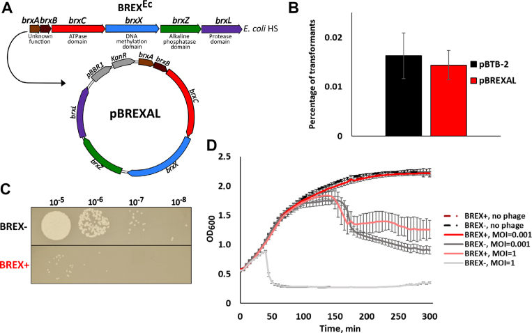
Closely related six-gene brxABCXZL clusters (Figure 1A) are present in multiple E. coli isolates (17). Escherichia coli HS, a natural isolate with the brxABCXZL cluster, was infected with several phages (λ, T4, T5 and T7). As controls, isogenic strains lacking the entire brxABCXZL cluster, or lacking the brxX putative methyltransferase gene, were used. All strains were found to be fully resistant to all phages tested. Thus, no conclusions about the contribution of brx cluster to phage resistance could be made. The entire E. coli HS brx cluster was therefore cloned, together with upstream sequences, on a low-copy E. coli pBTB-2 plasmid. The resulting plasmid was named pBREXAL (Figure 1A). Both pBTB-2 and pBREXAL plasmids transformed laboratory E. coli K12 strain BW25113 with equal efficiency (Figure 1B). Below, we refer to E. coli HS cluster as BREXEc; BW25113 cells carrying plasmid-borne BREXEc are referred to as BREX+; control cells carrying the pBTB-2 vector are referred to as BREX-.
Figure 1.
BREXEc protects cells from phage λ infection. (A) The BREX gene cluster from Escherichia coli HS is schematically shown; bioinformatically predicted putative functions of brx gene products are listed. The entire BREXEc cluster was cloned into the pBTB-2 vector to yield the pBREXAL plasmid. (B) Efficiency of transformation of the empty pBTB-2 vector and the pBREXAL plasmid into laboratory BW25113 E. coli to generate, correspondingly, BREX- and BREX+ cells. Mean values from three independent experiments are presented with standard deviations shown. P-value is 0.59. (C) Lawns formed by BREX+ and BREX- E. coli cells were spotted with indicated dilutions of phage λ lysate. Results of overnight growth at 37°C are shown. (D) Growth curves of BREX+ and BREX- E. coli cultures in the absence of infection, and during infection with λ phage at MOI of 0.001 and 1. Phage was added at t = 0. Each growth curve shows mean optical density values and standard deviations obtained from three independent experiments.
A modified λ cI857bor:Cm phage carrying a cI857 mutation in the cI gene coding for phage repressor, and marked with a chloramphenicol resistance gene that replaced the bor gene (encoding phage outer membrane lipoprotein) was obtained by thermal induction from E. coli MC4100 lysogen and tested for its ability to form plaques on lawns of BREX+ and BREX- cells or infect liquid cultures. As can be seen from Figure 1C, the efficiency of plaque formation was reduced ∼100-fold in the presence of BREXEc. In the absence of infection, the growth rates of BREX+ and BREX- cells in liquid culture were the same (Figure 1D). In cultures infected with phage λ at low multiplicity of infection (MOI) of 0.001, the BREX- culture collapsed ∼180 min post-infection. In contrast, the BREX+ culture continued to grow at the same rate as the uninfected culture (Figure 1D). At MOI of 1, BREX- cultures lysed 50 min post-infection, while BREX+ cultures continued to grow at the rate of uninfected control for ∼100 min. At later times the growth ceased and optical density slowly declined (Figure 1D). Overall, we conclude that similarly to the BREX system from B. cereus, BREXEc is functional in phage defense.
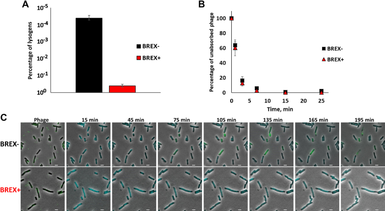
The experiments described above tested for the ability of BREXEc system to affect the lytic pathway of phage λ development that requires phage progeny formation. To determine if BREXEc also affects lysogenization, BREX+ and BREX- cultures were infected at high MOI and cells were plated on selective media containing chloramphenicol, where only lysogenized cells could form colonies. There was a strong (104-fold) suppression of chloramphenicol-resistant colony formation when BREX+ cultures were infected (Figure 2A). Since lysogenization does not require phage DNA replication (32), the result indicates that defensive action of BREXEc manifests itself either at the stage of phage adsorption or during injection of phage DNA. Measurements of the dynamics of unadsorbed phages in the course of lysogenization experiment revealed that the adsorption rate was the same in BREX+ and BREX- cultures (Figure 2B). A similar observation was made when phage adsorption to Bacillus subtilis cells with and without BREX system was monitored (17).
Figure 2.
BREXEc effects on lysogenization, adsorption and DNA injection by bacteriophage λ. (A) The bars show the numbers of chloramphenicol-resistant lysogenic colonies formed after high-MOI infection (MOI = 1) of BREX+ and BREX- cells with λ phage marked with a chloramphenicol resistance cassette. Mean values from three independent experiments are presented with standard deviations shown. (B) The experiment was conducted as in (A) but at an MOI of 0.01. At indicated times, cells were removed by centrifugation and unadsorbed infectious phage particles in the supernatant were determined on BREX- cells lawns. Mean values from three independent experiments are presented with standard deviations shown. P-value is 0.93. (C) Live microscopy of BREX- and BREX+ cells infected with phage λ. Images from a time-lapse movie show phage DNA injection. In the first picture, the fluorescent phage appears as a green dot on the cell surface. At 15 min, the SeqA-ECFP foci accumulate as one or two cyan dots, representing the ejected and replicated phage DNA respectively. Scale bar, 2 μm.
The process of infection was also monitored by live fluorescence microscopy using phage λ encoding capsid-decoration protein fused to EYFP. The host cells used for infection expressed a fused SeqA::ECFP protein and were dam−. SeqA specifically binds to DNA containing methylated or hemimethylated dam sites. As is shown elsewhere (29,33) this property allows one to monitor the process of infection by phage λ containing Dam-methylated DNA by observing fluorescent SeqA binding to injected phage genome, since bound SeqA forms distinct fluorescent foci on injected viral DNA. Immediately upon injection, one fluorescent dot is seen in infected cells. At later stages two dots corresponding to hemimethylated phage genomes are observed (29,33). Under our conditions (MOI = 1–5), phage DNA was injected in ∼53% of 853 BREX- cells analyzed as judged by the appearance of at least one SeqA focus. In some infected cells two SeqA foci appeared, followed by accumulation of yellow fluorescence due to the synthesis of capsid decoration protein::EYFP fusion. Initially diffuse, the EYFP fluorescence subsequently accumulated in speckles likely corresponding to phage capsids and cells lysed shortly thereafter (Figure 2C). BREX- cells where only one SeqA focus was observed survived and likely underwent lysogenic conversion. When BREX+ cells were infected, no ECFP foci or EYFP fluorescence accumulation was observed. The results suggest that phage DNA is either not injected in BREX+ cells or is rapidly degraded once inside the cells. Interestingly, the infected BREX+ cells stopped dividing and ∼18% of 911 cells examined became mildly elongated. Judging by the lack of propidium iodide staining, the membrane of infected BREX+ cells remained intact and no drop in CFU was observed when infected cells were deposited on agar plates, so activation of BREX, if it happens upon infection, does not kill cells.
BREXEc has no effect on lysogenic induction
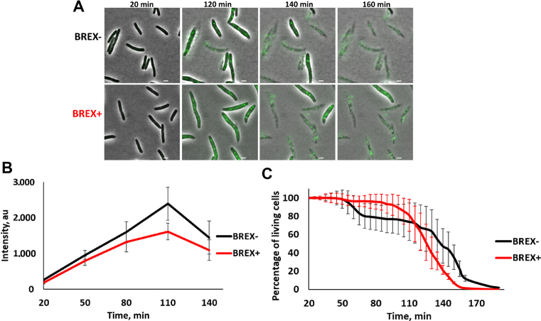
Escherichia coli cells lysogenized with λD-EYFP cI857bor::KanR were transformed with a plasmid with the BREXEc system or empty vector control. The resulting BREX+ and BREX- lysogenic cultures were shifted to 42°C to induce the prophage, transferred to 30°C and monitored over time using live fluorescent microscopy (Figure 3A). Preliminary experiments showed that BREX protection was as effective at 42°C as it was at 30 and 37°C (Supplementary Figure S2). Shortly after the induction both BREX+ and BREX- cells became fluorescent indicating accumulation of the decoration protein::EYFP fusion. At later times, fluorescent speckles corresponding to assembled phage capsids appeared inside the cells. The intensity of diffuse cytoplasmic fluorescence and the number of dots were nearly the same in BREX+ and BREX- cells (Figure 3B), indicating comparable phage yield. Most cells in both induced cultures lysed ∼3 h post-induction and the kinetics of cell lysis was the same in BREX+ and BREX- cultures (Figure 3C). We therefore conclude that BREXEc has no effect on lysogenic induction.
Figure 3.
Induction of λ prophage from BREX+ lysogens. (A) Images of cells from BREX- and BREX+ lysogenic cultures taken at indicated times after thermal induction. The induced phage encodes capsid decoration protein fused to EYFP. Scale bar, 2 μm. (B) Changes in EYFP fluorescence in induced cells are presented at various times post-induction. Mean numbers were calculated from fluorescence intensities obtained with ca. 300 cells. Standard deviations of mean numbers obtained in three independent experiments are shown. P-value is 0.46. (C) Quantification of a representative kinetic series showing decrease in live cells during microscopic observation of induced lysogenic cultures. Mean values are presented and standard deviations shown. P-value is 0.91.
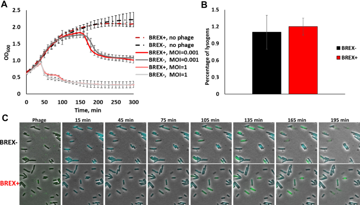
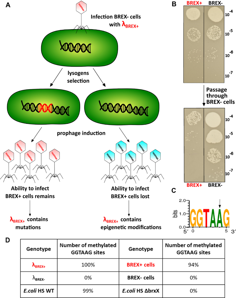
Phages collected after the induction of BREX+ lysogens were used to infect non-lysogenic BREX+ and BREX- cultures. Phage induced from BREX+ culture efficiently infected and lysed BREX+ cells in liquid cultures (Figure 4A). In addition, no difference in lysogenization efficiency of BREX+ and BREX- cultures with this phage was observed (Figure 4B). Live fluorescence microscopy experiments using cells expressing SeqA-ECFP fusion also demonstrated that phage induced from BREX+ culture infected both BREX+ and BREX- cells normally (Figure 4C). We conclude that BREX+ system is unable to protect cells from phages that were generated by the BREX+ host.
Figure 4.
λ phage induced from BREX+ lysogens is not subject to inhibitory action of BREXEc. (A) Growth curves of liquid cultures of BREX+ and BREX- cells in the absence of infection, and during infection with λ phage obtained after induction of BREX+ lysogens. Results of infections at MOI 0.001 and 1 are presented. In infected cultures, phage was added at t = 0. Mean values from three independent experiments are presented with standard deviations shown. (B) The bars show the number of chloramphenicol-resistant lysogenic colonies formed after infection (MOI = 5) of BREX+ and BREX- cells with phage λ phage obtained after induction of BREX+ lysogens (the phage is marked with a chloramphenicol resistance cassette). Mean values from three independent experiments are presented with standard deviations shown. P-value is 0.67. (C) Images of BREX+ and BREX- cultures infected (MOI = 1) with phage λ induced from BREX+ lysogens. Scale bar, 2 μm.
Phages that overcome BREX+ protection contain modified DNA
Progeny phage formed after infection of BREX- cells with phages induced from BREX+ lysogens (Figure 5A) was no longer able to infect BREX+ cells efficiently (Figure 5B). In contrast, progeny phage produced after the infection of BREX+ cells continued to infect BREX+ cells well (Figure 5B). The result suggests that phages induced from BREX+ lysogens contain an epigenetic modification. DNA prepared from phage virions induced from BREX+ lysogens was sequenced on a PacBio platform that detects a range of base modifications (34). The results showed that all 18 GGTAAG sites present in the λ genome were methylated at the fifth adenosine residue (Figure 5C and D). No such modification was present in DNA of phages induced from BREX- cells. Sequencing of BREX+ cells DNA showed that 94% of the 1708 genomic GGTAAG sites were also modified. No other BREX+ specific modifications were detected. It is worth noting that adenine in the CTTACC sequence complementary to the GGTAAG site remained unmodified. Genomic DNA was also isolated from E. coli HS strain and the brxX putative methyltraferase mutant. PacBio sequencing revealed that the fifth residue of the GGTAAG motif was modified in the wild-type strain while no modification was observed in the mutant (Figure 5D). We therefore conclude that (i) BREXEc methylates GGTAAG sites, (ii) the modification requires intact brxX and (iii) BREXEc-modified phages can productively infect BREX+ cells.
Figure 5.
BREX system epigenetically modifies phage λ. (A) The scheme shows two possible ways of appearance of phages that overcome BREX action. (B) At the top, lawns of BREX+ and BREX- cells were spotted with indicated dilutions of λ phage lysate obtained after induction of BREX+ lysogens. Results of overnight growth at 37°C are shown. Phages from a plaque obtained on BREX+ cells were used to re-infect BREX+ or BREX- cells. The results of spotting phage progeny from these infections on BREX− and BREX+ cell lawns are shown below. (C) The site modified in genomic DNA of Escherichia coli HS cells, E. coli BW25113 containing the pBREXAL plasmid and in the genome of phage λ induced from BREX+ lysogens. The arrow shows the site of BREX-dependent methylation. (D) Statistics of BREX site modification in BREX+ cells genome and in phage induced from these cells.
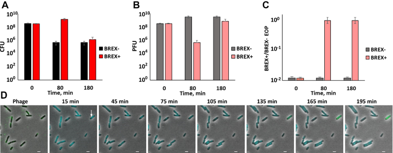
Given the epigenetic mechanism of overcoming BREX protection, we revisited infections of BREX+ cells with unmodified phage. When the number of surviving cells after infection at the MOI of 1 was monitored, there was ∼1000-times fewer survivor colonies in BREX- cultures compared to BREX+ cultures 80 min post-infection (Figure 6A). However, at later time points, the number of surviving cells became equally low in both cultures. Phage titer in infected cultures was also determined on lawns of BREX- and BREX+ cells. The ratio of these two titers, efficiency of plaquing, EOP, allows one to determine the state of phage DNA modification. Ca. 1% of initial phage used for infection was able to form plaques on BREX+ cells (see also Figure 1C). During infection of BREX- cells the overall phage titer increased but the proportion of phages able to form plaques of BREX+ lawns remained the same (Figure 6B and C). In BREX+ cells infection, the overall phage titer decreased dramatically (10 000 times) 80 min post-infection (for comparison, the burst time on BREX- cells at these conditions, when phage titer abruptly increases, is <40 min, Figure 1D). While the titer of phage in infected BREX+ cultures decreased, all phages were able to infect BREX+ cells. At later times, phage titer grew, while the ability to infect BREX+ cells with an EOP of 1 was retained. The experiment described above was conducted with λ cI857 bor:Cm phage used throughout this work and capable of lysogenic conversion. To exclude a possibility that phages that accumulate in infected BREX+ cultures appear through lysogenization followed by induction, we repeated the experiment using λvir virulent phage (31). An identical result was obtained (Supplementary Figure S3). We therefore conclude that during the infection of BREX+ culture with unmodified phage, modified phages appear in low frequency and then proceed to overtake the population. It is the appearance of these modified phages that must be responsible for eventual lysis of BREX+ cultures shown in Figure 1D. Live fluorescence microscopy analysis is consistent with this interpretation. While most BREX+ cells did not support productive infection, ECFP foci appeared in ∼1% of cells followed by subsequent accumulation of EYFP fluorescence and cell lysis, indicating productive infection (an example of such behavior can be seen in Figure 6D).
Figure 6.
Modified phage appears in the course of infection of BREX+ cells and overcomes protection. (A) The amount of colony forming units (CFU) in BREX+ and BREX- cultures infected at the MOI of 1 and t = 0 is shown 80 and 180 min post-infection. Mean values from three independent experiments are presented with standard deviations shown. (B) The amount of plaque forming units (PFUs) in BREX+ and BREX- cultures from panel (A). Mean values from three independent experiments are presented with standard deviations shown. (С) Efficiency of plaquing (EOP) of phages collected from infected cultures from panel (B) EOP was determined by calculating the ratio of phage titer on BREX+ and BREX- cell lawns. Mean values from three independent experiments are presented with standard deviations shown. (D) Live microscopy observation of BREX+ cells infected (MOI = 1) with phage λ. An arrow shows a productively infected cell. Scale bar, 2 μm.
The role of individual brx genes in phage protection and DNA modification
In agreement with data obtained using the brxX mutant of E. coli HS, deletion of brxX from the pBREXAL plasmid abolished phage protection and DNA modification (Table 1). These effects were complemented by the introduction of a compatible brxX expression plasmid. The role of other brx genes on phage protection/DNA modification was also investigated. Since deletions of brxZ or brxL genes in the context of the pBREXAL plasmid led to multiple frame-shifting deletions in other genes, we created two compatible plasmids separately expressing brxABC and brxXZL from arabinose inducible promoters. This two-plasmid system allowed, in the presence of arabinose, the same protection of cells from phage λ infection as pBREXAL. The genome of phage λ induced from lysogenic cells containing the full complement of brx genes on two plasmids and grown in the presence of arabinose was modified at GGTAAG sites (Table 1). As expected, deletion of the brxX gene in the context of the two-plasmid system led to the absence of both protection from infection and modification of phage/host DNA. Deletion of brxA had no effect on either protection from phage infection or on modification of phage/host DNA. Deletions of brxB, brxC, and brxZ abolished both protection and modification. Deletion of brxL abolished protection from infection but had a marginal effect on phage DNA modification (17 rather than 18 GGTAAG sites modified in phage genome). The result suggests that BrxL is either required for cell protection/limiting phage infection or that the presence of a single unmodified BREX site is not sufficient to allow the recognition of foreign DNA.
Table 1.
The state of methylation of host DNA in and ability to withstand phage infection by cells carrying BREX plasmids lacking indicated genes
| Genotype | BREX methylation | BREX defense |
|---|---|---|
| (BREX+)ΔX | − | − |
| BrxX only | − | − |
| ΔA | + | + |
| ΔB | − | − |
| ΔC | − | − |
| ΔX | − | − |
| ΔZ | − | − |
| ΔL | + | − |
| BREX+ (2 plasmids) | + | + |
The effect of removal of single brx genes was also studied by infecting cells at high MOI with unmodified phage λ. Phage progeny was collected and titered on BREX- and BREX+ cells. The results (Supplementary Table S3) were in complete agreement with lysogen induction experiments, showing that brxA and brxL are not necessary for the appearance of modified phages that are able to overcome BREX protection.
Individual expression of brxA, brxB, brxC and brxZ had no effect on cell growth and did not lead to protection from phage infection. Expression of brxX mildly inhibited cell growth and had no effect on phage infection. DNA prepared from these cells was not modified. Thus, BrxX alone is not sufficient for DNA modification by the BREX system. Interestingly, expression of brxL was highly toxic: cell growth ceased immediately after induction and the culture density gradually declined with time.
The data presented so far are consistent with a mechanism of BREX action that is similar to the classical R-M system action, though none of the brx genes products have a predicted nuclease function. When extracts of BREX+ cells were combined with unmodified λ DNA, no cleavage was observed, compared to BREX- cells lysates (Figure 7). In contrast, extract of cells containing a plasmid with the EcoRV Type II R-M system (21 recognition of sites in λ genome) readily cleaved λ DNA under these conditions.
Figure 7.
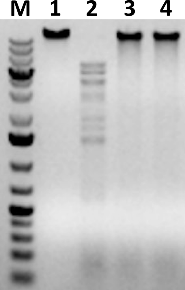
BREX+ cells extracts lack nuclease activity under conditions when restriction-endonuclease activity is detected. Cell extracts prepared from Escherichia coli cells harboring a plasmid containing EcoRV restriction-modification system genes (lane 2), pBREXAL plasmid (lane 3) or pBTB-2 control vector (lane 4) were combined with unmodified λ phage DNA, incubated and reaction products were resolved by agarose gel electrophoresis. Lane 1 is a control lane, phage genome incubated with buffer. M is a molecular weight marker.
BREX protection is circumvented by glycosylation of phage DNA
To determine the generality of the protective effect of BREXEc, BREX+ cells were infected with a set of different phages: M13, Qβ, T5, T4, T7, VR5, VR7 and VpaE1 (Table 2). No protection from infection with Qβ, an RNA phage, was observed. Likewise, there was no protection against infection with M13, a single-stranded DNA phage with a double-stranded replicative intracellular form. The rest of the phages tested have double-stranded DNA genomes. The extent of BREX protection from these phages varied significantly without apparent dependence of the number of GGTAAG sites in their genomes. The apparent lack of dependence of restriction on the number of GGTAAG methylation sites is striking, considering the site number dependence in tested restriction-modification systems (35). Similar to phage λ, ∼100-fold protection from T5 and T7 infection in plaque-forming assay was observed. On the other hand, no VR7 and VpaE1 plaques were formed on BREX+ lawns, indicating very high degree of protection. In stark contrast, no protection was observed from T4 and VR5 phage infection (Table 2). The DNA of T4 is hydroxymethylated at cytosines and is additionally glycosylated (36). Restriction analysis of the VR5 phage genomic DNA confirmed that this phage also has glycosylated DNA (Supplementary Figure S4). To test if glycosylation protects from BREX system action, we assayed T4147, a mutant of T4 bearing unglycosylated hydroxymethylcytosines in its genome (23). The extent of BREX protection from T4147 phage was similar to that observed for VR7 and VpaE1 phages, which have, respectively, modified and non-modified cytosines in their unglycosylated DNA (Table 2 and Supplementary Figure S5). We conclude that phages can overcome BREX system in at least two ways: (i) GGTAAG sites methylation, which requires brxX and brxBCZ, or (ii) glycosylation of their DNA, most probably at cytosines in the opposite strand of the GGTAAG sites.
Table 2.
Ability of BREXEc to protect cells from infections other than λ
| Phage | Genome | Number of GGTAAG sites | BREXEc protection* |
|---|---|---|---|
| Qβ | ssRNA | 3** | 100 |
| M13 | ssDNA | 3*** | 100 |
| T4 | dsDNA | 40 | 100 |
| T4147 | dsDNA | 39 | >1011 |
| T5 | dsDNA | 65 | 102 |
| T7 | dsDNA | 44 | 102 |
| VR5 | dsDNA | 85 | 100 |
| VR7 | dsDNA | 66 | >1011 |
| VpaE1 | dsDNA | 88 | >1010 |
| λ | dsDNA | 18 | 102 |
*The ratio of the number of phage plaques on the BREX- lawn to the number of plaques on the BREX+ lawn.
**GGUAAG sites.
***Positive strand.
DISCUSSION
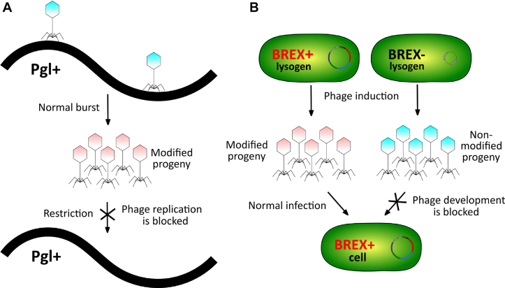
In this work, we demonstrate that E. coli BREX cluster protects cells from infection by diverse dsDNA phages. At least one of BREX genes, brxA, which codes for a protein of unknown function, is not essential for phage defense at our conditions. Of the remaining five brx genes, two are homologous to previously characterized Pgl system genes, encoding the putative methyltransferase BrxX (PglX) and alkaline phosphatase BrxZ (PglZ). The hallmark behavior of the Pgl system, decrease of phage yield during continuous infection but not in single-step infections, led to a proposal that the Pgl defense logic is inverted compared to that of R-M systems, where modification of phage DNA prevents restriction from happening, allowing productive infection (Figure 8A). It was further proposed the Pgl system consists of two functional modules, phage alteration/protective module pal and the defensive pgl module that blocks the development of modified phage.
Figure 8.
(A) Schematics of the Pgl mechanism based on the model by Chinenova et al. (13). (B) Schematics of the BREXEc function.
Here, we show that the pal module of E. coli BREX system functions by methylating a specific asymmetric site in phage DNA. The enzymatic activity of BrxX—in the presence of BrxB, BrxC, and BrxZ—is likely responsible for this modification. Site-specific methylation of phage DNA or global glycosylation of cytosine residues allows a phage to bypass BREX defense (Figure 8B). Thus, in this regard the E. coli BREX system functions similarly to R-M systems and is distinct from the Pgl system. Earlier work with BREX from B. cereus failed to observe modification of DNA of phage progeny collected after BREX+ infection (17). The result is probably due to very low yield of phage progeny production in these experiments, which caused the authors to analyze unabsorbed/non infecting phage particles, whose genomes naturally remained unmodified. While the sites of methylation in E. coli and B. cereus BREX systems have unrelated sequences (GGTAAG and TAGGAG, respectively), there are also important commonalities—they are asymmetric, and it is an adenine in the fifth position of the recognition site that is being methylated. The site of methylation by the Pgl system, if it exists, is unknown. Phylogenetic analysis indicates that BREX systems can be divided into six types based on the presence of characteristic genes. The E. coli and B. cereus BREX systems belong to the same type I (17) and may share a common mechanism of self-versus-non-self differentiation. The Pgl system belongs to a different type, type II, which may help explain the apparent differences in reported behavior of Pgl and the two type I systems.
Our results establish the mechanism responsible for self-protection of BREX carrying cells and the means by which phages can overcome the BREX defense. Overall, the logic appears to be similar to that observed for R-M systems, where rare modified genetic invaders appear due to stochastic events, multiply and eventually take over the population of initially protected cells. It is interesting that BREX systems may undergo phase variation due to a homopolymeric tract in the brxX coding sequence (17) and this strategy may allow the cells to alternate the proportion of BREX+ and BREX− cells in the population which may help withstand phage predation.
For most-studied Type II R-M systems, the palindromic nature of the recognition site ensures that epigenetic protective modification is heritable. The BREX site is asymmetric, and maintenance of epigenetic marks likely require interactions between different sites, as observed for Type I and Type III systems (37–39). Our observation that phages that contain a single unmodified BREX site are able to infect BREX+ cells is consistent with this idea.
The nature of defensive action of BREX remains elusive. The λ infection must be blocked at a very early stage after the absorption of the phage. In principle, either a block of DNA injection or rapid degradation of injected DNA prior to its replication is consistent with our live microscopy data. However, rapid degradation of unmodified DNA, a mechanism that occurs in cells protected by R-M systems, is expected to decrease the transformation efficiency of plasmids carrying the protective systems genes, into naïve cells. In the absence of proper regulation, plasmids harboring known R-M systems transform naïve cells very poorly due to premature synthesis of endonuclease (40). Plasmid expressing BREX genes is transformed into naïve cells with high efficiency. Moreover, unmodified phage DNA remains stable in extracts of BREX+ cells, again arguing against rapid degradation. Identification of the defense mechanism by BREX systems will require establishment of functional interactions between essential system components, since our mutagenesis results as well as earlier data collected with the Pgl system indicate that there must be several toxin-antitoxin type interactions that allow stable maintenance of the system. Interestingly, the likely toxin in BREX, the BrxL putative protease, is absent in Pgl, suggesting that these systems may employ multiple strategies to limit viral infections.
Supplementary Material
ACKNOWLEDGEMENTS
The authors thank P. Cohen and Prof. James B. Kaper for providing E. coli HS strain used in this study, R. Young for providing the lysogenic MC4100 strain and for discussions, and M. Zakharova for providing pEF42 plasmid and assistance in nuclease activity assays.
SUPPLEMENTARY DATA
Supplementary Data are available at NAR Online.
FUNDING
Skolkovo Institute of Science and Technology; Russian Foundation for Basic Research [18–34-00845]; National Institute of Health [R01GM107597, in part] and R01 GM10407; European Social Fund [09.3.3-LMT-K-712-01-0126]. Funding for open access charge: Skolkovo Institute of Science and Technology.
Conflict of interest statement. None declared.
REFERENCES
- 1. Suttle C. Marine viruses–major players in the global ecosystem. Nat. Rev. Microbiol. 2007; 5:801–812. [DOI] [PubMed] [Google Scholar]
- 2. Dammeyer T., Bagby S.C., Sullivan M.B., Chisholm S.W., Frankenberg-Dinkel N.. Efficient phage-mediated pigment biosynthesis in oceanic cyanobacteria. Curr. Biol. 2008; 18:442–448. [DOI] [PubMed] [Google Scholar]
- 3. Loenen W.A.M., Dryden D.T.F., Raleigh E.A., Wilson G.G., Murrayy N.E.. Highlights of the DNA cutters: a short history of the restriction enzymes. Nucleic Acids Res. 2014; 42:3–19. [DOI] [PMC free article] [PubMed] [Google Scholar]
- 4. Williams R.J. Restriction endonucleases: classification, properties, and applications. Mol. Biotechnol. 2003; 23:225–243. [DOI] [PubMed] [Google Scholar]
- 5. Marraffini L.A. CRISPR-Cas immunity in prokaryotes. Nature. 2015; 526:55–61. [DOI] [PubMed] [Google Scholar]
- 6. Koonin E.V., Makarova K.S.. CRISPR-Cas: an adaptive immunity system in prokaryotes. RNA Biol. 2009; 10:679–686. [DOI] [PMC free article] [PubMed] [Google Scholar]
- 7. Shmakov S., Smargon A., Scott D., Cox D., Pyzocha N., Yan W., Abudayyeh O.O., Gootenberg J.S., Makarova K.S., Wolf Y.I. et al. Diversity and evolution of class 2 CRISPR–Cas systems. Nat. Rev. Microbiol. 2017; 15:169–182. [DOI] [PMC free article] [PubMed] [Google Scholar]
- 8. Masuda H., Inouye M.. Toxins of prokaryotic toxin-antitoxin systems with sequence-specific endoribonuclease activity. Toxins (Basel). 2017; 9:E140. [DOI] [PMC free article] [PubMed] [Google Scholar]
- 9. Chopin M.C., Chopin A., Bidnenko E.. Phage abortive infection in lactococci: variations on a theme. Curr. Opin. Microbiol. 2005; 8:473–479. [DOI] [PubMed] [Google Scholar]
- 10. Blower T.R., Pei X.Y., Short F.L., Fineran P.C., Humphreys D.P., Luisi B.F., Salmond G.P.C.. A processed noncoding RNA regulates an altruistic bacterial antiviral system. Nat. Struct. Mol. Biol. 2011; 18:185–190. [DOI] [PMC free article] [PubMed] [Google Scholar]
- 11. Koonin E. V., Makarova K.S., Wolf Y.I.. Evolutionary genomics of defense systems in archaea and bacteria. Annu. Rev. Microbiol. 2017; 71:233–261. [DOI] [PMC free article] [PubMed] [Google Scholar]
- 12. Doron S., Melamed S., Ofir G., Leavitt A., Lopatina A., Keren M., Amitai G., Sorek R.. Systematic discovery of antiphage defense systems in the microbial pangenome. Science. 2018; 359:eaar4120. [DOI] [PMC free article] [PubMed] [Google Scholar]
- 13. Chinenova T.A., Mkrtumian N.M., Lomovskaia N.D.. Genetic characteristics of a new phage resistance trait in Streptomyces coelicolor A3(2). Genetika. 1982; 18:1945–1952. [PubMed] [Google Scholar]
- 14. Sumby P., Smith M.C.M.. Genetics of the phage growth limitation (Pgl) system of Streptomyces coelicolor A3(2). Mol. Microbiol. 2002; 44:489–500. [DOI] [PubMed] [Google Scholar]
- 15. Hoskisson P.A., Sumby P., Smith M.C.M.. The phage growth limitation system in Streptomyces coelicolor A(3)2 is a toxin/antitoxin system, comprising enzymes with DNA methyltransferase, protein kinase and ATPase activity. Virology. 2015; 477:100–109. [DOI] [PMC free article] [PubMed] [Google Scholar]
- 16. Makarova K.S., Wolf Y.I., Snir S., Koonin E. V.. Defense islands in bacterial and archaeal genomes and prediction of novel defense systems. J. Bacteriol. 2011; 193:6039–6056. [DOI] [PMC free article] [PubMed] [Google Scholar]
- 17. Goldfarb T., Sberro H., Weinstock E., Cohen O., Doron S., Charpak-Amikam Y., Afik S., Ofir G., Sorek R., Bikard D. et al. BREX is a novel phage resistance system widespread in microbial genomes. EMBO J. 2015; 34:169–183. [DOI] [PMC free article] [PubMed] [Google Scholar]
- 18. Datsenko K.A., Wanner B.L.. One-step inactivation of chromosomal genes in Escherichia coli K-12 using PCR products. Proc. Natl. Acad. Sci. U.S.A. 2000; 97:6640–6645. [DOI] [PMC free article] [PubMed] [Google Scholar]
- 19. Miller EM, Nickoloff JA. Escherichia coli electro transformation. Methods Mol. Biol. 1995; 147:105–113. [DOI] [PubMed] [Google Scholar]
- 20. Rasko D.A., Rosovitz M.J., Myers G.S.A., Mongodin E.F., Fricke W.F., Gajer P., Crabtree J., Sebaihia M., Thomson N.R., Chaudhuri R. et al. The pangenome structure of Escherichia coli: comparative genomic analysis of E. coli commensal and pathogenic isolates. J. Bacteriol. 2008; 190:6881–6893. [DOI] [PMC free article] [PubMed] [Google Scholar]
- 21. Jiang Y., Chen B., Duan C., Sun B., Yang J., Yang S.. Multigene editing in the Escherichia coli genome via the CRISPR-Cas9 system. Appl. Environ. Microbiol. 2015; 81:2506–2514. [DOI] [PMC free article] [PubMed] [Google Scholar]
- 22. Lynch M.D., Gill R.T.. Broad host range vectors for stable genomic library construction. Biotechnol. Bioeng. 2006; 94:151–158. [DOI] [PubMed] [Google Scholar]
- 23. Bair C.L., Black L.W.. A type IV modification dependent restriction nuclease that targets glucosylated hydroxymethyl cytosine modified DNAs. J. Mol. Biol. 2007; 366:768–778. [DOI] [PMC free article] [PubMed] [Google Scholar]
- 24. Kaliniene L., Klausa V., Truncaite L.. Low-temperature T4-like coliphages vB_EcoM-VR5, vB_EcoM-VR7 and vB_EcoM-VR20. Arch. Virol. 2010; 155:871–880. [DOI] [PubMed] [Google Scholar]
- 25. Šimoliūnas E., Vilkaitytė M., Kaliniene L., Zajančkauskaitė A., Kaupinis A., Staniulis J., Valius M., Meškys R., Truncaitė L.. Incomplete LPS core-specific felix01-like virus vB_EcoM_VpaE1. Viruses. 2015; 7:6163–6181. [DOI] [PMC free article] [PubMed] [Google Scholar]
- 26. Horton R.M., Ho S.N., Pullen J.K., Hunt H.D., Cai Z., Pease L.R.. Gene splicing by overlap extension. Methods Enzymol. 1993; 217:270–279. [DOI] [PubMed] [Google Scholar]
- 27. Hengen P.N. Methods and reagents. Preparing ultra-competent Escherichia coli. Trends Biochem. Sci. 1996; 21:75–76. [PubMed] [Google Scholar]
- 28. Zeng L., Skinner S.O., Sippy J., Feiss M., Golding I.. Decision making at a subcellular level determines the outcome of bacteriophage infection. Cell. 2010; 141:682–691. [DOI] [PMC free article] [PubMed] [Google Scholar]
- 29. Shao Q., Hawkins A., Zeng L.. Phage DNA dynamics in cells with different fates. Biophys. J. 2015; 108:2048–2060. [DOI] [PMC free article] [PubMed] [Google Scholar]
- 30. Wallace D.M. Large- and small-scale phenol extractions. Methods Enzymol. 1987; 152:33–41. [DOI] [PubMed] [Google Scholar]
- 31. Semenova E., Minakhin L., Bogdanova E., Nagornykh M., Vasilov A., Heyduk T., Solonin A., Zakharova M., Severinov K.. Transcription regulation of the EcoRV restriction-modification system. Nucleic Acids Res. 2005; 33:6942–6951. [DOI] [PMC free article] [PubMed] [Google Scholar]
- 32. Oppenheim A.B., Kobiler O., Stavans J., Court D.L., Adhya S.. Switches in bacteriophage lambda development. Annu. Rev. Genet. 2005; 39:409–429. [DOI] [PubMed] [Google Scholar]
- 33. Babic A., Lindner A.B., Vulic M., Stewart E.J., Radman M.. Direct visualization of horizontal gene transfer. Science. 2008; 319:1533–1536. [DOI] [PubMed] [Google Scholar]
- 34. Flusberg B.A., Webster D.R., Lee J.H., Travers K.J., Olivares E.C., Clark T.A., Korlach J., Turner S.W.. Direct detection of DNA methylation during single-molecule, real-time sequencing. Nat. Methods. 2010; 7:461–465. [DOI] [PMC free article] [PubMed] [Google Scholar]
- 35. Pleška M., Guet C.C.. Effects of mutations in phage restriction sites during escape from restriction–modification. Biol. Lett. 2017; 13:20170646. [DOI] [PMC free article] [PubMed] [Google Scholar]
- 36. Lehman I.R., Pratt E.A.. On the structure of the glucosylated hydroxymethylcytosine nucleotides of coliphages T2, T4, and T6. J. Biol. Chem. 1960; 235:3254–3259. [PubMed] [Google Scholar]
- 37. Dreier J., MacWilliams M.P., Bickle T.A.. DNA cleavage by the type IC restriction-modification enzyme EcoR124II. J. Mol. Biol. 1996; 264:722–733. [DOI] [PubMed] [Google Scholar]
- 38. Meisel A., Bickle T.A., Kriiger D.H., Schroeder C.. Type III restriction enzymes need two inversely oriented recognition sites for DNA cleavage. Nature. 1992; 355:467–469. [DOI] [PubMed] [Google Scholar]
- 39. Van Aelst K., Toth J., Ramanathan S.P., Schwarz F.W., Seidel R., Szczelkun M.D.. Type III restriction enzymes cleave DNA by long-range interaction between sites in both head-to-head and tail-to-tail inverted repeat. Proc. Natl. Acad. Sci. U.S.A. 2010; 107:9123–9128. [DOI] [PMC free article] [PubMed] [Google Scholar]
- 40. Johnston C., Martin B., Polard P., Claverys J.P.. Postreplication targeting of transformants by bacterial immune systems. Trends Microbiol. 2013; 10:516–521. [DOI] [PubMed] [Google Scholar]
Associated Data
This section collects any data citations, data availability statements, or supplementary materials included in this article.


