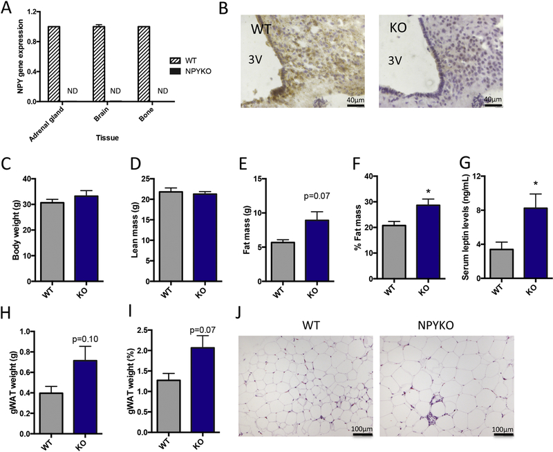
Figure 3: Validation of global NPYKO and evaluation of body composition.
Gene expression of NPY in adrenal gland, brain and bone (A), n = 3. Immunohistochemistry for NPY in arcuate nucleus (B). The third ventricle is marked (3V). Body weight of male mice at 14 weeks of age (C). Whole body DXA data: Lean mass (D), fat mass (E) and % fat mass (F). Serum leptin levels as determined by ELISA (G). Dissected gonadal white adipose tissue weight (H) and expressed as % of body weight (I). Representative images of haematoxylin and eosin staining of gonadal white adipose tissue (J). Mean±SEM. Statistical analysis: Students t-test (C-I), where *p<0.05. Body weight and DXA analyses: Male WT (n=5); Male NPYKO (n=7).

