Summary
The developmental origin of oligodendrocyte progenitors (OLPs) in the forebrain has been controversial. We now show, by Cre-lox fate mapping in transgenic mice, that the first OLPs originate in the medial ganglionic eminence (MGE) and anterior entopeduncular area (AEP) in the ventral forebrain. From there, they populate the entire embryonic telencephalon including the cerebral cortex before being joined by a second wave of OLPs from the lateral and/or caudal ganglionic eminences (LGE and CGE). Finally, a third wave arises within the postnatal cortex. When any one population is destroyed at source by targeted expression of Diphtheria toxin, the remaining cells take over and the mice survive and behave normally, with a normal complement of oligodendrocytes and myelin. Thus, functionally redundant populations of OLPs compete for space in the developing brain. Strikingly, the embryonic MGE- and AEP-derived population is eliminated during postnatal life, raising questions about the nature and purpose of the competition.
Introduction
Different subclasses of neurons and glia are generated sequentially in different parts of the ventricular zones (VZ) of the developing spinal cord and brain. For example, spinal motor neurons and oligodendrocytes (OLs, the myelin-forming cells) are derived from precursors that reside in a specialized domain of the ventral VZ called pMN, defined by expression of the transcription factor Olig21–4. From there, oligodendrocyte progenitors (OLPs) migrate all through the spinal cord before differentiating into myelin-forming OLs. Later, an additional source(s) of OLPs arises in the dorsal spinal cord, contributing 10-15% of the final OL population5–7. OLP generation in pMN depends on the signalling molecule Sonic hedgehog (Shh) while the dorsal source might be Shh-independent5–9.
OL generation in more anterior parts of the neural tube - particularly the forebrain - is not so well understood. The neuroepithelium of the medial ganglionic eminence (MGE) and anterior entopeduncular area (AEP) in the ventral forebrain expresses Shh and its receptor Patched (Ptc) as well as Olig2, suggesting that OLPs might be formed primarily in these regions10–12. Indeed, migratory OLPs, defined by expression of platelet-derived growth factor receptor-alpha (Pdgfra), can be seen streaming away from the MGE/AEP in all directions after embryonic day 12 (E12) in the mouse, apparently entering the developing cerebral cortex (dorsal telencephalon) around E1611,13. The number of OLPs in the cortex increases dramatically between E16 and birth (~E18) but it is not known whether this reflects continuing inward migration and proliferation of ventrally-derived OLPs or a new source of OLPs within the cortex. There is some evidence on both sides. For example, some OLPs in the cortex express the transcription factors Dlx1 and Dlx2, suggesting that they originate in the ventral telencephalon where Dlx1 and Dlx2 are first activated14,15. Moreover, in chick embryos expression of the oligodendrocyte-lineage markers myelin proteolipid protein (Plp-Dm20) and the O4 antigen is initially restricted to the AEP16 and chick-quail grafting experiments indicate that all cortical OLs are ventrally-derived in birds17. On the other hand, cell fate mapping using Emx1-Cre transgenic mice suggests that many OLs in the cortex are generated locally18. Furthermore, retroviral fate-mapping demonstrates that cortical OLs continue to be formed in the postnatal period from precursors in the subventricular zones (SVZ) located at the tips of the lateral ventricles at the cortico-striatal boundary19,20. It is important to resolve this uncertainty because OLs with different developmental origins, perhaps specified via different signalling pathways, might have distinct properties and functions in the mature brain.
To resolve the question of OL origins we used the Cre-lox approach in transgenic mice to follow the development of distinct ventral or dorsal precursor populations in the telencephalon. We found that development of OL lineage cells in the forebrain is unexpectedly complex and dynamic. There is an early wave of OLP generation from Nkx2.1-expressing precursors in the ventral forebrain; these first appear in the VZ of the ventral MGE and AEP around embryonic day 12.5 (E12.5) and subsequently migrate widely into all parts of the telencephalon, entering the cerebral cortex after E16. At first they are the only OLPs in the forebrain but soon their contribution to the total OLP population declines. By postnatal day 10 (P10) there are very few Nkx2.1-derived OLPs or OLs in the cortex, although they are still the major population in the ventral (sub-pallial) region. Instead, they are overtaken in the cortex by other populations of OLPs derived first from Gsh2-positive precursors in the LGE/CGE and later from endogenous Emx1-positive cortical precursors. Even in the ventral forebrain, close to their original source, the Nkx2.1-derived OLPs and OLs are gradually lost and replaced by other populations and are almost completely eliminated from the adult forebrain. These findings help to integrate the plethora of previous studies that report either ventral, dorsal or multiple OLP origins10–24.
Our results raise the question of whether the different OL lineages from MGE/AEP, LGE/CGE and cortex are functionally specialized. To address this we devised a genetic ablation strategy to eliminate each of the three OLP populations separately with diphtheria toxin A fragment (DTA)25. We found that when we killed any one of the populations the effect was benign; adjacent populations spread into the vacant territory, a normal distribution of OLPs was restored and the mice developed and survived normally. Therefore, we did not so far find evidence of functional heterogeneity among different OL lineages in the forebrain. Our data demonstrate that different regional sub-populations of OLPs compete for space in the developing brain. This might contribute to the ultimate demise of the embryonic MGE/AEP (Nkx2.1)-derived OLP population.
Results
The first OLPs are derived from the MGE-AEP
Previous immunohistochemical and transplant studies suggested that there is a source(s) of OL lineage cells in the ventral forebrain (MGE and AEP)11,13–17,21,22. To test directly whether OLPs are generated in the ventral telencephalon we generated PAC transgenics that express Cre recombinase under the transcriptional control of Nkx2.1, which encodes a homeodomain transcription factor expressed in the MGE, septum, AEP, pre-optic area and other more posterior ventral forebrain regions. Expression of the Cre transgene faithfully recapitulated the endogenous Nkx2.1 pattern (supplementary Fig. 1). We crossed the Nkx2.1-Cre mice to the Cre-dependent Rosa26R-GFP reporter line and examined embryonic offspring for the presence of Pdgfra-positive (Pdgfra+) OLPs that also expressed green fluorescent protein (GFP), indicating that they must have originated from Nkx2.1-expressing precursors.
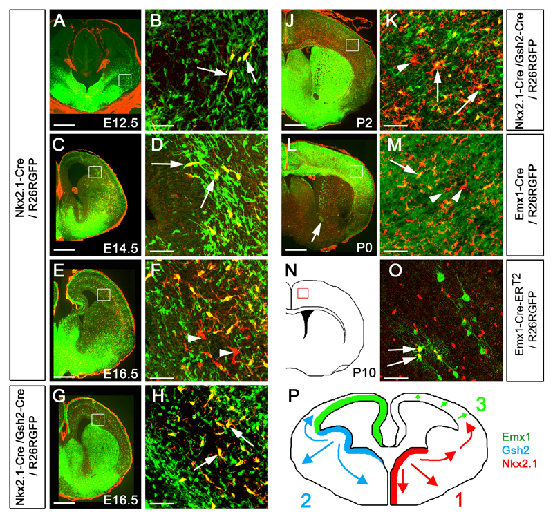
In most parts of the telencephalon the (GFP+, Pdgfra+) OLP population was only a small fraction of all GFP+ cells. In the cortex, the majority of GFP+ cells represent GABAergic interneurons that are known to originate in the MGE26–28. The first (GFP+, Pdgfra+) OLPs appeared in the ventral MGE and AEP at E11.5-E12.5 (Fig. 1a,b) and gradually spread throughout the telencephalon in a ventral-to-dorsal manner. Until E14.5 the great majority of Pdgfra+ OLPs in the ventral telencephalon as well as in the cerebral cortex co-expressed GFP, indicating that the main source of OLs in the embryonic forebrain lies within the Nkx2.1-expressing neuroepithelium (Fig. 1c,d). However, by E16.5 a new population of GFP-negative, Pdgfra+ OLPs appeared, mainly within the cortical intermediate zone (arrowheads, Fig.1e, f). These GFP-negative OLPs must originate outside the MGE/AEP.
Figure 1.
Three successive waves of OLs generated from distinct precursor populations at different times during forebrain development. (a, b) Generation of the first wave of Pdgfra+ OLPs begins at E12.5 from Nkx2.1-expressing precursors in the MGE/AEP. These OLPs express GFP (green) in Nkx2.1-Cre/ Rosa26R-GFP embryos (arrows in b). (c, d) By E14.5 Pdgfra+, GFP+ cells start to appear at the cortico-striatal boundary in Nkx2.1-Cre/ Rosa26R-GFP embryos (arrows in d), demonstrating migration of MGE/AEP-derived OLPs into the developing cortex. (e, f) By E16.5 a new wave of Pdgfra+, GFP-negative OLPs is observed in Nkx2.1-Cre/ R26R-GFP cortex; these must be generated from Nkx2.1-negative precursors (arrowheads in f). (g, h) All Pdgfra+ cells in the telencephalon co-express GFP in Nkx2.1-Cre/ Gsh2-Cre/ Rosa26R-GFP triple-transgenic embryos (arrows in h), indicating that all OLPs are derived from the ventral forebrain (MGE/AEP + LGE/CGE) at this stage. (i, j) Another wave of Pdgfra+, GFP-negative OLPs appears in the telencephalon of early postnatal Nkx2.1-Cre/ Gsh2-Cre/ Rosa26R-GFP mice (arrowheads in j). (k, l) Emx1-expressing cortical precursors begin to generate OLPs/OLs after birth (arrows in l). (m, n) Tamoxifen administered once to pregnant Emx1-CreERT2 females at E9.5, before invasion of the cortex by ventrally-derived OLPs, activates Cre recombination in the embryos and results in expression of the GFP reporter in a subset of Sox10+ OLPs (arrows in n)- which must therefore have been generated from endogenous cortical precursors. (o) Conclusion: three sequential waves of OLPs are generated from different parts of the telencephalic VZ: 1) from Nkx2.1-expressing precursors starting at E12.5, 2) from Gsh2-expressing LGE/CGE precursors starting at E15.5 and 3) from Emx1-expressing cortical precursors starting around birth (P0). Scale bars: (a, c, e, g, i, k) 500 μm; (b, d, f, h, j, l, n) 60 μm.
Subsequent waves of OLPs from the LGE/CGE and cortex
Another region of the telencephalon that expresses Olig2 and seemed likely to generate OLs is the LGE. To fate map the LGE we generated PAC transgenics that express Cre under Gsh2 control. Gsh2 is expressed strongly in the LGE/CGE but partially overlaps with Nkx2.1 in the MGE (supplementary Fig. 1). To map the overall contribution of the ventral forebrain we generated triple-transgenic Gsh2-Cre/Nkx2.1-Cre/Rosa26R-GFP mice. In E16.5 triple-transgenics, practically all Pdgfra+ OLPS in the telencephalon, including the lateral cortex, were also GFP+ (Fig. 1g,h). This shows that before birth all OLPs in the cortex are ventrally-derived, migrating first from the MGE/AEP and subsequently from the LGE and/or CGE. By the day of birth, however, GFP-negative OLPs once again had started to accumulate in the cortex of the triple-transgenics (arrowhead, Fig. 1i, j), indicating the presence of yet another source(s) of OLPs outside either the MGE/AEP or the LGE/CGE - possibly within the cortex itself.
To examine the fates of endogenous cortical precursors we generated Emx1-Cre transgenics (supplementary Fig. 1). In these mice Cre is expressed strongly in cortical precursors and activation of the GFP reporter in Emx1-Cre/Rosa26R-GFP animals is widespread in the postnatal cortex (supplementary Fig. 1). GFP was present throughout the cytoplasm of the cells and even along axons, so that fibre tracts projecting from the cortex to the ventral forebrain were GFP-labelled (arrow, Fig. 1k). Emx1-derived (GFP+, Pdgfra+) OLPs first appeared in the cortex around birth (Fig. 1k,l) and rapidly increased in number thereafter. Emx1-derived OLPs were never observed in the ventral telencephalon.
Our data suggested that cortical OLPs are composed of two immigrant populations from the MGE/AEP and LGE/CGE and a resident cortical population. To exclude the possibility that the Emx1-derived OLPs are ventrally-derived cells that start to express Emx1 de novo after they migrate into the cortical field, we generated another transgenic line that expresses the tamoxifen-inducible form of Cre recombinase (CreERT2) under Emx1 control (supplementary Fig. 1). We induced Cre activity transiently by administering tamoxifen once at E9.5, prior to the onset of gliogenesis. Since tamoxifen is cleared within 48 hours, this avoids Cre being activated in OLPs that migrate into the cortex from elsewhere after ~E11.5. We analyzed the tamoxifen-treated animals at P9. Pdgfra expression in the CNS is restricted to early OLPs and is down-regulated as soon as the cells exit the cell cycle and start to differentiate into OLs29,30. We therefore used an antibody against Sox10, which starts to be expressed in OLPs slightly later than Pdgfra but persists after Pdgfra is down-regulated, including in mature OLs31. CreERT2-mediated recombination was relatively inefficient compared to regular Cre, so that well separated GFP-labelled cells or clusters of cells were observed throughout the cortex. These included Sox10+ OLs/OLPs as well as cells with the morphology of neurons and astrocytes (Fig. 1m,n). The (GFP+, Sox10+) cells comprised around 20% of all GFP+ cells and were observed in all parts of the cortex (arrows, Fig. 1n). We conclude that they are formed from endogenous cortical precursors and not from inwardly migrating cells.
Postnatal eradication of the embryonic Nkx2.1-derived lineage
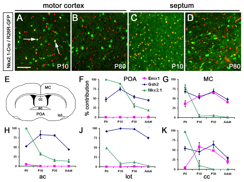
Our data demonstrate that there are three successive waves of OLP generation and migration in the embryonic telencephalon – from the MGE/AEP, LGE/CGE and cortex respectively (Fig. 1o). To determine how these three OLP populations contribute to the postnatal and adult telencephalon and to what extent they intermingle, we analyzed Nkx2.1-Cre/Rosa26R-GFP, Gsh2-Cre/Rosa26R-GFP and Emx1-Cre/Rosa26R-GFP mice from newborn (P0) to young adult (P80) (Fig. 2). The contribution of Nkx2.1-derived Sox10+ OLs/OLPs throughout the forebrain was high at birth, including dorsal regions such as the corpus callosum as well as ventral regions such as the septum and pre-optic area. Surprisingly, by P10 the proportion of Nkx2.1-derived OLs/OLPs had plummeted to a very low level in the cortex (Fig. 2a,b,g,j). Most remarkably, the Nkx2.1-derived population declined to a very small fraction of all OL lineage cells in most parts of the adult forebrain, even in ventral regions close to their original source (Fig. 2c,d,f,h,i). Thus, the Nkx2.1-expressing region of the ventral forebrain gives rise to migratory OLPs that populate the embryonic forebrain including the cortex but most of these are eliminated after birth.
Figure 2.
The embryonic Nkx2.1-derived OL lineage is rapidly eliminated during postnatal life. (a, b) In Nkx2.1-Cre/ Rosa26R-GFP mice GFP+ (green), Sox10+ (red) double-positive OLPs are already a minority in the P10 cortex (arrows in a), and their contribution drops to zero by P80. (c, d) At P10 practically all OLPs in ventral regions such as the septum are derived from Nkx2.1-expressing (MGE/AEP-derived) precursors (GFP, Sox10 double-positive; yellow nuclei in c) but even here they are replaced by other populations (GFP-negative) by P80. (e-j) The proportional contributions of the three different populations of Sox10+ OL lineage cells are presented as percentage of the total number of Sox10+ cells in each region (average± standard deviation). OLs/OLPs derived from Nkx2.1-expressing precursors are largely eliminated between birth and adulthood from most regions examined. OLs/OLPs generated from cortical precursors (Emx1-derived) remain within the cortex at all stages. Gsh2-expressing precursors give rise to OLs/OLPs that spread throughout the telencephalon. POA, preoptic area; MC, motor cortex; AC, anterior commissure; LOT, lateral olfactory tract; CC, corpus callosum. Scale bar: 100 μm.
From the data shown, it is clear that the majority of OLPs in most regions of the postnatal telencephalon is contributed by Gsh2-expressing precursors (Fig. 2e-j). The Emx1 precursor contribution is almost entirely restricted to the cortex throughout life, although in the adult a few Emx1-derived OLs are found in the septum. From P10 to P80 the cortex is populated by similar proportions of Emx1- and Gsh2-derived OLs/OLPs (Emx1 contribution declining from ~70% to ~30%).
The cortical Emx-derived and ventral telencephalic Nkx2.1- and Gsh2-derived OLs/OLPs account for nearly all OL-lineage cells in the early postnatal telencephalon. However, by adult stages the total contribution of telencephalic OLs/OLPs drops to 60-75%, especially in ventral territories such as the pre-optic area (Fig. 2f) and fibre tracts such as the corpus callosum, lateral olfactory tract and anterior commisure (Fig. 2h,i,j). Although we cannot exclude the possibility that this might be due to selective down-regulation of the Rosa26 promoter in some mature cells, we believe that it more likely reflects posterior-to-anterior migration of cells along fibre tracts. We have observed such influx of cells into the telencephalon from the diencephalon along axons of the internal capsule (data not shown).
Functional equivalence of OLs with different origins
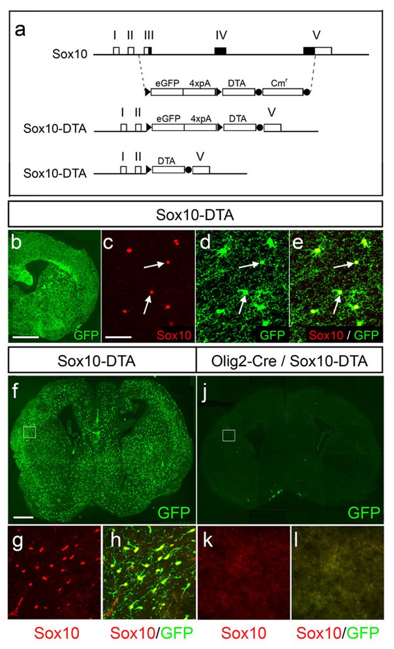
The presence of distinct OL populations within the telencephalon led us to question whether the different OL “lineages” are functionally specialized. To address this we devised a transgenic approach to eliminate OL lineage cells according to their site of origin, by targeted expression of the diphtheria toxin A fragment (DTA)32. A transgenic mouse line was produced using a PAC-based transgene with the structure Sox10-lox-GFP-STOP-lox-DTA (Fig. 3a). The transgene was faithfully regulated because Sox10 and GFP were co-expressed in the vast majority of OL lineage cells throughout the CNS (>98% co-expression in the P3 cortex) (Fig. 3b-e). Following Cre recombination, GFP should be excised along with the STOP cassette, thus activating DTA and killing the cells. Henceforth, we refer to this DTA line simply as Sox10-DTA.
Figure 3.
Design and activity of a Cre-inducible Diphtheria toxin (DTA) transgene under Sox10 transcriptional control. (a) Intron/exon structure of the Sox10 locus (top). The genomic region spanning exons III – V was replaced with the cassette lox-eGFP-polyA4-lox-DTA-frt-Cmr-frt by homologous recombination in bacteria. The Cmr cassette was then removed by transient activation of Flp recombinase in bacteria, producing a latent DTA transgene that expresses GFP under Sox10 transcriptional control (Sox10-DTA). In the presence of Cre recombinase the GFP-polyA cassette is removed, thus activating the DTA cassette and killing the expressing cells (further details on request). (b-e) Coronal section through the telencephalon of a Sox10-DTA transgenic mouse at P3 and high magnification images of the cortex from the same section. All Sox10+ OLPs/OLs in the Sox10-DTA mouse co-express GFP, demonstrating the veracity of transgene expression. (f-k) The DTA transgene was activated by crossing Sox10-DTA to an Olig2-Cre mouse, excising the GFP cassette in all Olig2-expressing precursors and their descendants, hence permitting expression of DTA and highly specific killing of all Sox10+ OL lineage cells. The effectiveness of this tool is demonstrated by the loss of all (Sox10+, GFP+) OL lineage cells in the double-transgenics (compare f-h with i-k). The Olig2-Cre/Sox10-DTA mice died shortly after birth, apparently from a motor deficit that prevented them from feeding. Scale bars: (b) 1 mm; (c-e) 50 μm; (f, i) 500 μm; (g-h, j-k) 80 μm.
To test our cell ablation strategy we crossed Sox10-DTA to an Olig2-Cre line that expresses Cre recombinase in all OL lineage cells, regardless of their embryonic origin (N.Kessaris, D.Rowitch and W.Richardson, unpublished). Olig2-Cre/Sox10-DTA neonates had neither GFP+ nor Sox10+ cells anywhere in their brain or spinal cord, confirming efficient excision of the GFP coding sequences and activation of DTA (Fig. 3f-k). These embryos appeared normal at birth but died within 24-48 hours. We do not yet know the cause of death but suspect a motor deficit.
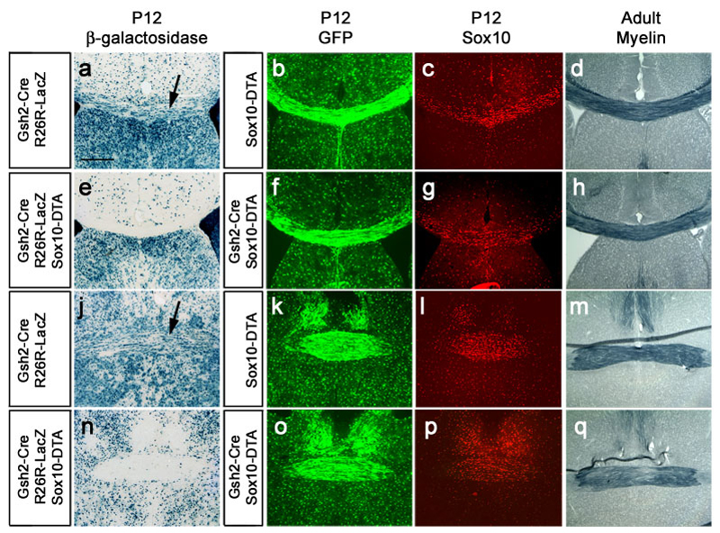
To ask whether the region-specific telencephalic OL populations are functionally distinct, we generated a series of transgenics that combined Sox10-DTA with either Emx1-Cre or Gsh2-Cre (Fig. 4). In the presence of Rosa26R-lacZ as an independent reporter for Cre activity, cells from the region under study should be positive for beta-galactosidase (βgal) (i.e. labeled blue). Therefore, if region-specific ablation is successful there should be an absence of blue OLs or OLPs while βgal-negative, GFP+ OLs/OLPs that are generated outside the region of interest should survive. This is illustrated most dramatically in white matter tracts such as the corpus callosum or the anterior commissure, where the majority of cell bodies represent OLs/OLPs or other glia (Fig. 4a-d). For example, at P12 there were many βgal+ (blue) cells in the corpus callosum and anterior commissure of double-transgenic Gsh2-Cre/Rosa26R-lacZ mice (Fig. 4a,c) but practically none in Gsh2-Cre/Rosa26R-lacZ/Sox10-DTA triple-transgenics (Fig. 4b,d). Nevertheless, there appeared to be just as many (GFP+, Sox10+) cells in the corpus callosum and anterior commissure of ablated mice (Gsh2-Cre/Sox10-DTA) as in non-ablated mice (Sox10-DTA) (Fig. 4e-h). For example, we counted (9.0 ± 1.5) x 104 Sox10-positive cells/mm3 in the corpus callosum of non-ablated mice (Fig. 4i) compared to (9.1 ± 1.2) x 104 cells in (Gsh2-Cre/Sox10-DTA) mice (Fig. 4j). Similarly, we counted (9.0 ± 1.4) x 104 Sox10-positive cells/mm3 in the anterior commissure of control mice (Fig. 4k) compared to (9.0 ± 0.8) x 104 cells in (Gsh2-Cre/Sox10-DTA) mice (Fig. 4l). [Data are presented as (mean ± standard deviation)]. We conclude that the loss of Gsh2-derived OLs/OLPs is compensated for by another population(s) - in the corpus callosum, presumably by Emx1-derived (cortical) cells and in the anterior commissure presumably by Nkx2.1-derived cells, although there could also have been invasion from further afield. Compensation was also seen in the corpus callosum when we ablated the Emx1-derived OL/OLP population (not shown). In none of these examples was there any obvious alteration to the amount of myelin in adult animals, as visualized by Sudan black histochemistry (Fig. 4m-p).
Figure 4.
Genetic ablation of region-specific OL populations reveals functional redundancy and compensation among the different lineages. (a-d) Gsh2-derived white matter glia (mainly OL lineage cells) were visualized in the corpus callosum (a-b) or anterior commissure (c-d) of P12 Gsh2-Cre/Rosa26R-lacZ (a,c) and Gsh2-Cre/ Sox10-DTA/ Rosa26-R-lacZ (b,d) mice (coronal sections, arrows). Many blue cells are visible in these tracts in Gsh2-Cre/Rosa26R-lacZ mice but are missing from the equivalent structures of Gsh2-Cre/ Sox10-DTA/ Rosa26-R-lacZ mice, showing that Gsh2-derived OLs have been effectively ablated in the latter through the action of DTA. (e-l) Despite the specific loss of Gsh2-derived OLs, there is no apparent reduction in the total complement of OLs in these white matter tracts as visualized by GFP staining (compare e with f, g with h) or Sox10 staining (compare i with j and k with l). (m-p) There is no apparent reduction in the amount of myelin in adult white matter tracts of ablated Gsh2-Cre/ Sox10-DTA mice as visualized by Sudan black histochemistry (compare m with n, o with p). Thus it appears that the loss of Gsh2-derived OLs is compensated for by the invasion of alternative OL lineages. Similar compensation was observed when the Emx1-derived OL lineage was ablated in Emx1-Cre/ Sox10-DTA mice (data available on request). The mice resulting from these crosses survived, behaved and reproduced normally until at least P60. These experiments demonstrate that the different regional OL lineages compete for territory in the normal developing CNS and further suggest that the different OL populations are functionally equivalent. Scale bar: 400 μm.
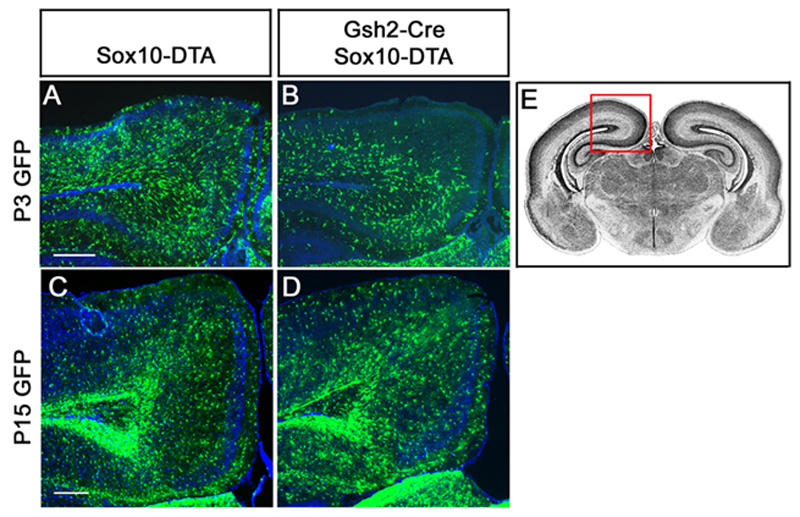
Although the number and distribution of (GFP+, Sox10+) OLs/OLPs in Emx1-Cre/Sox10-DTA, Gsh2-Cre/Sox10-DTA and Nkx2.1-Cre/Sox10-DTA mice were ultimately indistinguishable from normal controls, accumulation of OLPs was noticeably delayed in some regions (Fig. 5). For example, in Sox10-DTA/Gsh2-Cre mice there was a slight delay in the arrival of OLPs in the outer layers of the cortex (compare Fig. 5a,c), consistent with our observation that Emx1-derived OLPs start to be generated slightly later than Gsh2-derived OLPs. Despite the loss of Gsh2-derived OLPs, the mice recovered fully by P10 by which time there was a normal number and distribution of OLPs in the cortex (Fig. 5b,d). Even in the olfactory tracts in the ventral forebrain - normally populated solely by Gsh2-derived OLs - there was complete replenishment of OLs from another source(s). The animals all survived and reproduced normally with no obvious neurological symptoms. These and other data imply that the Gsh2-, Emx1- and Nkx2.1-derived OL lineages are functionally equivalent, in contrast to the different neuronal populations that are generated from the same germinal fields.
Figure 5.
Transient delay in accumulation of OLPs after ablation of the LGE/CGE-derived population in Gsh2-Cre/Sox10-DTA mice. Coronal forebrain sections of P3 or P15 mice were immunolabeled for GFP (green) and Sox10 (not shown). GFP+ cells are Sox10-expressing OLPs that have NOT undergone Cre recombination to excise GFP and activate DTA. (a, b) Posterior, dorso-medial cortex of non-ablated mice. GFP+ cells are more-or-less evenly distributed through the outer cortex of control mice, both at P3 and P15. (c, d) Ablated mice. Accumulation of OLPs is delayed in the outer cortex following ablation of the LGE/CGE-derived population (compare a, c). The distribution is normalized by P15 (compare b, d). Since LGE/CGE-derived OLPs are absent from the ablated animals (c, d), neighbouring populations must make up the difference. For orientation of sections see (e). Scale bars: 300 μm.
Discussion
We have examined the developmental origins of OLPs in the forebrain by Cre-lox fate mapping in transgenic mice. We discovered that OLPs are generated in most parts of the telencephalic VZ including MGE/AEP, LGE/CGE and cortex. There is a temporal gradient of production from ventral to dorsal, OLPs being generated first from the MGE/AEP at E11.5, then the LGE/CGE around E15 and finally the cortex after birth. The earliest-formed OLPs, from the MGE/AEP, are later eliminated during postnatal development. These findings help resolve the confusion surrounding previous reports that have emphasized either ventral, dorsal or multiple origins, depending on the developmental stage examined and/or the experimental approach10–24.
Although we have defined three region-specific OLP populations based on the expression of three regionally-restricted transcription factors (Nkx2.1, Gsh2, Emx1), it seems possible that OLP generation is not itself regionally restricted but proceeds in a “Mexican wave” from ventral to dorsal. This harks back to the early idea that the entire neuroepithelium undergoes a switch from neurogenesis to gliogenesis as part of a constitutive maturation programme33. However, the pioneering early studies were necessarily speculative to a degree, because with the methods available at that time it was difficult to identify glial cells unambiguously and it was not possible to detect long range OLP migration or other dynamic cell behaviour.
Our data seem to contradict previous fate-mapping studies in chick-quail and chick-mouse chimeras, which pointed to a purely ventral source of cortical OLPs17. It seems possible that there is a real difference between birds and rodents in this respect - perhaps additional, more dorsal sources of OLPs evolved in mammals to cope with their increased cortical volume. Alternatively, late (dorsal) waves of OLP generation might have been missed in the avian transplant studies.
Surprisingly, the earliest-forming population of OL lineage cells – from Nkx2.1-expressing precursors in the MGE/AEP – was almost completely eradicated by adulthood in most parts of the forebrain. This might be partly explained by a dilution effect caused by the ~20-fold increase in cerebral cortical volume between E16 and P10 (N.Kessaris unpublished data). However, this cannot explain the removal of MGE/AEP-derived OLs from the ventral telencephalon. It is possible that MGE/AEP-derived OLPs/OLs compete less effectively than their counterparts for survival factors in the postnatal CNS or that they have some other inherent disadvantage. Alternatively, there might be a steady rate of turnover and replacement of all OLs during postnatal life and the Nkx2.1-derived OLs might be gradually substituted by another lineage(s). This is a plausible explanation because we have observed in the course of our fate mapping studies that the postnatal SVZ at the tips of the lateral ventricles – known to be a source of neurons and glia after birth19,20 – is derived from the embryonic LGE/CGE and cortex but not the MGE/AEP (not shown). Note that not all Nkx2.1-derived cells are eliminated in the adult; cortical interneurons and basal ganglia neurons persist long-term.
Specification of OLPs in the ventral neural tube relies on morphogenetic signals, including Sonic hedgehog (Shh), from local organizing centres. For example, Shh is secreted from the notochord and floor plate at the ventral midline of the spinal cord34,35. In the telencephalon, it is synthesized by ventral neuroepithelial cells in the pre-optic area, AEP and MGE11,36. In both spinal cord and forebrain, Shh expression is required for the production of ventrally-derived neurons and OLPs10,11,22,36–40. Some OLPs still form in the spinal cord and forebrain of Shh–/– mice5,22 and there is clear in vitro evidence for the existence of a Hedgehog (Hh)-independent route to OLP production8,9. Fibroblast growth factor (FGF) has been shown to specify OLPs in cultures of embryonic dorsal spinal cord or cerebral cortex in the presence of cyclopamine, which blocks all hedgehog signalling by binding to the Hh co-receptor Smoothened (Smo)8,9. The existence of a Hh-independent pathway has also been demonstrated by the FGF-mediated induction of OL lineage markers in cultured Smo–/– embryonic stem cells5. It is possible that the postnatal wave of OLPs that we observe within the cerebral cortex is triggered by this Hh-independent, FGF-dependent pathway but it is also possible that late activation of Shh or another Hh family member within the cortex might be responsible.
Different OLP “lineages” have been described in the forebrain that either do or do not express Pdgfra10,21. However, there is currently no evidence for a Sox10-independent OL lineage so our quantitative work, which is based on Sox10-staining, describes the origin of all OLs/OLPs regardless of their “lineage” as defined by expression of other markers. Additional genetic studies will be required to resolve the issue of a Pdgfra-independent lineage because Pdgfra-negative OLPs might represent cells that have started to differentiate into OLs and consequently down-regulated Pdgfra, rather than a distinct type of progenitor cell per se. Morphological and biochemical subtypes of myelinating OLs have also been described41–43 but we do not yet know whether these are generated equally from all parts of the telencephalon. In any case, it appears that the different populations of OLPs in the forebrain are functionally equivalent because, when one population was substituted by another, the mice survived and behaved normally. We did not subject the animals to detailed behavioural analysis so it remains possible that there might be subtle performance differences among the various OL populations or differences will emerge as the animals age.
It is striking that when any one OLP population was ablated, neighbouring populations quickly expanded to fill the available space – demonstrating that OLPs normally compete with one another for territory in the developing CNS. What is the nature of this competition? How do OLPs sense the absence of their neighbours and invade the empty space? We previously presented evidence that OLPs in the spinal cord compete for limiting quantities of Pdgf-AA and proliferate until their rate of PDGF consumption (by receptor binding and internalization) exactly balances the rate of supply from other cells (neurons and astrocytes)44,45. Since the rate and pattern of supply is fixed so is the final number and distribution of OLPs, no matter where they originate. It seems likely that similar principles apply in the developing forebrain.
The existence of both ventral and dorsal OLP origins in the telencephalon mirrors the situation in the spinal cord. In the cord there is a major source of OLPs in the pMN domain of the ventral VZ1–4. These OLPs start to be produced around E12.5 in the mouse. However, a minority (~10-15%) of spinal cord OLPs are generated from more dorsal territories that come into play later, around E16.55–7. The dorsally-derived OLPs are normally held in check by their ventrally-derived counterparts, because when the ventral source is suppressed in Nkx6.1/Nkx6.2 double knockout mice, the dorsal OLPs proliferate more than they usually do and can generate up to 30% of the normal number of OLPs before birth7. It is conceivable that dorsally-derived OLPs might even re-populate the cord completely, given enough time, but the Nkx6 null mice die at birth. Thus, dorsally- and ventrally-derived OLPs compete for space in the spinal cord just as they do in the forebrain. Under normal circumstances the ventrally-derived population predominates in the spinal cord, whereas the more dorsally-derived populations prevail in the forebrain. This compensatory redundancy might serve to ensure rapid, fail-safe myelination of the complex mammalian brain and/or a flexible response to demyelinating damage or disease.
Methods
Generation of transgenic mice
PAC transgenic mice expressing Cre under control of Nkx2.1, Gsh2 or Emx1 were generated as previously described6. The genomic DNA fragments used to generate each of the three mice spanned approximately 180 kb of genomic DNA (for Nkx2.1 and Emx1), and 110 kb for Gsh2. Details of the genomic PACs are available on request. In all three cases the codon-improved Cre recombinase (iCre) with a nuclear localization signal46 was fused to the translation initiation codon using a PCR-based approach. This was followed by an SV40 polyadenylation signal. The coding region of Nkx2.9 was removed from the genomic Nkx2.1 PAC by homologous recombination. To generate the Sox10-lox-GFP-STOP-lox-DTA mouse we used a 120 kb NotI fragment from a Sox10 genomic PAC. This region included 60 kb upstream and 50 kb downstream of Sox10. We replaced the entire Sox10 open reading frame with a floxed eGFP-polyA cassette, followed by the Diphtheria toxin A fragment coding sequences, which encode an attenuated G383A version of DTA (kindly provided by Ian Maxwell)47. All PACs were purchased from the UK HGMP Resource centre. PAC modification was carried out in a bacterial system48. Cre reporter mice used in this work were Rosa26R-eGFP49 for GFP immunohistochemistry and Rosa26R-lacZ50 for enzymatic detection of β-galactosidase activity.
In situ hybridization and immunohistochemistry
Tissues were prepared for in situ hybridization and immunohistochemistry as described6. The probe used to detect expression of the Cre transgene was a 900 bp iCre fragment spanning the entire iCre ORF. Antibodies used in this study include rabbit anti-GFP at 1:8000 dilution (AbCam); guinea-pig anti-Sox10 at 1:2000 and rat anti-Pdgfra at 1:500 dilution (BD Biosciences). Myelin was stained with Sudan Black according to standard protocols.
Cell counts
For quantification of OLs/OLPs derived from different telencephalic regions (Fig. 2), we counted GFP-negative and GFP-positive Sox10+ OLs/OLPs in confocal microscope images taken with a 20x objective (at least three fields from three or more 20 μm sections taken from one or two animals). The average number of Sox10+ cells per field ranged from ~50 (e.g. in the pre-optic area at P0) to over 450 (e.g. in the adult anterior commissure). For each experiment the data are presented as the proportion of total Sox10+ cells that were also GFP+.
To quantify the total density of OLs/OLPs in control mice relative to mice in which one telencephalic OL/OLP population had been ablated (e.g. in Gsh2-Cre/Sox10-DTA mice) (Fig. 5), we counted (GFP+, Sox10+) cells in white matter tracts (corpus callosum and anterior commissure) in micrographs taken with a 5x objective (five or six fields in five or six sections from each of two mice). The numbers were normalized and quoted in the text as Sox10+ cells per mm3.
Induction of CreERT2 by tamoxifen
Tamoxifen was dissolved in corn oil by sonication for 30 minutes. A single dose of 4 mg (100 μl at 40 mg/ml) was administered to pregnant females by oral gavage. At this dose spontaneous abortion was usually not a problem. All pups were delivered by cesarean section at E18.5, reared by a foster mother and examined at P9.
Supplementary Material
Acknowledgements
We thank our colleagues in the Wolfson Institute for Biomedical Research and elsewhere for discussion, technical help and advice — especially M. Fruttiger, U. Dennehy and R. Taveira-Marques. We thank I. Maxwell for supplying the DTA plasmid. This work was funded by the UK Medical Research Council, the Wellcome Trust Functional Genomics Initiative and a Wellcome Trust Prize Studentship to M. Fogarty.
Footnotes
Competing Interests statement
The authors declare that they have no competing financial interests.
References
- 1.Sun T, Pringle NP, Hardy AP, Richardson WD, Smith HK. Pax6 influences the time and site of origin of glial precursors in the ventral neural tube. Mol Cell Neurosci. 1998;12:228–239. doi: 10.1006/mcne.1998.0711. [DOI] [PubMed] [Google Scholar]
- 2.Lu QR, et al. Common developmental requirement for oligodendrocyte lineage gene (Olig) function indicates a motor neuron/oligodendrocyte lineage connection. Cell. 2002;109:75–86. doi: 10.1016/s0092-8674(02)00678-5. [DOI] [PubMed] [Google Scholar]
- 3.Takebayashi H, et al. The basic helix-loop-helix factor Olig2 is essential for the development of motoneuron and oligodendrocyte lineages. Curr Biol. 2002;12:1157–1163. doi: 10.1016/s0960-9822(02)00926-0. [DOI] [PubMed] [Google Scholar]
- 4.Zhou Q, Anderson DJ. The bHLH transcription factors OLIG2 and OLIG1 couple neuronal and glial subtype specification. Cell. 2002;109:61–73. doi: 10.1016/s0092-8674(02)00677-3. [DOI] [PubMed] [Google Scholar]
- 5.Cai J, et al. Generation of oligodendrocyte precursor cells from mouse dorsal spinal cord independent of Nkx6 regulation and Shh signaling. Neuron. 2005;45:41–53. doi: 10.1016/j.neuron.2004.12.028. [DOI] [PubMed] [Google Scholar]
- 6.Fogarty M, Richardson WD, Kessaris N. A subset of oligodendrocytes generated from radial glia in the dorsal spinal cord. Development. 2005;132:1951–1959. doi: 10.1242/dev.01777. [DOI] [PubMed] [Google Scholar]
- 7.Vallstedt A, Klos JM, Ericson J. Multiple dorsoventral origins of oligodendrocyte generation in the spinal cord and hindbrain. Neuron. 2005;45:55–67. doi: 10.1016/j.neuron.2004.12.026. [DOI] [PubMed] [Google Scholar]
- 8.Chandran S, et al. FGF-dependent generation of oligodendrocytes by a hedgehog-independent pathway. Development. 2004;130:6599–6609. doi: 10.1242/dev.00871. [DOI] [PubMed] [Google Scholar]
- 9.Kessaris N, Jamen F, Rubin L, Richardson WD. Cooperation between sonic hedgehog and fibroblast growth factor/MAPK signalling pathways in neocortical precursors. Development. 2004;131:1289–1298. doi: 10.1242/dev.01027. [DOI] [PubMed] [Google Scholar]
- 10.Spassky N, et al. Sonic hedgehog-dependent emergence of oligodendrocytes in the telencephalon: evidence for a source of oligodendrocytes in the olfactory bulb that is independent of PDGFRalpha signaling. Development. 2001;128:4993–5004. doi: 10.1242/dev.128.24.4993. [DOI] [PubMed] [Google Scholar]
- 11.Tekki-Kessaris N, et al. Hedgehog-dependent oligodendrocyte lineage specification in the telencephalon. Development. 2001;128:2545–2554. doi: 10.1242/dev.128.13.2545. [DOI] [PubMed] [Google Scholar]
- 12.Fuccillo M, Rallu M, McMahon AP, Fishell G. Temporal requirement for hedgehog signaling in ventral telencephalic patterning. Development. 2004;131:5031–5040. doi: 10.1242/dev.01349. [DOI] [PubMed] [Google Scholar]
- 13.Pringle NP, Richardson WD. A singularity of PDGF alpha-receptor expression in the dorsoventral axis of the neural tube may define the origin of the oligodendrocyte lineage. Development. 1993;117:525–533. doi: 10.1242/dev.117.2.525. [DOI] [PubMed] [Google Scholar]
- 14.He W, Ingraham C, Rising L, Goderie S, Temple S. Multipotent stem cells from the mouse basal forebrain contribute GABAergic neurons and oligodendrocytes to the cerebral cortex during embryogenesis. J Neurosci. 2001;21:8854–8862. doi: 10.1523/JNEUROSCI.21-22-08854.2001. [DOI] [PMC free article] [PubMed] [Google Scholar]
- 15.Marshall CA, Goldman JE. Subpallial Dlx2-expressing cells give rise to astrocytes and oligodendrocytes in the cerebral cortex and white matter. J Neurosci. 2002;22:9821–9830. doi: 10.1523/JNEUROSCI.22-22-09821.2002. [DOI] [PMC free article] [PubMed] [Google Scholar]
- 16.Perez Villegas EM, et al. Early specification of oligodendrocytes in the chick embryonic brain. Dev Biol. 1999;216:98–113. doi: 10.1006/dbio.1999.9438. [DOI] [PubMed] [Google Scholar]
- 17.Olivier C, et al. Monofocal origin of telencephalic oligodendrocytes in the chick embryo : the entopeduncular area. Development. 2000;128:1757–1769. doi: 10.1242/dev.128.10.1757. [DOI] [PubMed] [Google Scholar]
- 18.Gorski JA, et al. Cortical excitatory neurons and glia, but not GABAergic neurons, are produced in the Emx1-expressing lineage. J Neurosci. 2002;22:6309–6314. doi: 10.1523/JNEUROSCI.22-15-06309.2002. [DOI] [PMC free article] [PubMed] [Google Scholar]
- 19.Levison SW, Goldman JE. Both oligodendrocytes and astrocytes develop from progenitors in the subventricular zone of postnatal rat forebrain. Neuron. 1993;10:201–212. doi: 10.1016/0896-6273(93)90311-e. [DOI] [PubMed] [Google Scholar]
- 20.Luskin MB, McDermott K. Divergent lineages for oligodendrocytes and astrocytes originating in the neonatal forebrain subventricular zone. Glia. 1994;11:211–226. doi: 10.1002/glia.440110302. [DOI] [PubMed] [Google Scholar]
- 21.Spassky N, et al. Multiple restricted origin of oligodendrocytes. J Neurosci. 1998;18:8331–8343. doi: 10.1523/JNEUROSCI.18-20-08331.1998. [DOI] [PMC free article] [PubMed] [Google Scholar]
- 22.Nery S, Wichterle H, Fishell G. Sonic hedgehog contributes to oligodendrocyte specification in the mammalian forebrain. Development. 2001;128:527–540. doi: 10.1242/dev.128.4.527. [DOI] [PubMed] [Google Scholar]
- 23.Wichterle H, Turnbull DH, Nery S, Fishell G, Alvarez-Buylla A. In utero fate mapping reveals distinct migratory pathways and fates of neurons born in the mammalian basal forebrain. Development. 2001;128:3759–3771. doi: 10.1242/dev.128.19.3759. [DOI] [PubMed] [Google Scholar]
- 24.Ivanova A, et al. Evidence for a second wave of oligodendrogenesis in the postnatal cerebral cortex of the mouse. J Neurosci Res. 2003;73:581–592. doi: 10.1002/jnr.10717. [DOI] [PubMed] [Google Scholar]
- 25.Palmiter RD, et al. Cell lineage ablation in transgenic mice by cell-specific expression of a toxin gene. Cell. 1987;50:435–443. doi: 10.1016/0092-8674(87)90497-1. [DOI] [PubMed] [Google Scholar]
- 26.Corbin JG, Nery S, Fishell G. Telencephalic cells take a tangent: non-radial migration in the mammalian forebrain. Nat Neurosci. 2001;4:1177–1182. doi: 10.1038/nn749. [DOI] [PubMed] [Google Scholar]
- 27.Parnavelas JG. The origin and migration of cortical neurones: new vistas. Trends Neurosci. 2000;23:126–131. doi: 10.1016/s0166-2236(00)01553-8. [DOI] [PubMed] [Google Scholar]
- 28.Marin O, Rubenstein JL. A long, remarkable journey: tangential migration in the telencephalon. Nat Rev Neurosci. 2001;2:780–790. doi: 10.1038/35097509. [DOI] [PubMed] [Google Scholar]
- 29.Hart IK, Richardson WD, Heldin C-H, Westermark B, Raff MC. PDGF receptors on cells of the oligodendrocyte-type-2 astrocyte (O-2A) cell lineage. Development. 1989;105:595–603. doi: 10.1242/dev.105.3.595. [DOI] [PubMed] [Google Scholar]
- 30.Hall A, Giese NA, Richardson WD. Spinal cord oligodendrocytes develop from ventrally derived progenitor cells that express PDGF alpha-receptors. Development. 1996;122:4085–4094. doi: 10.1242/dev.122.12.4085. [DOI] [PubMed] [Google Scholar]
- 31.Stolt CC, et al. Terminal differentiation of myelin-forming oligodendrocytes depends on the transcription factor Sox10. Genes Dev. 2002;16:165–170. doi: 10.1101/gad.215802. [DOI] [PMC free article] [PubMed] [Google Scholar]
- 32.Breitman ML, et al. Genetic ablation: targeted expression of a toxin gene causes microphthalmia in transgenic mice. Science. 1987;238:1563–1565. doi: 10.1126/science.3685993. [DOI] [PubMed] [Google Scholar]
- 33.Altman J. Proliferation and migration of undifferentiated precursor cells in the rat during postnatal gliogenesis. Exp Neurol. 1966;16:263–278. doi: 10.1016/0014-4886(66)90063-x. [DOI] [PubMed] [Google Scholar]
- 34.Roelink H, et al. Floor plate and motor neuron induction by vhh-1, a vertebrate homolog of hedgehog expressed by the notochord. Cell. 1994;76:761–775. doi: 10.1016/0092-8674(94)90514-2. [DOI] [PubMed] [Google Scholar]
- 35.Echelard Y, et al. Sonic hedgehog, a member of a family of putative signaling molecules, is implicated in the regulation of CNS polarity. Cell. 1993;75:1417–1430. doi: 10.1016/0092-8674(93)90627-3. [DOI] [PubMed] [Google Scholar]
- 36.Ericson J, et al. Sonic hedgehog induces the differentiation of ventral forebrain neurons: a common signal for ventral patterning within the neural tube. Cell. 1995;81:747–756. doi: 10.1016/0092-8674(95)90536-7. [DOI] [PubMed] [Google Scholar]
- 37.Poncet C, et al. Induction of oligodendrocyte precursors in the trunk neural tube by ventralizing signals: effects of notochord and floor plate grafts, and of sonic hedgehog. Mech Dev. 1996;60:13–32. doi: 10.1016/s0925-4773(96)00595-3. [DOI] [PubMed] [Google Scholar]
- 38.Pringle NP, et al. Determination of neuroepithelial cell fate: induction of the oligodendrocyte lineage by ventral midline cells and Sonic hedgehog. Dev Biol. 1996;177:30–42. doi: 10.1006/dbio.1996.0142. [DOI] [PubMed] [Google Scholar]
- 39.Ericson J, et al. Pax6 controls progenitor cell identity and neuronal fate in response to graded Shh signaling. Cell. 1997;90:169–180. doi: 10.1016/s0092-8674(00)80323-2. [DOI] [PubMed] [Google Scholar]
- 40.Orentas DM, Hayes JE, Dyer KL, Miller RH. Sonic hedgehog signaling is required during the appearance of spinal cord oligodendrocyte precursors. Development. 1999;126:2419–2429. doi: 10.1242/dev.126.11.2419. [DOI] [PubMed] [Google Scholar]
- 41.Bjartmar C, Hildebrand C, Loinder K. Morphological heterogeneity of rat oligodendrocytes: electron microscopic studies on serial sections. Glia. 1994;11:235–244. doi: 10.1002/glia.440110304. [DOI] [PubMed] [Google Scholar]
- 42.Butt AM, Ibrahim M, Ruge FM, Berry M. Biochemical subtypes of oligodendrocyte in the anterior medullary velum of the rat as revealed by the monoclonal antibody Rip. Glia. 1995;14:185–197. doi: 10.1002/glia.440140304. [DOI] [PubMed] [Google Scholar]
- 43.Butt AM, Ibrahim M, Berry M. The relationship between developing oligodendrocyte units and maturing axons during myelinogenesis in the anterior medullary velum of neonatal rats. J Neurocytol. 1997;27:327–338. doi: 10.1023/a:1018556702353. [DOI] [PubMed] [Google Scholar]
- 44.Calver AR, et al. Oligodendrocyte population dynamics and the role of PDGF in vivo. Neuron. 1998;20:869–882. doi: 10.1016/s0896-6273(00)80469-9. [DOI] [PubMed] [Google Scholar]
- 45.van Heyningen P, Calver AR, Richardson WD. Control of progenitor cell number by mitogen supply and demand. Curr Biol. 2001;11:232–241. doi: 10.1016/s0960-9822(01)00075-6. [DOI] [PubMed] [Google Scholar]
- 46.Shimshek DR, et al. Codon-improved Cre recombinase (iCre) expression in the mouse. Genesis. 2002;32:19–26. doi: 10.1002/gene.10023. [DOI] [PubMed] [Google Scholar]
- 47.Maxwell F, Maxwell IH, Glode LM. Cloning, sequence determination, and expression in transfected cells of the coding sequence for the tox 176 attenuated diphtheria toxin A chain. Mol Cell Biol. 1987;7:1576–1579. doi: 10.1128/mcb.7.4.1576. [DOI] [PMC free article] [PubMed] [Google Scholar]
- 48.Lee EC, et al. A highly efficient Escherichia coli-based chromosome engineering system adapted for recombinogenic targeting and subcloning of BAC DNA. Genomics. 2001;73:56–75. doi: 10.1006/geno.2000.6451. Lee, E. C., Yu, D., Martinez, D. V, Tessarollo, L., Swing, D. A., Court, D. L, Jenkins, N. A., and Copeland, N. G. [DOI] [PubMed] [Google Scholar]
- 49.Mao X, Fujiwara Y, Chapdelaine A, Yang H, Orkin SH. Activation of EGFP expression by Cre-mediated excision in a new ROSA26 reporter mouse strain. Blood. 2001;97:324–326. doi: 10.1182/blood.v97.1.324. [DOI] [PubMed] [Google Scholar]
- 50.Soriano P. Generalized lacZ expression with the ROSA26 Cre reporter strain. Nat Genet. 1999;21:70–71. doi: 10.1038/5007. [DOI] [PubMed] [Google Scholar]
Associated Data
This section collects any data citations, data availability statements, or supplementary materials included in this article.


