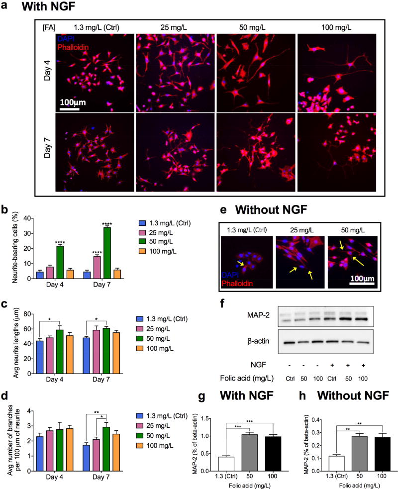
Figure 3.
In vitro effects of folic acid supplementation on differentiation of PC-12 cells. A) Representative immunohistochemical images of PC-12 cells treated with different concentrations of folic acid (mg/L) in the presence of NGF for four and seven days. B–D) Percentage of neurite-bearing cells (B), average neurite lengths (C), and average number of branches per 100 µm of a neurite (D) in PC-12 cells treated with different concentrations of folic acid (mg/L) in the presence of NGF for four and seven days. Data are shown as mean ± S.E.M., n=200. E) Representative immunohistochemical images of PC-12 cells stimulated solely with folic acid (mg/L) in the absence of NGF (arrows indicate extended neurites). F) Western blotting results showing protein expressions (MAP-2 and load control, β-actin) in PC-12 cells treated with various concentrations of folic acid (mg/L) in the absence or presence of 50 ng/mL NGF for 14 days. G, H) Quantitative comparison of MAP-2 expressions (F) in PC-12 cells treated with different concentrations of folic acid in the presence of NGF (G) and in the absence of NGF (H) for 14 days. (****P<0.0001, ***P<0.001, **P<0.01, *P<0.05).

