Probiotics represent a promising alternative strategy to control infection and disease caused by marine pathogens of aquaculturally important species. Generally, the beneficial effects of probiotics include improved water quality, control of pathogenic bacteria and their virulence, stimulation of the immune system, and improved animal growth. Previously, we isolated a probiotic bacterium, Phaeobacter inhibens S4Sm, which protects oyster larvae from Vibrio coralliilyticus RE22Sm infection. We also demonstrated that both antibiotic secretion and biofilm formation play important roles in S4Sm probiotic activity. Here, we report that P. inhibens S4Sm, an alphaproteobacterium and member of the Roseobacter clade, also secretes secondary metabolites that hijack the quorum sensing ability of V. coralliilyticus RE22Sm, suppressing virulence gene expression. This finding demonstrates that probiotic bacteria can exert their host protection by using a multipronged array of behaviors that limit the ability of pathogens to become established and cause infection.
KEYWORDS: N-acyl homoserine lactones, Phaeobacter inhibens, Vibrio coralliilyticus, marine pathogens, metalloprotease, oyster disease, probiotic, quorum sensing
ABSTRACT
Phaeobacter inhibens S4Sm acts as a probiotic bacterium against the oyster pathogen Vibrio coralliilyticus. Here, we report that P. inhibens S4Sm secretes three molecules that downregulate the transcription of major virulence factors, metalloprotease genes, in V. coralliilyticus cultures. The effects of the S4Sm culture supernatant on the transcription of three genes involved in protease activity, namely, vcpA, vcpB, and vcpR (encoding metalloproteases A and B and their transcriptional regulator, respectively), were examined by reverse transcriptase quantitative PCR (qRT-PCR). The expression of vcpB and vcpR were reduced to 36% and 6.6%, respectively, compared to that in an untreated control. We constructed a V. coralliilyticus green fluorescent protein (GFP) reporter strain to detect the activity of inhibitory compounds. Using a bioassay-guided approach, the molecules responsible for V. coralliilyticus protease inhibition activity were isolated from S4Sm supernatant and identified as three N-acyl homoserine lactones (AHLs). The three AHLs are N-(3-hydroxydecanoyl)-l-homoserine lactone, N-(dodecanoyl-2,5-diene)-l-homoserine lactone, and N-(3-hydroxytetradecanoyl-7-ene)-l-homoserine lactone, and their half maximal inhibitory concentrations (IC50s) against V. coralliilyticus protease activity were 0.26 μM, 3.7 μM, and 2.9 μM, respectively. Our qRT-PCR data demonstrated that exposures to the individual AHLs reduced the transcription of vcpR and vcpB. Combinations of the three AHLs (any two or all three AHLs) on V. coralliilyticus produced additive effects on protease inhibition activity. These AHL compounds may contribute to the host protective effects of S4Sm by disrupting the quorum sensing pathway that activates protease transcription of V. coralliilyticus.
IMPORTANCE Probiotics represent a promising alternative strategy to control infection and disease caused by marine pathogens of aquaculturally important species. Generally, the beneficial effects of probiotics include improved water quality, control of pathogenic bacteria and their virulence, stimulation of the immune system, and improved animal growth. Previously, we isolated a probiotic bacterium, Phaeobacter inhibens S4Sm, which protects oyster larvae from Vibrio coralliilyticus RE22Sm infection. We also demonstrated that both antibiotic secretion and biofilm formation play important roles in S4Sm probiotic activity. Here, we report that P. inhibens S4Sm, an alphaproteobacterium and member of the Roseobacter clade, also secretes secondary metabolites that hijack the quorum sensing ability of V. coralliilyticus RE22Sm, suppressing virulence gene expression. This finding demonstrates that probiotic bacteria can exert their host protection by using a multipronged array of behaviors that limit the ability of pathogens to become established and cause infection.
INTRODUCTION
Infections caused by pathogenic marine bacteria are a major problem for both the shellfish and finfish aquaculture industries, causing severe disease and high mortality, which seriously affect aquaculture production and cause significant economic loss (1, 2). Opportunistic pathogens from the Vibrionaceae family frequently cause disease in a variety of shellfish (3, 4). For example, Vibrio coralliilyticus RE22 (formerly Vibrio tubiashii RE22 [5, 6]), a reemerging pathogen of larval bivalve mollusks, causes invasive and toxigenic disease and has been responsible for high mortalities among oysters on the west coast of the United States (3). Vibriosis is characterized by a rapid and large reduction in larval motility, detached vela, and necrotic soft tissue, which result in high mortality within 1 day of infection (7).
While V. coralliilyticus virulence certainly involves several factors, the hemolysin and extracellular protease activities are thought to play important roles during pathogenesis in oysters (4, 8). V. coralliilyticus RE22 harbors two metalloproteases, VcpA and VcpB (formerly VtpA and VtpB) (4) and contains at least one hemolysin gene locus, vchBA (formerly vthBA) (8). The extracellular metalloproteases facilitate bacterial invasion and the infection process, acting to enhance tissue permeability and leading to necrotic tissue damage and cytotoxicity in the host (9). Mutations in the two metalloprotease genes resulted in significantly reduced protease activity of the V. coralliilyticus RE22 supernatant and toxicity to oyster larvae; thus, these two proteases are considered major virulence factors in V. coralliilyticus (4). Although the regulation of the virulence factors in V. coralliilyticus RE22 is not fully understood, Hasegawa and Hase (10) reported that VcpR is a member of the TetR family of transcriptional regulators and positively regulates the expression of several virulence genes, including vcpA, vcpB, and vchBA in V. coralliilyticus RE22. VcpR was also found to exhibit strong homology to several quorum sensing regulators, including orthologs LuxR (Vibrio harveyi, identity 84%) and HapR (Vibrio cholerae, identity 75%), suggesting that VcpR functions as a quorum sensing regulator in V. coralliilyticus RE22. However, the mechanism(s) of VcpR regulation of various virulence factors in V. coralliilyticus RE22 has not been fully investigated.
Phaeobacter inhibens is a member of the Alphaproteobacteria and of the Roseobacter clade (11–13). Previously, we isolated P. inhibens S4 from the inner shell surface of a healthy oyster and showed that S4Sm (a spontaneous streptomycin-resistant mutant) functions as a probiotic treatment for oyster larvae. S4Sm exhibited strong antipathogen activity and increased host survival against V. coralliilyticus RE22 and Alliroseovarius (Roseovarius) crassostreae challenge (14). We also demonstrated that the production of tropodithietic acid (TDA), a broad-spectrum antibiotic active against many marine pathogens, and biofilm formation play important roles in S4Sm probiotic activity (15). However, the complete mechanisms for the probiotic activities of P. inhibens S4Sm are not fully understood.
In addition to antibiotic production, probiotic bacteria are able to secrete various types of secondary metabolites, some of which antagonize pathogenic bacteria. Holmstrøm and Gram (16) reported that Pseudomonas fluorescens AH2 secretes siderophores into the culture supernatant, which efficiently chelate iron, resulting in the cessation of growth due to iron deprivation for the pathogen Vibrio anguillarum. Li et al. (17) reported that Lactobacillus reuteri produces cyclic dipeptides, which are able to quench agr-mediated expression of toxic shock syndrome toxin-1 in staphylococci. Bayoumi and Griffiths (18) discovered that bioactive molecules produced by Bifidobacterium bifidum possess inhibitory activity against regulatory and virulence genes in the major pathogenicity islands of Salmonella enterica serovar Typhimurium and Escherichia coli O157:H7.
In this report, the ability of P. inhibens S4Sm to produce secondary metabolites that repress the expression of extracellular protease activity, a major virulence factor, in V. coralliilyticus RE22Sm is described. The compounds were identified as N-acyl homoserine lactones (AHLs). The active AHLs function to suppress the transcription of vcpR, the quorum sensing regulator that positively regulates protease (VcpB) production. These results contribute to a better understanding of interspecies cell-to-cell communication between P. inhibens and V. coralliilyticus and provide an additional mechanism by which probiotic bacteria may attenuate virulence factor production by bacterial pathogens.
RESULTS
P. inhibens S4Sm supernatant inhibited protease activity of V. coralliilyticus RE22Sm but not growth.
We were curious whether P. inhibens S4Sm produced any compounds other than TDA that might affect virulence factors such as protease activity. When a V. coralliilyticus RE22Sm culture (∼1 × 10⁸ CFU/ml) was incubated in fresh yeast extract-peptone plus marine salts (mYP30) medium containing an equal volume of a sterile-filtered culture supernatant from stationary-phase P. inhibens S4Sm culture, the protease activity of V. coralliilyticus RE22Sm was suppressed (Fig. 1A). Specifically, at 1 h, no protease activity was detected in the V. coralliilyticus RE22Sm culture incubated with the P. inhibens S4Sm supernatant, while the V. coralliilyticus RE22Sm control culture had 149 ± 33 U of protease activity. At later times, protease activity from the S4Sm-treated sample increased but was still significantly lower than the control. In contrast, the growth rates of V. coralliilyticus RE22Sm in mYP30 (control) and in mYP30 supplemented with the P. inhibens S4Sm supernatant were nearly identical throughout the 3 h of incubation (Fig. 1B).
FIG 1.
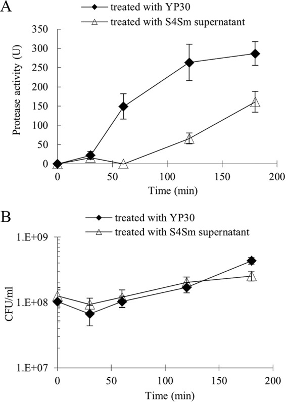
Effects of P. inhibens S4Sm culture supernatant on protease activity and growth of V. coralliilyticus RE22Sm. (A) Determination of RE22Sm protease activities after addition of S4Sm supernatant or fresh mYP30 medium (control) by measuring OD442 of azopeptide from azocasein degradation caused by protease activity. (B) Growth of RE22Sm cells treated with S4Sm supernatant or fresh mYP30 medium (control). Data are averages from two independent experiments and each independent experiment had three replicates. Error bars represent one standard deviation.
P. inhibens S4Sm supernatant inhibited transcription of metalloprotease gene vcpB and its transcriptional regulator, vcpR.
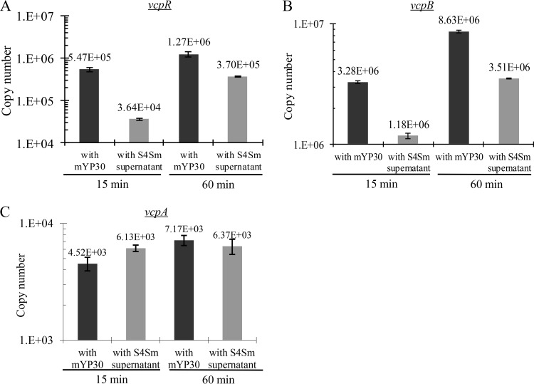
Since the S4Sm culture supernatant suppressed V. coralliilyticus RE22Sm protease activity, we examined whether there was a protease enzyme inhibitor in the S4Sm culture supernatant. Our data demonstrated that the mixture of S4Sm and V. coralliilyticus RE22Sm cell-free culture supernatants (1:1 mixture) had the same protease activity as the mixture of fresh mYP30 medium and the V. coralliilyticus RE22Sm supernatant (1:1 mixture) (see Fig. S1 in the supplemental material), indicating that there was no direct protease enzyme inhibitor in the S4Sm supernatant. Next, we used real-time reverse transcriptase quantitative PCR (qRT-PCR) to probe the inhibition of the transcription of the vcpR, vcpB, and vcpA genes. The transcription of vcpR and vcpB was reduced to 6.6% and 36%, respectively, after 15 min in the treated RE22Sm cultures compared to that in the control (Fig. 2A and B). After 60 min, the transcription of vcpR and vcpB was 29% and 41%, respectively, of the control. In contrast, there was no significant difference between the transcription levels of vcpA in treated and control cultures (Fig. 2C). These data suggested that one or more compounds present in the S4Sm culture supernatant inhibited V. coralliilyticus RE22Sm protease activity by suppressing transcription from the vcpR transcriptional regulator.
FIG 2.
Effects of P. inhibens S4Sm stationary-phase culture supernatant on transcription of vcpA, vcpB, and vcpR in V. coralliilyticus RE22Sm. Expression of vcpR (A), vcpB (B), and vcpA (C) determined by qRT-PCR analysis of RE22Sm during late-logarithmic-phase growth (∼108 CFU/ml) after the addition of either the P. inhibens S4Sm supernatant or sterile mYP30 culture medium. Data are representative of two independent experiments. Each value is the average from three replicates. Error bars represent one standard deviation.
Previously, Hasegawa and Hase (10) identified VcpR as a member of the TetR family of transcriptional regulators and found that it exhibited high homology to several quorum sensing regulators, including LuxR (V. harveyi, identity, 84%; E value, 1e−126) and HapR (V. cholerae, identity, 75%; E value, 2e−121), suggesting that VcpR functions as a quorum sensing regulator in V. coralliilyticus RE22 and that elements of the RE22 quorum sensing pathway might be involved in protease production in RE22. It should be noted that genes encoding homologs of the AHL quorum sensing (QS) circuit components, including luxM, luxN, luxO, and luxU, have been detected in the draft genome of in V. coralliilyticus RE22Sm (accession no. LGLS01000000 [6]). The luxM gene was identified as encoding the acyl-homoserine-lactone synthase, and luxN was identified as encoding the AHL receptor protein the in RE22Sm genome. Deletion mutations in luxM, luxN, or vcpR genes resulted in the loss of at least 50% of the protease activity in RE22Sm (see Fig. S2). This indicates that the quorum sensing pathway regulates protease production in RE22.
Isolation of active compounds antagonizing the expression of V. coralliilyticus protease.
We first confirmed that the pure TDA antibiotic (0.5 to 1.0 μg/ml) did not inhibit protease activity but did inhibit V. coralliilyticus RE22Sm growth at 1.0 μg/ml (see Fig. S3). This observation strongly suggested that other molecules, not TDA, secreted by P. inhibens S4Sm were responsible for the inhibition of protease transcription and activity. Furthermore, the induction of protease activity in V. coralliilyticus RE22Sm was delayed for 2 h when the cells were exposed to either the S4Sm supernatant or an ethyl acetate extract of the S4Sm supernatant (see Fig. S4A). The growth of RE22Sm in all treatment groups was unaffected (Fig. S4B). These results suggested that compounds that inhibit V. coralliilyticus RE22Sm protease expression were in the ethyl acetate extract and are likely nonpolar molecules. To guide the isolation of the active compounds, a reporter strain, V. coralliilyticus WZ112, was constructed (Table 1). WZ112 harbors a plasmid that contains the promoter region of vcpB fused to a promoterless gfp. WZ112 was cultivated in 96-well plates in the presence of test fractions. The wells containing compounds that inhibit VcpB protease induction had lower fluorescence signals. Further purification of ethyl acetate fractions by using successive reversed-phase medium-pressure chromatography, LH-20 size exclusion chromatography, and C18 high-pressure liquid chromatography (HPLC) resulted in the isolation of three active compounds, herein designated 1, 2 and 3.
TABLE 1.
Bacterial strains and plasmids used in this study
| Strain or plasmid | Description | Resistance | Reference or source |
|---|---|---|---|
| Strains | |||
| P. inhibens | |||
| S4 | Previously named Phaeobacter sp. strain S4; wild-type isolate from the inner shell of oysters | 14 | |
| S4Sm | Spontaneous Smr mutant of S4 | Smr | 15 |
| V. coralliilyticus | |||
| RE22 | Wild-type isolate strain | 5, 6 | |
| RE22Sm | Spontaneous Smr mutant of RE22 | Smr | 15 |
| WZ112 | Reporter strain, RE22Sm tagged by pSUP203-PvcpB-gfp | Smr Kmr Apr | This study |
| CS01 | luxN deletion mutant | Smr | This study |
| CS03 | vcpR insertional merodiploid mutant | Smr Kmr | This study |
| CS04 | luxM deletion mutant | Smr | This study |
| V. anguillarum | |||
| M93sm | Spontaneous Smr mutant of M93 (serotype J-O-1) | Smr | 46 |
| E. coli | |||
| Sm10 | thi thr leu tonA lacY supE recA RP4-2 Tc::Mu::Km (λpir) | Kmr | 47 |
| WS203 | Sm10(pSUP203) | Cmr Kmr Apr | This study |
| WS203-vcpB | Sm10(pSUP203-PvcpB-gfp) | Kmr Apr | This study |
| JB525 | MT102(pJBA132) | Tcr | 48 |
| JM109L | JM109(pSB1075) | Apr | 49 |
| Plasmids | |||
| pSUP203 | E. coli-V. anguillarum shuttle vector | Apr Kmr | This study |
| pSUP203-PvcpB-gfp | Derivative of pSUP203 | Apr Kmr | This study |
| pJBA132 | luxR-PluxI-RBSII-gfp (ASV) in pME6031 (detects C6- to C8-AHLs) | 48 | |
| pSB1075 | luxCDABE fused to lasRI′ in pUC18 (detects C10- to C12-AHLs) | 49 | |
| pDM5 | Suicide vector for construction of deletion mutant; sacBR oriR6K | Cmr Kmr | This study |
Structure determination of the inhibitors.
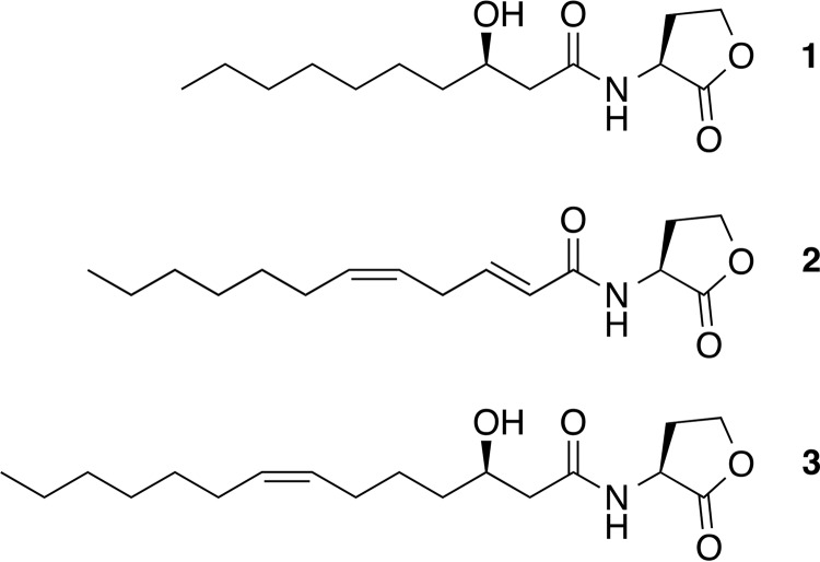
The structures of compounds 1, 2, and 3 were identified as N-acyl-homoserine lactones (AHLs) (Fig. 3) using a combination of analyses emphasizing nuclear magnetic resonance (NMR) spectroscopic and mass spectrometric data. Spectral data revealed compound 1 to be the previously reported Phaeobacter AHL (3R)-N-(3-hydroxydecanoyl)-l-homoserine lactone (see Fig. S5A and B) (19, 20). The molecular formula of compound 2 was determined as C16H25NO3 by high-resolution electrospray ionization-mass spectrometry (HRESI-MS; AB SCIEX TripleTOF 4600) at m/z 302.1729 [M+Na]+ (calculated for C16H25NO3Na, 302.1732). Compound 2 showed similar 1H and 13C NMR features as compound 1 (see Fig. S6A and B, respectively); the difference between them resided in the acyl chain. Four olefinic protons signal at δ 6.80 (1 H, dt, J = 15.6, 6.3 Hz, H-3), 5.95 (1 H, dt, J = 15.6, 1.7 Hz, H-2), 5.55 (1 H, m, H-6), and 5.41 (1 H, m, H-5) were observed in the 1H NMR spectrum, indicating the presence of two double bonds in the side chain. An analysis of two dimensional (2D) NMR (1H-1H correlation spectroscopy [COSY] and heteronuclear multiple bond correlation [HMBC]) (Fig. S6C to E) data enabled the assignment of the positions of the double bonds. The 2E,5Z configurations were determined by the coupling constant (J2,3 = 15.6 Hz) and the nuclear Overhauser effect spectroscopy (NOESY) correlation between H-5 and H-6. Thus, compound 2 was elucidated as the new AHL (2E,5Z)-N-(dodecanoyl-2,5-diene)-l-homoserine lactone. Compound 3 was identified as (3R,7Z)-N-(3-hydroxytetradecanoyl-7-ene)-l-homoserine lactone by comparing the NMR and MS data with those previously reported for this molecule (Fig. S5C and D) (21).
FIG 3.
Chemical structures of N-acyl homoserine lactones produced by P. inhibens S4Sm. The compounds are 1, (3R)-N-(3-hydroxydecanoyl)-l-homoserine lactone; 2, (2E,5Z)-N-(dodecanoyl-2,5-diene)-l-homoserine lactone; and 3, (3R,7Z)-N-(3-hydroxytetradecanoyl-7-ene)-l-homoserine lactone.
Exposure to the individual AHL reduced transcription of vcpR and vcpB.
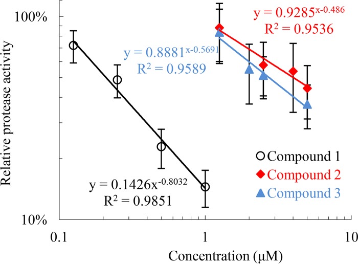
All three AHLs showed concentration-dependent inhibition of protease activity on late exponential-phase V. coralliilyticus RE22Sm (Fig. 4). The half maximal inhibitory concentrations (IC50s) of 1, 2, and 3 were determined to be 0.26 μM, 3.7 μM, and 2.9 μM, respectively. None of the molecules demonstrated growth effects against V. coralliilyticus RE22Sm at the highest concentration tested (5 μM) (see Fig. S7).
FIG 4.
Concentration-response analyses of the three AHLs. Series of different concentrations of each AHL were used to treat V. coralliilyticus RE22Sm and the protease activities were measured. Concentration-response curves (trend lines) of each AHL were obtained. Data are the averages from two independent experiments and each independent experiment had three replicates. Error bars represent one standard deviation.
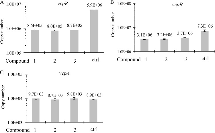
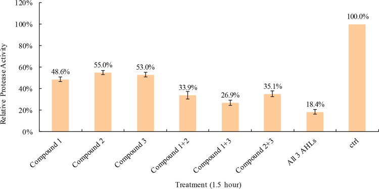
We then asked if the AHLs also altered the transcription of genes involved in V. coralliilyticus RE22Sm protease production. The data presented in Fig. 5 demonstrate that when compounds 1, 2, and 3 were added to cultures at their calculated IC50 concentrations, the transcription levels of vcpR and vcpB were reduced to ∼14% to 15% and ∼42% to 51%, respectively, of the untreated control. In contrast, the transcription of vcpA was unaffected by AHL exposure (98% to 110% of the untreated control). Furthermore, while treatment with any single AHL compound at its IC50 suppressed V. coralliilyticus RE22Sm protease activity by ∼50% of the untreated control (Fig. 6), the effects of combinations of these AHLs were additive. The treatment of V. coralliilyticus RE22Sm cells with a combination of any two AHLs (each at IC50) decreased protease activities further: 32% for compounds 1 plus 2, 27% for compounds 1 plus 3, and 34% for compounds 2 plus 3. The treatment of V. coralliilyticus RE22Sm cells with all three AHLs (each at IC50) resulted in the greatest inhibition of protease activity, with the treated cells having only 18% of the control cell activity (Fig. 6). These data suggest that the three AHLs produced by P. inhibens S4Sm inhibit the transcription of the transcriptional regulator, VcpR, to downregulate transcription of the metalloprotease VcpB.
FIG 5.
Effects of single AHL treatment on transcription of vcpA, vcpB, and vcpR in V. coralliilyticus RE22Sm. Expression of vcpR (A), vcpB (B), and vcpA (C) determined by qRT-PCR analysis of V. coralliilyticus RE22Sm treated with individual AHLs (at their own IC50) for 15 min during late-logarithmic-phase growth (∼108 CFU/ml). Data are representative of two independent experiments. Each value is the average from three replicates. Error bars represent one standard deviation.
FIG 6.
Effects of different combinations of three AHLs on protease activity of V. coralliilyticus RE22Sm. Determination of protease activities of V. coralliilyticus RE22Sm strain treated with a single AHL (at the IC50), various combinations of AHLs (each at the calculated IC50), or an appropriate amount of methanol (ctrl) by measuring OD442 of azopeptide from azocasein degradation caused by protease activity. Data are representative of two independent experiments. Each value is the average from three replicates. Error bars represent one standard deviation.
Inhibitory effects of P. inhibens S4Sm AHLs against V. coralliilyticus RE22Sm protease production were specific.
Previously, Ke et al. (22) showed that in Vibrio harveyi long-chain AHLs acted as antagonists of N-[(R)-3-hydroxybutanoyl]-l-homoserine lactone (3OH-C4-HSL) binding to LuxN, causing a suppression of bioluminescence. We tested whether other AHL molecules, which share the same acyl-chain lengths as the P. inhibens S4Sm AHLs but with different modifications, would show similar inhibitory activity against V. coralliilyticus protease transcription. V. coralliilyticus RE22Sm cells were treated with the synthetic AHLs N-decanoyl-dl-homoserine lactone, N-dodecanoyl-dl-homoserine lactone, or N-tetradecanoyl-dl-homoserine lactone at concentrations of 0.26 μM, 3.7 μM, or 2.9 μM (the IC50s of compounds 1, 2, and 3), respectively, to determine if the inhibition of the transcription of vcpR and vcpB was specific to AHLs produced by P. inhibens S4Sm. The data presented in Fig. S8 show that the transcription levels of vcpB and vcpR were reduced to 60% to 100% and 44% to 88%, respectively, of the untreated control and were not statistically significant. Additionally, these AHLs did not affect the growth of V. coralliilyticus RE22Sm (Fig. S8C).
Our data suggest that AHLs from P. inhibens S4Sm act as antagonists to quorum sensing in V. coralliilyticus. We were interested to determine whether the P. inhibens S4Sm AHLs act to inhibit the transcription of vanT (a vcpR homolog; the two proteins, VanT and VcpR, are 83% identical, 90% similar, E value = 3e−130) in V. anguillarum M93Sm. Using two AHL indicator strains, E. coli JB525 (detects C6- to C8-HSLs) and E. coli JM109L (detects C10- to C12-HSLs) (23), we found that V. anguillarum M93Sm secreted both C6- to C8-HSLs and C10- to C12-HSLs (corresponding to 3-OH-C6-HSL and 3-oxo-C10-HSL reported by Croxatto et al. [24] in V. anguillarum NB10), while V. coralliilyticus RE22Sm and P. inhibens S4Sm secrete C10- to C12-HSLs but not C6- to C8-HSLs. When the effect of the P. inhibens S4Sm culture supernatant on V. anguillarum M93Sm vanT expression was examined, we found that the expression of vanT was not affected (see Fig. S9). These data further support the idea that the P. inhibens S4Sm AHL antagonism to quorum sensing is highly specific.
DISCUSSION
In this study, we present data that P. inhibens S4Sm produces AHLs, which block virulence factor production and may contribute to P. inhibens probiotic activity against the marine pathogen V. coralliilyticus. Specifically, we found that the stationary-phase culture supernatant of P. inhibens S4Sm repressed protease activity, but not growth, of V. coralliilyticus RE22. Our data suggest, but do not conclusively demonstrate, that the repression of protease activity resulted from a repression of the transcriptional activator vcpR and the subsequent repression of the protease gene vcpB. A vcpB-gfp reporter strain of V. coralliilyticus was constructed to help identify three AHLs as the repressors of vcpR and vcpB. Since the active compounds are AHLs, this suggests that they function as quorum sensing (QS) compounds for P. inhibens S4Sm and as quorum quenching (QQ) compounds against V. coralliilyticus RE22.
Quorum sensing has been demonstrated to regulate diverse physiological activities in Gram-positive and Gram-negative bacteria, including virulence, symbiosis, competence, conjugation, antibiotic production, motility, and biofilm formation (25). Gram-negative bacteria generally use AHLs as autoinducers for QS. Quorum quenching is a disruption of QS pathways by any of several mechanisms, including antagonist binding to the native AHL receptor, degradation of native AHL signals, suppression of AHL synthetase, and receptor activities (25). Thus, QQ can interfere with QS-regulated bacterial physiological functions (23, 26). For example, synthetic antagonists are able to block pathogenic bacterial quorum sensing in Pseudomonas aeruginosa and Agrobacterium tumefaciens (27). Additionally, McClean et al. (28) found that in Chromobacterium violaceum, exogenously added AHLs (C10- to C14-HSLs) inhibited the production of violacein in the presence of inducing concentrations of the native QS AHL (C6-HSL) and may represent QQ by competitive inhibition. Hemolytic activity (downregulation of streptolysin S) and viability of Streptococcus pyogenes were inhibited by oxo-C12-HSL and oxo-C14-HSL and by AHLs secreted by Pseudomonas aeruginosa (29). This inhibition appears to be an example of interspecies signaling or eavesdropping. In contrast, some bacteria, such as Bacillus and Streptomyces strains, are able to produce AHL-degrading enzymes, such as lactonase (30) and acylase (31). Other bacteria have been shown to secrete non-AHL secondary metabolites that interfere with QS-controlled processes (32). The interference with QS-regulated virulence has been proposed as a new direction for the development of novel antimicrobial chemotherapies (32–34).
The protease inhibition activity of the P. inhibens S4Sm supernatant was transient, as it gradually diminished during the experiment (Fig. 1A; see also Fig. S3A in the supplemental material). One possibility for this observation is the competitive inhibition of V. coralliilyticus RE22-AHL-mediated QS regulation of vcpR gene expression by P. inhibens S4Sm AHLs. Specifically, as V. coralliilyticus RE22Sm cells grow, increasing concentrations of native AHLs are produced, diluting the exogenous P. inhibens S4Sm-AHLs and resulting in decreased antagonizing effects from P. inhibens S4Sm-AHL molecules toward V. coralliilyticus RE22Sm protease activity. Additionally, the inhibitory effects of the three P. inhibens S4Sm AHLs on V. coralliilyticus RE22Sm protease activity were additive. This suggests that each of the three S4Sm AHLs is able to competitively bind the V. coralliilyticus RE22Sm LuxN to interfere with its function as a LuxU-PO4 phosphatase and cause QQ effects. It should be noted that a search of the V. coralliilyticus RE22Sm genome (accession no. LGLS01000000) revealed only one luxN gene. Additional studies comparing the AHLs of P. inhibens S4Sm and V. coralliilyticus RE22, as well as AHL binding to LuxN, need to be carried out in order to better define this QQ mechanism.
Previously, Berger et al. (19) showed that P. inhibens produces N-3-hydroxydecanoylhomoserine lactone, which is involved in the QS induction of the TDA biosynthesis genes in P. inhibens. Similarly, N-(3-hydroxytetradecanoyl-7-ene)-L-homoserine lactone is produced by Rhizobium leguminosarum, where it is regulates several QS-mediated pathways, including growth inhibition, adaptation to stationary phase, and shorter-chain AHL production (35). To our knowledge, the production of N-(dodecanoyl-2,5-diene)-l-homoserine lactone has not been previously reported and is a newly discovered AHL. No previous studies have described any biological functions of either N-(dodecanoyl-2,5-diene)-l-homoserine lactone or N-(3-hydroxytetradecanoyl-7-ene)-l-homoserine lactone in P. inhibens. The synthetic AHLs did not have an inhibitory effect on V. coralliilyticus RE22Sm vcpR and vcpB transcription, suggesting that the inhibition from P. inhibens S4Sm AHLs on V. coralliilyticus RE22Sm protease production is specific.
Generally, AHL molecules contain common homoserine lactone moieties and a specific acyl side chains. The side chains vary among different species, and the specificity is determined by such factors as length and sites of oxidation (36). It was previously demonstrated that LuxN of V. harveyi, which detects N-[(R)-3-hydroxybutanoyl]-l-homoserine lactone (3OH-C4-HSL), can be antagonized by AHLs that lack the hydroxyl group on C-3 if the AHLs have a long acyl chain (N-hexanoyl-l-homoserine lactone [C6-HSL], N-octanoyl-l-homoserine lactone [C8-HSL], N-decanoyl-l-homoserine lactone [C10-HSL], and N-dodecanoyl-l-homoserine lactone [C12-HSL]) (22). In contrast, our data demonstrate that the expression levels of vcpR and vcpB were much lower in V. coralliilyticus RE22Sm cells exposed to P. inhibens S4Sm AHLs than in V. coralliilyticus RE22Sm cells exposed to saturated long-chained AHLs. These observations suggest that the three AHLs produced by P. inhibens S4Sm are specific inhibitors of the QS system of the oyster pathogen V. coralliilyticus RE22, which regulates the transcription of the master regulator gene vcpR and, in turn, the transcription of the metalloprotease gene vcpB.
Previously, we demonstrated that P. inhibens S4Sm is an effective probiotic organism because of its ability to form a biofilm and secrete an antibiotic (TDA) (14, 15). In this study, we showed that S4Sm produces antivirulence agents, AHLs, that appear to cause QQ in a specific pathogen, V. coralliilyticus RE22, which shares the same environmental niche. This finding further demonstrates that probiotic bacteria can exert their host protection by using a multipronged array of behaviors that limit the ability of pathogens to become established and cause harm.
MATERIALS AND METHODS
Bacterial strains and growth conditions.
All bacterial strains and plasmids used in this study are listed in Table 1. P. inhibens S4 was isolated from the inner surface of an oyster shell (14). P. inhibens strains and V. coralliilyticus strains were routinely grown in yeast extract (0.5%)-peptone (0.1%) broth plus 3% marine salts (mYP30) (14), supplemented with the appropriate antibiotic, in a shaking water bath at 27°C. Cell densities were estimated by the optical density at 600 nm (OD600) and more accurately determined by serial dilution and spot plating. The specific conditions for each experiment are described in the text. Escherichia coli strains were routinely grown in Luria-Bertani broth plus 1% NaCl (LB10) (37). Antibiotics were used at the following concentrations: streptomycin 200 μg/ml (Sm200) and chloramphenicol 5 μg/ml (Cm5) for P. inhibens, Sm200, ampicillin 100 μg/ml (Ap100), Cm5, and kanamycin 50 μg/ml (Km50) for Vibrio strains, and Ap100, Cm20, Km50, and tetracycline 15 μg/ml (Tc15) for E. coli. The genes were renamed to reflect the reclassification of V. tubiashii RE22 to V. coralliilyticus RE22 (e.g., vtpA to vcpA, vtpB to vcpB, and vtpR to vcpR) (38).
P. inhibens supernatant challenge assay.
The P. inhibens supernatant challenge assay was a modification of the method described previously by Holmstrøm and Gram (16). P. inhibens S4Sm supernatant challenge experiments were initially conducted by using exponentially growing V. coralliilyticus cultures (OD600 of 0.6 to 0.8) and transferring 1 ml of a culture to 4 ml of mYP30 medium containing 2.5 ml of S4Sm supernatant and 1.5 ml of fresh mYP30. For the control, 1 ml of an exponentially growing V. coralliilyticus culture was added to 4 ml of fresh mYP30. Cell densities (CFU/ml) were determined at different times after challenge. Also, 1 ml V. coralliilyticus RE22Sm supernatant and mRNA were collected at each time point and stored at −20°C and −74°C, respectively, for future use. All experiments were repeated at least twice.
Detection and quantification of protease activity.
The protease activity of the culture supernatants was quantified using the azocasein method of Windle and Kelleher (39) as modified by Denkin and Nelson (40). V. coralliilyticus filtered supernatant (100 μl) was incubated for 30 min at 27°C with 100 μl of azocasein solution. The reactions were terminated by the addition of trichloroacetic acid (TCA; 10% [wt/vol]) to a final concentration of 6.7% (wt/vol). The mixture was allowed to stand for 2 min and centrifuged (12,000 × g for 8 min) to remove unreacted azocasein, and the supernatant containing azopeptides was suspended in 700 μl of 525 mM NaOH. The absorbance of the azopeptide supernatant was measured at 442 nm. Protease activity units (U) were calculated with the following equation: U = [1,000 (OD442)/CFU] × 109, where OD442 is the optical density at 442 nm.
mRNA extraction.
mRNA extraction was performed using the protocol previously described by Li et al. (41). V. coralliilyticus cells under different treatments were treated with RNA Protect bacterial reagent (Qiagen, Valencia, CA) according to the manufacturer’s instructions. Total RNA was isolated using an RNeasy kit and QIAcube (Qiagen) according to the manufacturer’s instructions and stored at −74°C for future use.
Real-time qRT-PCR.
Real-time reverse transcriptase quantitative PCR (qRT-PCR) was performed using the protocol previously described by Li et al. (41). qRT-PCR was used to quantify various mRNAs by use of a Roche480 Multiplex quantitative PCR system and Brilliant II SYBR green single-step qRT-PCR master mix (Agilent Technologies, Wilmington, DE) with 10 ng of total RNA in 20-μl reaction mixtures. The thermal profile was 50°C for 30 min, 95°C for 15 min, and then 40 cycles of 95°C for 30 s and 60°C for 30 s. The qRT-PCR primers are listed in Table 2. Fluorescence was measured at the end of the 60°C step during every cycle. The PCR products for each gene were obtained and purified according to a PCR cleanup kit protocol (Monarch PCR & DNA cleanup kit, T1030S; NEB). A standard curve using the purified PCR products was generated for each gene as described by the qRT-PCR protocol from the Genomics and Sequencing Center of the University of Rhode Island (42). A suspension of ∼1 × 10⁸ copies of a gene template was obtained and then diluted in a 10-fold dilution series (in nuclease-free water containing 100 ng/μl yeast tRNA; Sigma-Aldrich, St. Louis, MO) to obtain ∼1 × 101 copies of the template. The standard curve generated for each gene was used to ascertain primer efficiency and calculate absolute copy numbers (42). Samples were run in triplicates along with no-RT and no-template controls. All experiments were repeated at least twice.
TABLE 2.
Primers used in this study
| Primer | Sequence (5′→3′)a | Description |
|---|---|---|
| pw200 | GCGGTAAAGCTTGAACACGTAGAAAGCCAGTCC | Amplification of kan ORF, forward, with HindIII site |
| pw201 | CTATATGGATCCCGTTTCTGGGTGAGCAAAAAC | Amplification of kan ORF, reverse, with BamHI site |
| pw204 | GATGTTGGTACCTGTGGATCTCTGCCATTGGCT | Amplification of vcpB promoter region, forward, with KpnI site |
| pw205 | CTGCATGGTACCTATGATTCTCCTTATCGTGGC | Amplification of vcpB promotor region, reverse, with KpnI site |
| pw178b | GACATCTTTAAATCTGAAGGTGGTCTAC | qRT-PCR amplicon of partial vcpA, forward |
| pw179b | GTAGTATTGAGAAGCATGGTCGATAGAG | qRT-PCR amplicon of partial vcpA, reverse |
| pw184b | GGTTTCTGTTGCTACAGTTTTTAACTACTTC | qRT-PCR amplicon of partial vcpR, forward |
| pw185b | ATATTATCTGATAGGAAGTTTGAGAACTGAC | qRT-PCR amplicon of partial vcpR, reverse |
| pw190b | CTAAAAGCATCGTTTGATTCTGGTAGCACTAA | qRT-PCR amplicon of partial vcpB, forward |
| pw191b | GTAAGAAACAATGTCACCATCGCTATCACTAC | qRT-PCR amplicon of partial vcpB, reverse |
| luxN 5′fp | tgtggaatcccgggagagctAACAGTTATTTTCATACAGATCTTC | Amplification of luxN 5′ fragment, forward |
| luxN 5′rp | ctcttattgtAAATCATGTAAATTACACGAGTTC | Amplification of luxN 5′ fragment, reverse |
| luxN 3′fp | tacatgatttACAATAAGAGTGGCGGCAG | Amplification of luxN 3′ fragment, forward |
| luxN 3′rp | gcatgcgggtaacctgagctTTCTCCCGATTCACCTGATAG | Amplification of luxN 3′ fragment, reverse |
| vcpR 5′fp | tgtggaatcccgggagagctTATACAACTCAATTGGCAAGG | Amplification of vcpR 5′ fragment, forward |
| vcpR 5′rp | gattgctttgATGCGATTTCCATCAGTTG | Amplification of vcpR 5′ fragment, reverse |
| vcpR 3′fp | gaaatcgcatCAAAGCAATCGAGCGTGG | Amplification of vcpR 3′ fragment, forward |
| vcpR 3′rp | gcatgcgggtaacctgagctTCTAGGTAGCTTTGTGTCAG | Amplification of vcpR 3′ fragment, reverse |
| luxM fp | GCCGAGCTCCACAACATGCAGAAATACTC | Amplification of luxM ORF, forward |
| luxM rp | GCCTCTAGAAACTCAATTTATGGCGTTCT | Amplification of luxM ORF, reverse |
| pr25cs | CCTCCTGTTCAGCTACTGACGGGGTG | For sequencing of the DNA fragment inserted in pDM4 multicloning site |
Primer design according to the Gibson assembly protocol (44). Lowercase nucleotides for 5′fp and 3′rp primers are homologous to the pDM4 vector backbone and to each other for proper Gibson assembly. Underlined sequences are engineered restriction sites.
Annealing temperature is 60°C.
Challenge of V. coralliilyticus cells with ethyl acetate extract of P. inhibens S4Sm supernatant.
The P. inhibens cell-free supernatant was extracted twice with ethyl acetate (1:1 volume ratio), and the combined organic layers were concentrated using a rotary evaporator and stored at −20°C until use. The extract was redissolved in fresh mYP30 medium and added to V. coralliilyticus RE22Sm cells (∼108 CFU/ml). The CFU/ml of RE22Sm cells was determined at 1-, 2-, and 3-h time points. The V. coralliilyticus RE22Sm supernatant was collected at each time point and stored at −20°C.
Construction of reporter strain V. coralliilyticus WZ112 and gfp reporter assay.
V. coralliilyticus RE22Sm was tagged by pSUP203-PvcpB-gfp. The kanamycin resistance gene, cloned from pCR2.1 TOPO, replaced the tetracycline region in pSUP202-gfp. The resulting plasmid was designated pSUP203-gfp. The promoter region of vcpB was cloned and fused with the gfp region in pSUP203-gfp. The primers for the amplification of clones are described in Table 2. E. coli Sm10 harboring plasmid pSUP203-PvcpB-gfp was conjugated with V. coralliilyticus RE22Sm by using procedures described previously (43). The transconjugants were confirmed by antibiotic selection and fluorescence microscopy and designated strain WZ112. Aliquots (50 μl) of test compounds from the P. inhibens S4Sm supernatant (see below) were transferred into a 96-well microtiter plate, dried in vacuo, and then resuspended in 200 μl of V. coralliilyticus WZ112 culture (OD600 of 0.3). After 1.5 h of incubation at 27°C, OD600 and fluorescence signals were measured (Mx3005 multiplex quantitative PCR system, plate read function). The relative fluorescence (RF) was calculated as fluorescence/OD600.
Identification of compounds that repress transcription of vcpR and vcpB.
P. inhibens S4Sm (50 × 1 liter) was cultured in mYP30 medium. The cultures were grown to stationary phase (shaking at 175 rpm for 96 h at 28°C). The cells were pelleted by centrifugation (10,000 × g for 10 min), and the resulting supernatants were acidified to pH 3 with formic acid. The acidified supernatants were twice extracted with equal volumes of acidified ethyl acetate (0.1% formic acid), and the organic phases were concentrated in vacuo at 27°C. The resulting extract of P. inhibens S4Sm supernatant was separated by Sephadex LH-20 column chromatography (3 cm by 80 cm) using chloroform-methanol (1:1 [vol/vol]) to provide 60 fractions. Fractions (13 to 27) active in the WZ112 gfp reporter assay were pooled (fraction A, 123 mg) and further separated by medium-pressure liquid chromatography (prepacked C18 column, 2 cm by 30 cm; Star Instruments, Manassas, VA, USA), eluting with a linear gradient of 10% to 100% methanol in water at 3 ml/min. The active fractions (21 to 29 of 29) were combined (fraction B, 48.5 mg) and further purified by HPLC (Hitachi Elite LaChrom system consisting of an L2130 pump, L-2200 autosampler, L-2455 diode array detector, and a Phenomenex Luna C18 column [250 mm by 10 mm, S-5 μm], operated by EZChrom Elite software) and eluting with methanol-water (40% to 100% methanol in H2O over 25 min, 3 ml/min) to provide compounds 1 (2.0 mg), 2 (600 μg), and 3 (500 μg).
Concentration-response analyses of three AHL compounds.
Exponentially growing V. coralliilyticus cultures (OD600 of 0.6 to 0.8) were treated with serial dilutions of the purified AHL molecules (0.125 to 5.0 μM) from P. inhibens. One volume of V. coralliilyticus culture was added to 4 volumes of fresh mYP30 medium containing a specific concentration of AHL (dissolved in methanol, 0.4% final concentration). Medium containing only 0.4% methanol (final concentration) was used as a control. V. coralliilyticus RE22Sm supernatants were collected at 1.5 h and used to examine the protease activity. A series of concentration-response (protease activity) data were obtained and plotted. According to the concentration-response curve, the half maximal inhibitory concentration (IC50) was calculated for each purified AHL.
Treatment of V. coralliilyticus cells with purified AHLs.
V. coralliilyticus cultures (OD600 of 0.6 to 0.8) were treated with compounds 1 to 3 at their corresponding IC50s, both individually and in combination with the other AHL molecules, as described above in “Concentration-response analyses of three AHL compounds.” Cell densities (CFU/ml) were determined at 15 min and 90 min after the addition of the AHL(s), and V. coralliilyticus RE22Sm culture supernatants and mRNA were collected at each time point and stored at −20°C and −74°C, respectively.
Treatment of V. coralliilyticus cells with synthetic AHLs.
V. coralliilyticus cultures (OD600 of 0.6) were individually treated with N-decanoyl-dl-homoserine lactone, N-dodecanoyl-dl-homoserine lactone, or N-tetradecanoyl-dl-homoserine lactone (Sigma-Aldrich, St. Louis, MO) at a concentration of 0.26 μM, 3.7 μM, or 2.9 μM, respectively, as described above in “Treatment of V. coralliilyticus cells with purified AHLs.” Cell densities (CFU/ml) were determined at 0 min and 90 min after the addition of the AHL(s). V. coralliilyticus RE22Sm culture supernatants and mRNA were collected after the 90-min challenge and stored at −20°C and −74°C, respectively.
Construction of V. coralliilyticus RE22Sm merodiploid and allelic exchange mutants.
The modified pDM4 plasmid containing a kanamycin (Km) resistance gene, pDM5, was used to construct the allelic exchange mutants (Table 1) as described by Gibson et al. (44). The quorum sensing genes luxN, vcpR, and luxM were amplified by PCR from V. coralliilyticus RE22Sm genomic DNA, PCR purified, and inserted into pDM5 via the Gibson assembly reaction. The plasmid, pDM5, was linearized at the SacI restriction enzyme site within the multicloning region (MCR) for all mutation-destined Gibson assemblies. The Gibson assembly ligation mixture was introduced into E. coli Sm10 by electroporation with the Bio-Rad Gene Pulser II. Transformants were selected for on LB20 Cm20 agar plates, and confirmed by PCR. The mobilizable suicide vector, pDM5, was transferred from E. coli Sm10 into V. coralliilyticus RE22Sm by conjugation (45). Transconjugates were selected by utilizing the kanamycin resistance gene located on the suicide plasmid. The incorporation of the target gene fragments into the suicide vector was confirmed by PCR analysis using the appropriate 5′ forward primer and a check primer, pr25cs. The subsequent recombination resulted in insertional, single-crossover deletion merodiploid mutants. Next, double-crossover resolution of the merodiploids through sucrose counterselection yielded stable allelic exchange mutants. The double-crossover transconjugants were selected for by growth on mYP10 Sm200 plus 10% sucrose agar plates for a second crossover event. Sucrose was used as the counterselective agent, because pDM5 contains the sacBR gene cassette, which encodes levansucrase, ultimately converting sucrose to toxic levan (45). Putative allelic exchange mutants were screened for kanamycin sensitivity. Kanamycin-sensitive recombinants were then screened by PCR using the appropriate Gibson assembly primers and gel electrophoresis for the deletion mutation.
Statistical analysis.
Two-tailed Student’s t tests assuming unequal variances were used for statistical analyses for all experiments. P values of <0.05 were considered statistically significant.
Supplementary Material
ACKNOWLEDGMENTS
We thank Ralph Elston for providing Vibrio coralliilyticus RE22. We thank the D.N. group for helpful discussion and critical comments. We also thank Xiangyu Mou for providing training on multiple techniques used in this study, as well as for providing his insight and expertise that greatly assisted the research.
This work was supported by grants from the USDA (2016-67016-24905) and a Rhode Island Sea Grant (N00AR170076) to D.C.R. and D.R.N. The instruments used for chemical analyses were supported by an Institutional Development Award (IDeA) from the National Institute of General Medical Sciences of the National Institutes of Health (grant number 2 P20 GM103430). NMR data were acquired at a research facility supported in part by the National Science Foundation EPSCoR Cooperative Agreement (EPS-1004057). The funders had no role in study design, data collection and interpretation, or the decision to submit the work for publication.
W.Z. and D.R.N. designed the study. W.Z. created the strains used in this study. W.Z. performed all experiments under the supervision of D.R.N. with the following exceptions: C.P. and W.Z. performed extraction of P. inhibens S4Sm supernatant, C.P. and T.Y. performed the fractionation of P. inhibens S4Sm supernatant and structuring of active compounds under the supervision of D.C.R., and E.J.S. performed the experiments with the synthetic AHLs. W.Z. and D.R.N. wrote the paper with contributions and editing by C.P., T.Y., E.J.S., C.W.S., and D.C.R. The formatting of the paper was performed by W.Z. and D.R.N. All authors read and approved the final version of the manuscript.
Footnotes
Supplemental material for this article may be found at https://doi.org/10.1128/AEM.01545-18.
REFERENCES
- 1.Jackson JB, Kirby MX, Berger WH, Bjorndal KA, Botsford LW, Bourque BJ, Bradbury RH, Cooke R, Erlandson J, Estes JA, Hughes TP, Kidwell S, Lange CB, Lenihan HS, Pandolfi JM, Peterson CH, Steneck RS, Tegner MJ, Warner RR. 2001. Historical overfishing and the recent collapse of coastal ecosystems. Science 293:629–637. doi: 10.1126/science.1059199. [DOI] [PubMed] [Google Scholar]
- 2.Lenihan HS, Peterson CH. 1998. How habitat degradation through fishery disturbance enhances impacts of hypoxia on oyster reefs. Ecol Appl 8:128–140. doi: 10.1890/1051-0761(1998)008[0128:HHDTFD]2.0.CO;2. [DOI] [Google Scholar]
- 3.Elston RA, Hasegawa H, Humphrey KL, Polyak IK, Hase CC. 2008. Re-emergence of Vibrio tubiashii in bivalve shellfish aquaculture: severity, environmental drivers, geographic extent and management. Dis Aquat Organ 82:119–134. doi: 10.3354/dao01982. [DOI] [PubMed] [Google Scholar]
- 4.Hasegawa H, Lind EJ, Boin MA, Hase CC. 2008. The extracellular metalloprotease of Vibrio tubiashii is a major virulence factor for pacific oyster (Crassostrea gigas) larvae. Appl Environ Microbiol 74:4101–4110. doi: 10.1128/AEM.00061-08. [DOI] [PMC free article] [PubMed] [Google Scholar]
- 5.Wilson B, Muirhead A, Bazanella M, Huete-Stauffer C, Vezzulli L, Bourne DG. 2013. An improved detection and quantification method for the coral pathogen Vibrio coralliilyticus. PLoS One 8:e81800. doi: 10.1371/journal.pone.0081800. [DOI] [PMC free article] [PubMed] [Google Scholar]
- 6.Spinard E, Kessner L, Gomez-Chiarri M, Rowley DC, Nelson DR. 2015. Draft genome sequence of the marine pathogen Vibrio coralliilyticus RE22. Genome Announc 3:e01432-15. doi: 10.1128/genomeA.01432-15. [DOI] [PMC free article] [PubMed] [Google Scholar]
- 7.Sindermann CJ, Lightner DV. 1988. Vibriosis of larval oysters, p 271–274. In Disease diagnosis and control in North American aquaculture, vol 17 Elsevier, Amsterdam, Netherlands. [Google Scholar]
- 8.Hasegawa H, Hase CC. 2009. The extracellular metalloprotease of Vibrio tubiashii directly inhibits its extracellular haemolysin. Microbiology 155:2296–2305. doi: 10.1099/mic.0.028605-0. [DOI] [PubMed] [Google Scholar]
- 9.Miyoshi S, Shinoda S. 2000. Microbial metalloproteases and pathogenesis. Microbes Infect 2:91–98. doi: 10.1016/S1286-4579(00)00280-X. [DOI] [PubMed] [Google Scholar]
- 10.Hasegawa H, Hase CC. 2009. TetR-type transcriptional regulator VtpR functions as a global regulator in Vibrio tubiashii. Appl Environ Microbiol 75:7602–7609. doi: 10.1128/AEM.01016-09. [DOI] [PMC free article] [PubMed] [Google Scholar]
- 11.Bruhn JB, Nielsen KF, Hjelm M, Hansen M, Bresciani J, Schulz S, Gram L. 2005. Ecology, inhibitory activity, and morphogenesis of a marine antagonistic bacterium belonging to the Roseobacter clade. Appl Environ Microbiol 71:7263–7270. doi: 10.1128/AEM.71.11.7263-7270.2005. [DOI] [PMC free article] [PubMed] [Google Scholar]
- 12.Geng H, Bruhn JB, Nielsen KF, Gram L, Belas R. 2008. Genetic dissection of tropodithietic acid biosynthesis by marine roseobacters. Appl Environ Microbiol 74:1535–1545. doi: 10.1128/AEM.02339-07. [DOI] [PMC free article] [PubMed] [Google Scholar]
- 13.Brinkhoff T, Giebel HA, Simon M. 2008. Diversity, ecology, and genomics of the Roseobacter clade: a short overview. Arch Microbiol 189:531–539. doi: 10.1007/s00203-008-0353-y. [DOI] [PubMed] [Google Scholar]
- 14.Karim M, Zhao W, Nelson DR, Rowley D, Marta G-C. 2013. Probiotic strains for shellfish aquaculture: protection of Eastern oyster, Crassostreac virginica, larvae and juveniles against bacterial challenge. J Shellfish Res 32:401–108. doi: 10.2983/035.032.0220. [DOI] [Google Scholar]
- 15.Zhao W, Dao C, Karim M, Gomez-Chiarri M, Rowley D, Nelson DR. 2016. Contributions of tropodithietic acid and biofilm formation to the probiotic activity of Phaeobacter inhibens. BMC Microbiol 16:1. doi: 10.1186/s12866-015-0617-z. [DOI] [PMC free article] [PubMed] [Google Scholar]
- 16.Holmstrøm K, Gram L. 2003. Elucidation of the Vibrio anguillarum genetic response to the potential fish probiont Pseudomonas fluorescens AH2, using RNA-arbitrarily primed PCR. J Bacteriol 185:831–842. doi: 10.1128/JB.185.3.831-842.2003. [DOI] [PMC free article] [PubMed] [Google Scholar]
- 17.Li J, Wang W, Xu SX, Magarvey NA, McCormick JK. 2011. Lactobacillus reuteri-produced cyclic dipeptides quench agr-mediated expression of toxic shock syndrome toxin-1 in staphylococci. Proc Natl Acad Sci U S A 108:3360–3365. doi: 10.1073/pnas.1017431108. [DOI] [PMC free article] [PubMed] [Google Scholar]
- 18.Bayoumi MA, Griffiths MW. 2012. In vitro inhibition of expression of virulence genes responsible for colonization and systemic spread of enteric pathogens using Bifidobacterium bifidum secreted molecules. Int J Food Microbiol 156:255–263. doi: 10.1016/j.ijfoodmicro.2012.03.034. [DOI] [PubMed] [Google Scholar]
- 19.Berger M, Neumann A, Schulz S, Simon M, Brinkhoff T. 2011. Tropodithietic acid production in Phaeobacter gallaeciensis is regulated by N-acyl homoserine lactone-mediated quorum sensing. J Bacteriol 193:6576–6585. doi: 10.1128/JB.05818-11. [DOI] [PMC free article] [PubMed] [Google Scholar]
- 20.Thiel V, Kunze B, Verma P, Wagner-Döbler I, Schulz S. 2009. New structural variants of homoserine lactones in bacteria. Chembiochem 10:1861–1868. doi: 10.1002/cbic.200900126. [DOI] [PubMed] [Google Scholar]
- 21.Schripsema J, de Rudder KE, van Vliet TB, Lankhorst PP, de Vroom E, Kijne JW, van Brussel AA. 1996. Bacteriocin small of Rhizobium leguminosarum belongs to the class of N-acyl-l-homoserine lactone molecules, known as autoinducers and as quorum sensing co-transcription factors. J Bacteriol 178:366–371. doi: 10.1128/jb.178.2.366-371.1996. [DOI] [PMC free article] [PubMed] [Google Scholar]
- 22.Ke X, Miller LC, Bassler BL. 2015. Determinants governing ligand specificity of the Vibrio harveyi LuxN quorum-sensing receptor. Mol Microbiol 95:127–142. doi: 10.1111/mmi.12852. [DOI] [PMC free article] [PubMed] [Google Scholar]
- 23.Tang K, Zhang XH. 2014. Quorum quenching agents: resources for antivirulence therapy. Mar Drugs 12:3245–3282. doi: 10.3390/md12063245. [DOI] [PMC free article] [PubMed] [Google Scholar]
- 24.Croxatto A, Chalker VJ, Lauritz J, Jass J, Hardman A, Williams P, Camara M, Milton DL. 2002. VanT, a homologue of Vibrio harveyi LuxR, regulates serine, metalloprotease, pigment, and biofilm production in Vibrio anguillarum. J Bacteriol 184:1617–1629. doi: 10.1128/JB.184.6.1617-1629.2002. [DOI] [PMC free article] [PubMed] [Google Scholar]
- 25.Miller MB, Bassler BL. 2001. Quorum sensing in bacteria. Annu Rev Microbiol 55:165–199. doi: 10.1146/annurev.micro.55.1.165. [DOI] [PubMed] [Google Scholar]
- 26.Hirakawa H, Tomita H. 2013. Interference of bacterial cell-to-cell communication: a new concept of antimicrobial chemotherapy breaks antibiotic resistance. Front Microbiol 4:114. doi: 10.3389/fmicb.2013.00114. [DOI] [PMC free article] [PubMed] [Google Scholar]
- 27.Passador L, Tucker KD, Guertin KR, Journet MP, Kende AS, Iglewski BH. 1996. Functional analysis of the Pseudomonas aeruginosa autoinducer PAI. J Bacteriol 178:5995–6000. doi: 10.1128/jb.178.20.5995-6000.1996. [DOI] [PMC free article] [PubMed] [Google Scholar]
- 28.McClean KH, Winson MK, Fish L, Taylor A, Chhabra SR, Camara M, Daykin M, Lamb JH, Swift S, Bycroft BW, Stewart GS, Williams P. 1997. Quorum sensing and Chromobacterium violaceum: exploitation of violacein production and inhibition for the detection of N-acylhomoserine lactones. Microbiology 143:3703–3711. doi: 10.1099/00221287-143-12-3703. [DOI] [PubMed] [Google Scholar]
- 29.Saroj SD, Holmer L, Berengueras JM, Jonsson AB. 2017. Inhibitory role of acyl homoserine lactones in hemolytic activity and viability of Streptococcus pyogenes M6 S165. Sci Rep 7:44902. doi: 10.1038/srep44902. [DOI] [PMC free article] [PubMed] [Google Scholar]
- 30.Dong YH, Wang LH, Xu JL, Zhang HB, Zhang XF, Zhang LH. 2001. Quenching quorum-sensing-dependent bacterial infection by an N-acyl homoserine lactonase. Nature 411:813–817. doi: 10.1038/35081101. [DOI] [PubMed] [Google Scholar]
- 31.Park SY, Kang HO, Jang HS, Lee JK, Koo BT, Yum DY. 2005. Identification of extracellular N-acylhomoserine lactone acylase from a Streptomyces sp. and its application to quorum quenching. Appl Environ Microbiol 71:2632–2641. doi: 10.1128/AEM.71.5.2632-2641.2005. [DOI] [PMC free article] [PubMed] [Google Scholar]
- 32.Teasdale ME, Liu J, Wallace J, Akhlaghi F, Rowley DC. 2009. Secondary metabolites produced by the marine bacterium Halobacillus salinus that inhibit quorum sensing-controlled phenotypes in Gram-negative bacteria. Appl Environ Microbiol 75:567–572. doi: 10.1128/AEM.00632-08. [DOI] [PMC free article] [PubMed] [Google Scholar]
- 33.Teasdale ME, Donovan KA, Forschner-Dancause SR, Rowley DC. 2011. Gram-positive marine bacteria as a potential resource for the discovery of quorum sensing inhibitors. Mar Biotechnol (NY) 13:722–732. doi: 10.1007/s10126-010-9334-7. [DOI] [PubMed] [Google Scholar]
- 34.Hsiao A, Ahmed AM, Subramanian S, Griffin NW, Drewry LL, Petri WA Jr, Haque R, Ahmed T, Gordon JI. 2014. Members of the human gut microbiota involved in recovery from Vibrio cholerae infection. Nature 515:423–426. doi: 10.1038/nature13738. [DOI] [PMC free article] [PubMed] [Google Scholar]
- 35.Lithgow JK, Wilkinson A, Hardman A, Rodelas B, Wisniewski-Dye F, Williams P, Downie JA. 2000. The regulatory locus cinRI in Rhizobium leguminosarum controls a network of quorum-sensing loci. Mol Microbiol 37:81–97. doi: 10.1046/j.1365-2958.2000.01960.x. [DOI] [PubMed] [Google Scholar]
- 36.LaSarre B, Federle MJ. 2013. Exploiting quorum sensing to confuse bacterial pathogens. Microbiol Mol Biol Rev 77:73–111. doi: 10.1128/MMBR.00046-12. [DOI] [PMC free article] [PubMed] [Google Scholar]
- 37.Varina M, Denkin SM, Staroscik AM, Nelson DR. 2008. Identification and characterization of Epp, the secreted processing protease for the Vibrio anguillarum EmpA metalloprotease. J Bacteriol 190:6589–6597. doi: 10.1128/JB.00535-08. [DOI] [PMC free article] [PubMed] [Google Scholar]
- 38.Santos Ede O, Alves N Jr, Dias GM, Mazotto AM, Vermelho A, Vora GJ, Wilson B, Beltran VH, Bourne DG, Le Roux F, Thompson FL. 2011. Genomic and proteomic analyses of the coral pathogen Vibrio coralliilyticus reveal a diverse virulence repertoire. ISME J 5:1471–1483. doi: 10.1038/ismej.2011.19. [DOI] [PMC free article] [PubMed] [Google Scholar]
- 39.Windle HJ, Kelleher D. 1997. Identification and characterization of a metalloprotease activity from Helicobacter pylori. Infect Immun 65:3132–3137. [DOI] [PMC free article] [PubMed] [Google Scholar]
- 40.Denkin SM, Nelson DR. 2004. Regulation of Vibrio anguillarum empA metalloprotease expression and its role in virulence. Appl Environ Microbiol 70:4193–4204. doi: 10.1128/AEM.70.7.4193-4204.2004. [DOI] [PMC free article] [PubMed] [Google Scholar]
- 41.Li L, Mou X, Nelson DR. 2011. HlyU is a positive regulator of hemolysin expression in Vibrio anguillarum. J Bacteriol 193:4779–4789. doi: 10.1128/JB.01033-10. [DOI] [PMC free article] [PubMed] [Google Scholar]
- 42.Nelson DR, Johnson P, Atoyan AJ. 2010. Helpful tips for successful QPCR and QRT-PCR using SYBR green. https://web.uri.edu/gsc/procedures/.
- 43.Li L, Mou X, Nelson DR. 2013. Characterization of Plp, a phosphatidylcholine-specific phospholipase and hemolysin of Vibrio anguillarum. BMC Microbiol 13:271. doi: 10.1186/1471-2180-13-271. [DOI] [PMC free article] [PubMed] [Google Scholar]
- 44.Gibson DG, Young L, Chuang RY, Venter JC, Hutchison CA III, Smith HO. 2009. Enzymatic assembly of DNA molecules up to several hundred kilobases. Nat Methods 6:343–345. doi: 10.1038/nmeth.1318. [DOI] [PubMed] [Google Scholar]
- 45.Milton DL, O'Toole R, Horstedt P, Wolf-Watz H. 1996. Flagellin A is essential for the virulence of Vibrio anguillarum. J Bacteriol 178:1310–1319. doi: 10.1128/jb.178.5.1310-1319.1996. [DOI] [PMC free article] [PubMed] [Google Scholar]
- 46.Denkin SM, Sekaric P, Nelson DR. 2004. Gel shift analysis of the empA promoter region in Vibrio anguillarum. BMC Microbiol 4:42. doi: 10.1186/1471-2180-4-42. [DOI] [PMC free article] [PubMed] [Google Scholar]
- 47.Simon R, Priefer U, Puhler A. 1983. A broad host range mobilization system for in vivo genetic engineering: transposon mutagenesis in Gram negative bacteria. Nat Biotechnol 1:784–791. doi: 10.1038/nbt1183-784. [DOI] [Google Scholar]
- 48.Andersen JB, Heydorn A, Hentzer M, Eberl L, Geisenberger O, Christensen BB, Molin S, Givskov M. 2001. gfp-based N-acyl homoserine-lactone sensor systems for detection of bacterial communication. Appl Environ Microbiol 67:575–585. doi: 10.1128/AEM.67.2.575-585.2001. [DOI] [PMC free article] [PubMed] [Google Scholar]
- 49.Winson MK, Swift S, Fish L, Throup JP, Jorgensen F, Chhabra SR, Bycroft BW, Williams P, Stewart GS. 1998. Construction and analysis of luxCDABE-based plasmid sensors for investigating N-acyl homoserine lactone-mediated quorum sensing. FEMS Microbiol Lett 163:185–192. doi: 10.1111/j.1574-6968.1998.tb13044.x. [DOI] [PubMed] [Google Scholar]
Associated Data
This section collects any data citations, data availability statements, or supplementary materials included in this article.


