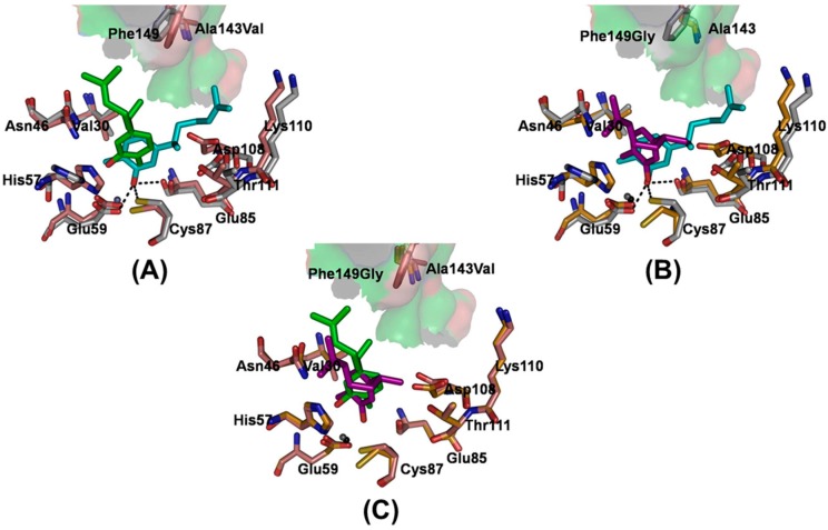
Figure 4.
A superimposition of structures of the mutants relative to wild type complex. Representative structures of the mutants (A) A143V and (B) F149G aligned to the WT structure for a 20-ns snapshot taken from the complex trajectories. The carbon atoms of both mutants are colored salmon (A143V) and bright orange (F149G), and the carbon atoms of the WT system are colored light gray. The carbon atoms of xanthorrhizol are colored green (A143V) and purple (F149G). Xanthorrhizol and amino acid residues are displayed as sticks; (C) Representative structure of the A143V mutant aligned to the F149G structure. The xanthorrhizol (cyan) and the involved amino acid residues (light gray) are displayed as sticks in the structure of the xanthorrhizol and wild-type EcTadA complex. The hydrogen bonds are displayed as black dashed lines.

