Key Clinical Message
Lambda sign is a valuable sign in the diagnosis of sarcoidosis, and FDGPET/CT has been used for planning therapy, monitoring treatment response, and for the follow‐up of patients with chronic persistent sarcoidosis.
Keywords: lambda sign, PET/CT, sarcoidosis
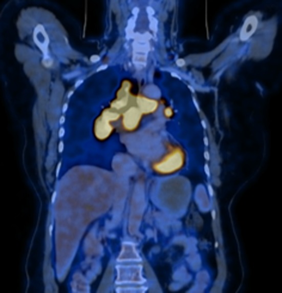
Sixty‐year‐old lady presented with lacrimal and parotid gland enlargement, along with chronic cough for 1 year. Her preliminary investigation showed elevated inflammatory markers, angiotensin‐converting enzyme (ACE) level of 140 U/L (normal value 8‐53 U/L) and chest X‐ray showing hilar adenopathy. Positron emission tomography with 2‐deoxy‐2‐[fluorine‐18] fluoro‐D‐glucose integrated with computed tomography (18F‐FDG PET/CT) was done for the staging, identification of occult sites, and for identifying suitable site for biopsy. 18F‐FDG PET/CT showed a lambda sign (λ), which is secondary to increased FDG uptake in right paratracheal and bilateral hilar lymph nodes (Figure 1). These imaging findings were suggestive of sarcoidosis, and transbronchial biopsy specimen confirmed the diagnosis of sarcoidosis. 18F‐FDG PET/CT appearance of hypermetabolic paratracheal and bilateral hilar lymphadenopathy in sarcoidosis is comparable to the lambda sign of 67 Ga‐citrate scintigraphy.1 With the advent of PET/CT, 67 Ga‐citrate scintigraphy is not routinely used in the evaluation of sarcoidosis, but lambda sign is still a valuable sign in the diagnosis of sarcoidosis. The advantage of FDG PET/CT over scintigraphy is that it can be used for planning therapy, monitoring treatment response, and for the follow‐up of patients with chronic persistent sarcoidosis.
Figure 1.

PET/CT—showing lambda sign—increased FDG uptake in right paratracheal and bilateral hilar lymph nodes
CONFLICT OF INTEREST
None declared.
AUTHOR CONTRIBUTION
All authors : Conceived and wrote paper.
Thomas J, Shagos GS, Firuz I. Lambda sign in sarcoidosis using PET/CT. Clin Case Rep. 2019;7:236–237. 10.1002/ccr3.1937
REFERENCES
- 1. Krüger S, Buck AK, Mottaghy FM, et al. Use of integrated FDG PET/CT in sarcoidosis. Clin Imaging. 2008;32:269‐273. [DOI] [PubMed] [Google Scholar]


