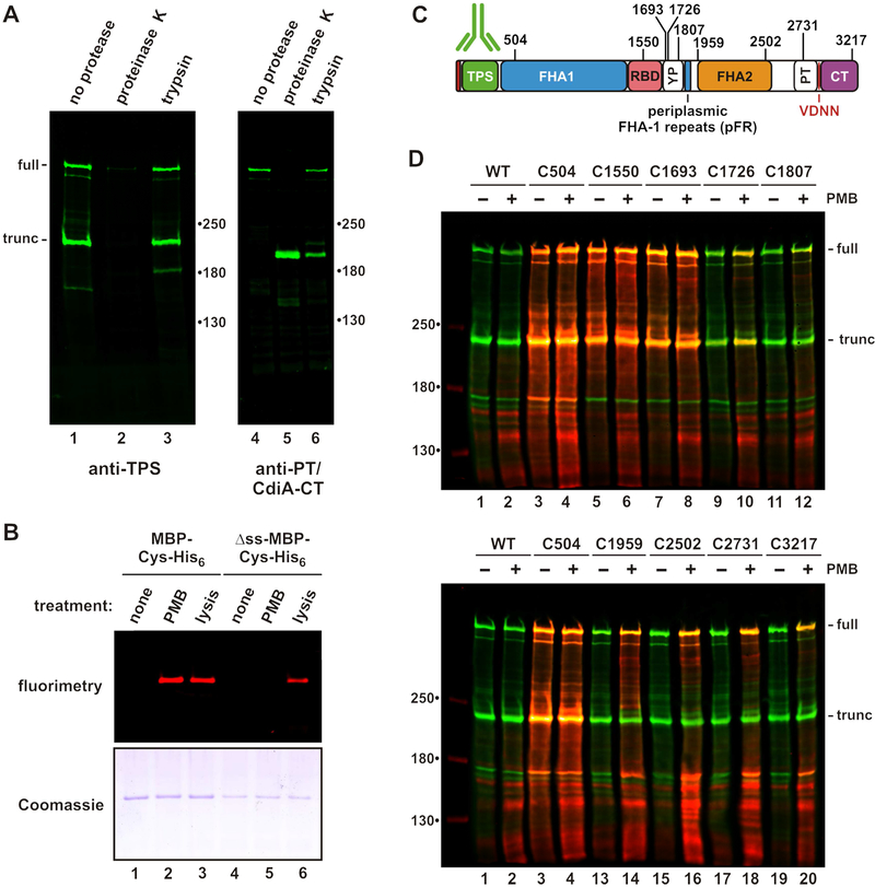
Figure 2. CdiA surface topology.
A) Protease protection assays. Immunoblot analysis with antisera the TPS and PT/CdiA-CT domains of CdiASTECO31. B) Polymyxin B (PMB) permeabilizes the outer membrane. Cells expressing periplasmic or cytoplasmic (Δss) maltose-binding protein (MBP) were incubated with maleimide-dye and permeabilized with PMB or lysed as indicated. Purified MBP was analyzed by fluorimaging (top) and SDS-PAGE (bottom). C) Cys substitutions in CdiASTECO31. The periplasmic FHA-1 repeat (pFR) cluster and major domains are indicated. See Figure S2. D) Immunoblot analysis of dye-labeled CdiA. CdiA expressing cells were incubated with maleimide-dye and permeabilized with PMB where indicated. CdiA was analyzed by immunoblotting with TPS antisera. Dye fluorescence (red) is superimposed onto antibody fluorescence (green).

