(Stem Cell Reports 6, 456–465; April 12, 2016)
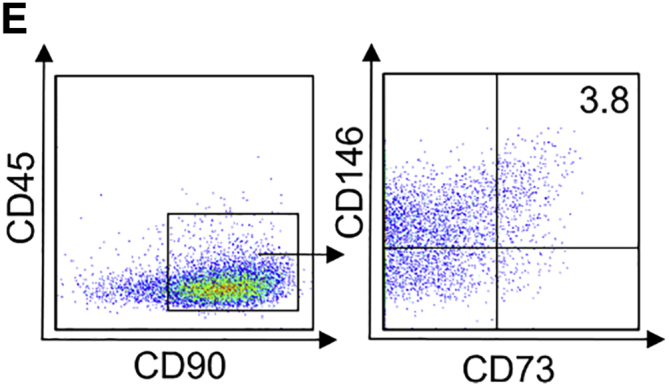
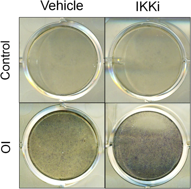
In the originally published Figures 1E, 2H and 3H, the CD73/CD146 FACS plots of the control cells were inadvertently duplicated from the same CD45/CD90 plot with different gating conditions, as multiple samples were being analyzed simultaneously. We have reanalyzed our FACS data, and the values were similar to those previously reported. The correct CD73/CD146 plots and histograms now appears below. Subsequently, the statement in the Results section should read: “…We were able to obtain 3.8% CD73+CD90+CD146+CD90- MSCs of the total differentiated cells from H1 hESCs (Figure 1E).” In the originally published version of Figure S3A, the control staining panels of Vehicle and IKKi-treated cells were inadvertently duplicated. This error was partly attributed to the similarity of the images. The correct control image for IKKi-treated cells has been replaced below. We sincerely apologize for this oversight, which does not affect any of our original conclusions.
Figure 1E.

Spontaneous Differentiation of hESCs without the Feeder Cell Layer
Figure 2H.
Effect of IKKi Treatment on Mesenchymal Lineage Specification of hESCs
Figure 3H.
Effect of p65 Knockdown on Differentiation and Mesenchymal Lineage Specification of hESCs
Figure S3A.
Osteogenic and Chondrogenic Differentiation Potential of Differentiated hESCs with or without IKKi Treatment, Related to Figure 2, corrected
Contributor Information
Christine Hong, Email: chong@dentistry.ucla.edu.
Cun-Yu Wang, Email: cwang@dentistry.ucla.edu.


