. 2018 Oct 3;11(1):72–88. doi: 10.1039/c8nr05520c

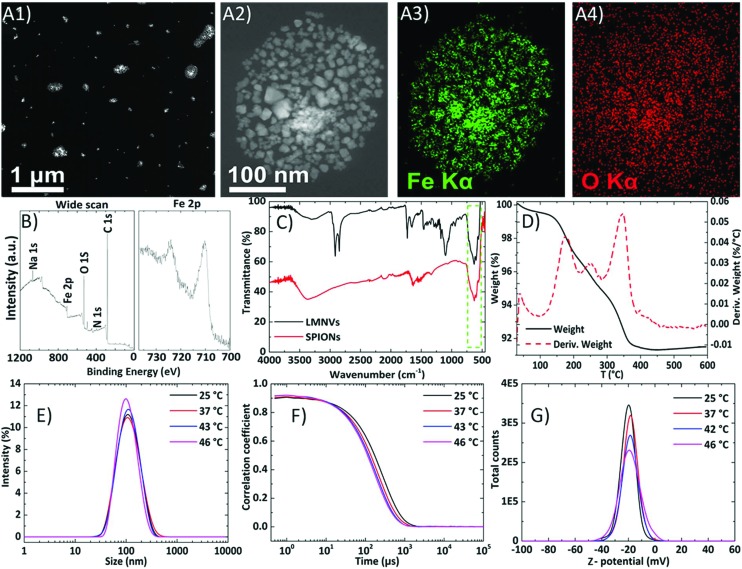
Fig. 1. Morphological and physicochemical characterization of LMNVs: (A1) high-angle annular dark field-scanning transmission electron microscopy (HAADF-STEM) image of LMNVs, (A2) magnified HAADF-STEM image of one unstained LMNV where the distribution of the encapsulated SPIONs is presented, (A3) and (A4) EDS maps of Fe and O over single LMNV, respectively; (B) XPS graph demonstrating the elements present on the surface of the LMNVs; (C) FT-IR spectra of plain SPIONs (bottom) and LMNVs (top) showing peaks attributed to the SPIONs (Fe–O), depicted inside the green rectangular area, and to the lipid components; (D) TGA/DTA graph presenting the weight reduction of the LMNVs during the melting of the lipids and of the polymeric components of the LMNVs (black curve). The red dashed line represents the derivative weight in which each peak represents transition temperatures (melting points) attributed to both the lipid matrix and the incorporated PEG; (E) Gaussian distributions of the hydrodynamic diameter (100 nm) of the LMNVs at various temperatures; (F) correlograms of the LMNVs at various temperatures demonstrating the stability of the LMNVs; (G) surface charge (about –19 mV) of the fabricated LMNVs at various temperatures.

Fig. 1