Abstract
The cryopreservation of ovarian tissue with subsequent transplantation of the tissue represents an established method of fertility protection for female patients who have to undergo gonadotoxic therapy. The procedure can be performed at any point in the cycle and thus generally does not lead to any delay in oncological therapy. With the aid of this procedure, more than 130 births to date worldwide have been able to be recorded. The birth rate is currently approximately 30% and it can be assumed that this will increase through the further optimisation of the cryopreservation and surgical technique. The concept paper presented here is intended to provide guidance for managing cryopreservation and transplantation of ovarian tissue to German-speaking reproductive medicine centres.
Key words: Ferti PROTEKT , fertility protection, ovarian tissue, cryopreservation, transplantation
Zusammenfassung
Die Kryokonservierung von Ovarialgewebe mit anschließender Transplantation des Gewebes stellt eine etablierte Methode der Fertilitätsprotektion für Patientinnen dar, die sich einer gonadotoxischen Therapie unterziehen müssen. Das Verfahren kann zu jedem Zykluszeitpunkt erfolgen und führt somit meistens zu keiner Verzögerung der onkologischen Therapie. Mithilfe dieses Verfahrens konnten bis dato mehr als 130 Geburten weltweit verzeichnet werden. Die Geburtenrate beträgt derzeit ca. 30% und es ist von einer Steigerung durch eine weitere Optimierung der Kryokonservierungs- und Operationstechnik auszugehen. Das hier vorgelegte Konzeptpapier soll Handlungsempfehlungen für den Umgang mit der Kryokonservierung und Transplantation von Ovarialgewebe an deutschsprachigen reproduktionsmedizinischen Zentren geben.
Schlüsselwörter: Ferti PROTEKT , Fertilitätsprotektion, Ovarialgewebe, Kryokonservierung, Transplantation
Introduction
Based on advancements in systemic drug (chemotherapy, targeted therapy) and locoregional therapy (surgery, radiation therapy), the survival rate in malignant and non-malignant diseases has significantly improved. However, the treatments frequently lead to a partial or complete impairment of gonad function with possible germ cell loss. Therefore it is important to counsel female patients prior to gonadotoxic therapy with regard to preserving their fertility. By now, there are a number of fertility-protecting techniques which offer a realistic chance of a later pregnancy. In addition to gonadal transposition prior to radiotherapy, drug-based gonad protection using GnRH analogues and cryopreservation of fertilised and unfertilised oocytes, the removal and cryopreservation of ovarian tissue represents a promising option for girls and women 1 , 2 , 3 .
The cryopreservation of ovarian tissue with subsequent transplantation is a method of fertility preservation which can be performed very shortly before cytotoxic therapy. It can take place at any point in the cycle and thus generally does not lead to any delay in oncological therapy 4 , 5 . Since the first births following ovarian tissue transplantation in 2004 worldwide 6 and in 2011 in Germany 7 , the method has been increasingly used in routine clinical practice. In the Ferti PROTEKT network (Germany, Austria and Switzerland), 163 transplantations were performed in 126 women by January 2018 8 . The figures from Germany are comparable with the successes internationally. In total, there have been more than 130 births following orthotopic transplantation of cryopreserved ovarian tissue to date worldwide 8 , 9 , 10 , 11 , 12 . The birth rate is currently approximately 30 – 35% and it can be assumed that this will increase further through optimisation of cryopreservation and surgical technique 13 . The transplantation of cryopreserved ovarian tissue is thus an established option for fertility protection for oncological patients 14 , 15 , 16 .
The concept paper presented here is intended to provide recommendations for action for the removal, transport, cryopreservation and transplantation of ovarian tissue in German-speaking reproductive medical centres and thus at centres of the Ferti PROTEKT network. This is to ensure the quality of the cryopreservation and transplantation of ovarian tissue at the individual centres with the objective of offering an optimal and consistent standard.
These are not recommendations in the sense of a guideline but rather a joint decision by leading experts on the subjects of removal, cryopreservation and transplantation of ovarian tissue. For this reason, the nomenclature does not correspond to the recommendation classifications of a guideline.
Removal of Ovarian Tissue
From whom and up to which age should ovarian tissue be removed?
The chances for a later pregnancy are greater the higher the ovarian reserve is, that is, the follicular density in the cryopreserved and transplanted ovarian tissue 17 , 18 . The Edinburgh criteria indicate an age limit of 35 years 19 , while other international publications mention a maximum age of 40 years 20 . In a publication by van der Ven et al., the pregnancy rates following transplantation of ovarian tissue were 33% in women who were under age 35 at the time of cryopreservation, in comparison to 18% in women who were over age 35 at the time of cryopreservation. In women over age 40, no pregnancy was able to be achieved 8 .
The cryopreservation of ovarian tissue can also be performed on pre- and peripubertal girls 21 . There are case reports indicating that an induction of puberty can be achieved through the transplantation of prepubertal ovarian tissue 22 , 23 . In addition, there is a case report of a birth following transplantation of cryopreserved premenarchal tissue 24 . This shows that transplantation following prepubertal cryopreservation of ovarian tissue can lead to pregnancies. However, in this regard, it must be borne in mind that the patient was already 13 years old with the onset of thelarche at the time the tissue was removed 24 . In addition, there is a press release of a birth following transplantation of ovarian tissue which had been cryopreserved prior to menarche at the age of 9 years 25 . The indication for cryopreservation of ovarian tissue in pre- and peripubertal girls is currently still unclear and requires an individual assessment of the type of therapy and also of the gonadotoxic dose 1 .
On which chemotherapy and from which level of risk for premature ovarian insufficiency (POI) should ovarian tissue be removed?
The cryopreservation of ovarian tissue is recommended for women in whom the preservation of ovarian function is directly or indirectly threatened due to an underlying oncological, haematological, or other illness 4 . This particularly applies in the case of upcoming gonadotoxic chemotherapy or radiation in the lesser pelvis.
The removal and cryopreservation of ovarian tissue should in particular be offered to all women with a high and intermediate risk of premature ovarian insufficiency (POI). However, even in women with a low risk of permanent amenorrhea, this option should be discussed with the patient. In doing so, it should be taken into account that a risk of POI of ≤ 20% means that one out of every five women who receives one of these chemotherapy regimens is at risk of suffering POI. The ovariotoxic effect of various chemotherapeutic agents and radiation can be found in the tables ( Tables 1 and 2 ) of the S2k guideline “Fertility preservation in oncological therapies” 1 .
Table 1 Ovariotoxic effect of various chemotherapeutic agents 1 .
| Risk | Regimen/substance |
|---|---|
| High risk (> 80% risk of permanent amenorrhoea) |
|
| Intermediate risk (40 – 60% risk of permanent amenorrhoea) |
|
| Low risk (< 20% risk of permanent amenorrhoea) |
|
| Very low or no risk of permanent amenorrhoea |
|
Table 2 Gonadotoxicity in the case of radiation of the ovary and risk of ovarian insufficiency (modified according to 26 ).
| Effective sterilising dose (ESD) | Ovarian radiation dose (Gy) |
|---|---|
| No relevant effects | 0.6 |
| No relevant effects at < 40 years | 1.5 |
| 0 years | 20.3 |
| 10 years | 18.4 |
| 20 years | 16.5 |
| 30 years | 14.3 |
| 40 years | 6 |
How should the tissue be removed?
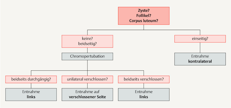
The gold standard for the removal of ovarian tissue is laparoscopy. A laparotomy should only be performed if this is necessary within the scope of the underlying oncological diseases. In the case of removal of ovarian tissue, chromopertubation should be performed under certain conditions (see Fig. 1 ). If unilateral tubal occlusion is seen, the ovarian tissue should be taken on this side in order to preserve the potentially still functioning ovarian tissue on the side with the patent tube. If no POI occurs, an optimal site is available there later for a transplantation. However, it should preferably be ensured that ovarian tissue is not taken on the side of a corpus luteum or a preovulatory follicle or in the case of existing ovarian cysts, since the follicular density of the ovarian tissue to be cryopreserved may be limited here and the cortex quality in any case.
Fig. 1.
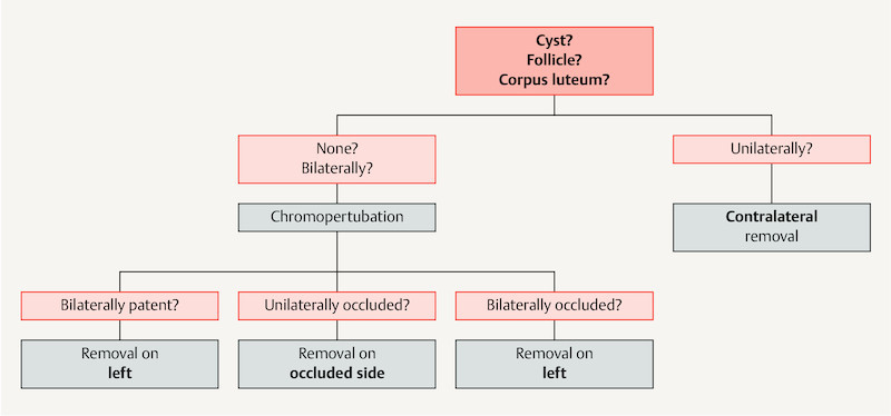
Algorithm for the removal of ovarian tissue.
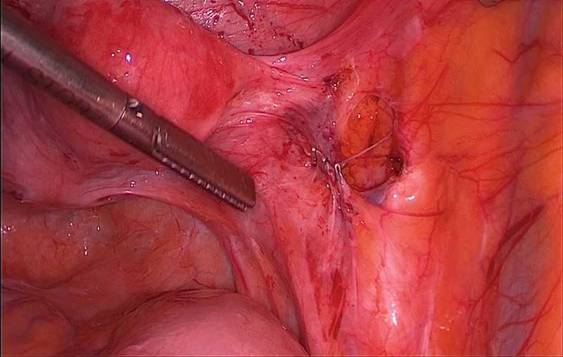
The ovarian tissue should be detached using scissors without electrical coagulation 27 , 28 , 29 . Haemostasis of the wound surface should be performed on the remaining ovary without forming additional areas of necrosis. There are no evidence-based studies available on the various techniques (coagulation, sutures) ( Fig. 2 ).
Fig. 2.

Removal of ovarian tissue via laparoscopy (photo from Department of Obstetrics and Gynaecology, Erlangen).
The surgical risks of removing ovarian tissue are low 27 , 28 , 29 . There is an increased risk of infections and bleeding which is only dependent on the oncological disease. The removal of ovarian tissue is anticipated to involve one complication per 500 laparoscopies which necessitates surgical revision 30 .
A histological examination of a reference sample of the ovarian tissue taken to rule out malignant cells and to identify follicles (a tissue fragment from the ovarian tissue taken measuring at least 2 × 2 mm) should be performed and documented by the centre at which the ovarian tissue was removed. The risks of ovarian metastasis and the resultant consequences can be found in the S2k guideline “Fertility preservation in oncological diseases” 1 .
How much ovarian tissue should be taken?
At least ½ to ⅔ of the ovary should be taken antimesenterically 27 , 28 , 29 ( Fig. 2 ). Bilateral removal at the ovaries should be considered to be an exception (for example, in the case of bilateral ovarian cysts). Due to the small amount of material, biopsies should not be performed 27 , 28 , 29 . However, the patient should also be informed of the possibility for a unilateral ovariectomy, particularly in the case of a high risk of POI. Data show that after a unilateral ovariectomy, menopause can occur approximately one year earlier 31 .
Transport of Ovarian Tissue
How and at which temperature should ovarian tissue be transported after removal and prior to cryopreservation?
Those facilities which do not have possibilities for performing cryopreservation of ovarian tissue themselves can send the ovarian tissue for tissue processing to specialised centres with an affiliated cryobank 32 .
For most organ and tissue transport (for example, kidney, liver), low temperatures are generally selected. The data regarding the transport of ovarian tissue are limited. In the few investigations conducted, the transport temperature varied between 4 – 37 °C (10 – 13). Most studies show that the best conditions are found in the case of low temperatures, around 4 °C. Low temperatures ensure that the cellular metabolism is decreased in the corresponding tissue/organ. In this connection, the need for and, at the same time, the consumption of oxygen are also reduced.
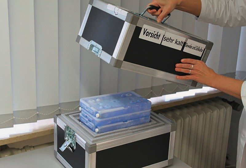
There are suitable transport contains with special pairs of cold packs for the transport of ovarian tissue. The transport container ensures continuous cooling of the tissue during transport at 4 °C and is provided by the respective cryobank ( Fig. 3 ).
Fig. 3.
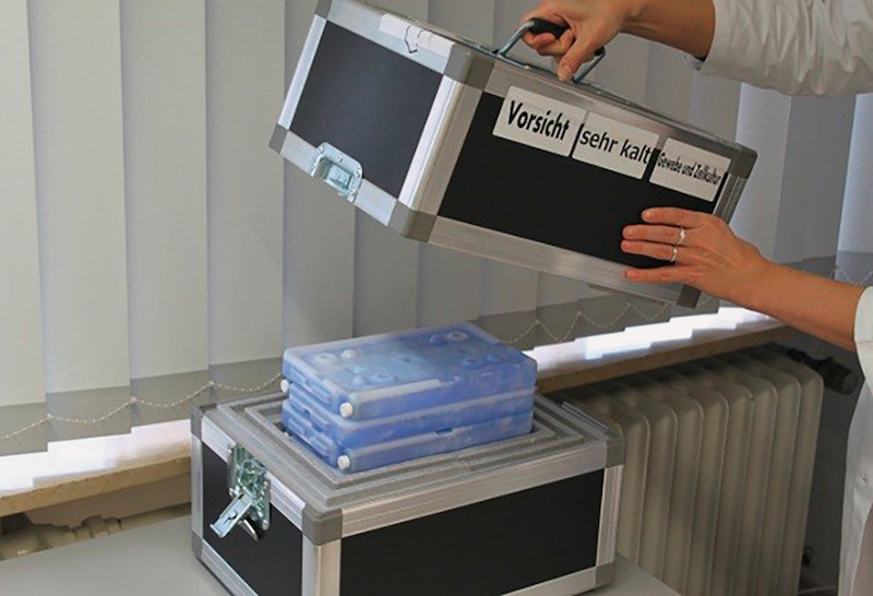
Transport container for ovarian tissue.
How long can ovarian tissue be transported prior to cryopreservation (transport period)?
The transport box can be shipped during the day via daytime courier service or overnight via express courier service. The maximum transport time of the tissue without a significant loss of follicles anticipated is currently not yet sufficiently proven. Investigations show that hypothermia of the ovarian tissue during various transport periods can last up to 24 hours without lasting damage to the follicular reserve 33 , 34 , 35 , 36 .
Which transport medium should be used?
Various transport media are described by different working groups in the literature 35 , 36 , 37 , 38 . There are no adequate investigations which show which transport medium is most suitable. In the case of organ and tissue transplants, Custodiol ® is a certified and approved medium which must be used under chilled conditions (4 – 8 °C) 39 .
Cryopreservation of Ovarian Tissue
Slow freezing vs. vitrification?
In general, two freezing techniques are used in reproductive medicine: the slow freezing method, a cryopreservation method with equilibration, and vitrification, a freezing method without equilibration 32 . Both methods are used worldwide in the cryopreservation of ovarian tissue. In routine practice, because of the better pool of data available, the slow-freezing method is currently recommended 1 , 5 , 40 . Apart from two published births following transplantation of vitrified ovarian tissue, the tissue in the case of all other births was slowly frozen 41 , 42 . The future significance of vitrification cannot yet be estimated due to the current data and validations are needed for universal clinical use 43 .
Slow freezing with cooling rates of 0.3 °C/min requires the use of freezers. In a computer-controlled slow-freezing method, the samples are cooled down according to a modified programme 44 such that they can subsequently be stored indefinitely in a nitrogen gas phase (at − 190 °C). Clear identification of the stored samples must be ensured, for example, through the use of the single European code or a comparable coding system.
How should the ovarian tissue be processed?
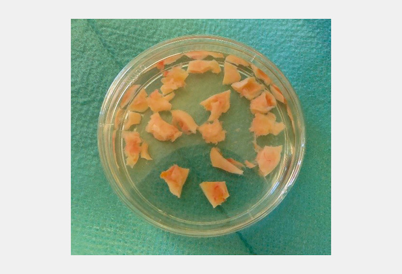
According to the standards of an IVF laboratory, a suitable room with a sterile class II laminar air flow unit in which surgical preparation can take place should be available for processing the ovarian tissue prior to freezing. In the case of chilled preparation, the ovarian medulla is to be carefully dissected away from the ovarian cortex and in doing so, a thin portion of residual stroma should be left to ensure that there is an optimal starting point for neovascularisation of the transplants later 43 . Depending on the size and quality, pieces of ovarian tissue measuring 3 × 2 × 1 mm up to a maximum of 4 × 8 × 1 mm should be cut from the prepared cortex ( Fig. 4 ). The size of the pieces should be listed in the transplantation record.
Fig. 4.
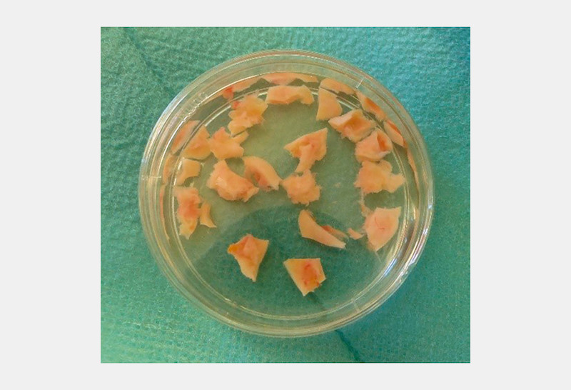
Cut ovarian tissue pieces.
Which freezing medium should be used?
The choice of the cryoprotective used is an important point which is largely responsible for whether cryopreservation is successful. Glycerine, ethylene glycol, DMSO and propanediol in combination with corresponding carrier media and a protein supplement are available for this 34 , 36 , 45 , 46 . Using the cryoprotectives ethylene glycol, propanediol or DMSO, similarly good survival rates of the germ cells as well as intact tissue morphologies and cell structures are demonstrated following cryopreservation and subsequent thawing; glycerine performed the worst in investigations 34 , 36 , 45 , 46 . Internationally, DMSO is the most frequently used and best established cryoprotective for ovarian tissue 41 , 47 , 48 .
Vitality testing of the ovarian tissue?
The prepared ovarian tissue is examined for quality assurance prior to cryopreservation and also during preparation for a transplantation in the case of premature ovarian failure following cytotoxic therapy. The examinations ensure that the removal of the tissue, its transport/processing and also the cryopreservation and thawing have not led to any relevant damage to the tissue – likewise, a possible cortex potential (number of primordial follicles in a particular area) of each patient is individually determined.
Small, standardised biopsy punches from various peripheral areas are methodically produced for this as part of the preparation of the ovarian tissue taken; following tissue digestion and calcein-acetoxymethyl ester staining or a live-dead assay, these punches are analysed for the number of vital primordial follicles. The concentration of vital primordial follicles and the anti-Müller hormone level in the serum, the antral follicle count and the age of the patient (all determined at the time that the ovarian tissue is removed or before the start of cytotoxic therapy) are considered to be parameters for determining the fertility reserve – but also for later determination of the number of ovarian cortex pieces to be transplanted.
For the validation of a facility which processes ovarian tissue, the Ferti PROTEKT network recommends that cryopreserving centres have their freezing technique checked through a xenotransplantation of ovarian tissue on immunodeficient severe-combined-immunodeficiency (SCID) mice before offering cryopreservation of ovarian tissue in routine clinical practice. This is able to show that the follicles have a qualitative development potential/follicular growth following cryopreservation and thawing.
Transplantation of Ovarian Tissue
When should ovarian tissue be transplanted and up to which age?
Transplantation of ovarian tissue should take place after successful oncological therapy in female patients with a manifest desire for a child and in the case of failure of ovarian function, for the resumption of folliculogenesis and to lead to a pregnancy 49 . A transplantation can be considered even in patients who still have a regular cycle but limited ovarian reserve. Transplantation to restore longstanding ovarian function which would, where applicable, replace necessary hormone replacement therapy should not be primarily recommended due to the limited activity of the transplanted tissue 50 . Prior to transplantation, especially if there is no POI and nevertheless a transplantation is being considered, the couple should undergo a basic clarification of causes of infertility to rule out or correct other causes of infertility or this should be included in the further treatment planning.
The age of 45 years should not be exceeded as an age limit for a transplantation 51 . If a transplantation is to be recommended at the earliest following oncological treatment, this should be determined depending on the underlying oncological disease and individually together with the attending oncologists, taking the age of the patient and the oncological prognosis into account.
How should the ovarian tissue be transplanted (surgical technique and site of transplantation)?
The transplantation of ovarian tissue is generally performed via laparoscopy 27 , 28 , 29 . In routine clinical practice, orthotopic transplantation is primarily used. In an orthotopic transplantation, the ovarian tissue is transplanted either in the pelvic wall caudodorsal to the ovaries or in/on the ovary. Internationally, various orthotopic transplantation techniques are described 27 , 28 , 29 . It is still unclear which of the techniques and which locations lead to the greatest chances for pregnancy.
The better circulation in the pelvic wall and the easier surgical feasibility (and the associated low surgical risk for the patient) are reasons for transplantation under the pelvic peritoneum as compared to the situation in the generally significantly atrophied ovary.
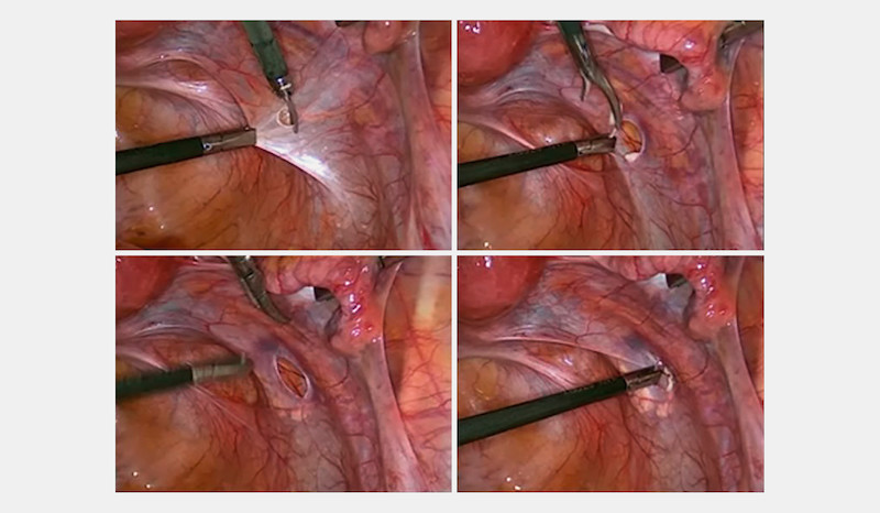
The parietal peritoneum of the ovarian fossa is incised for this purpose over a length of approximately 0.5 – 1 cm. A subperitoneal pocket is created by blunt dissection and the tissue pieces are implanted in the pocket. The tissue pieces should preferably lie side by side and not on top of each other ( Fig. 5 ). If the ovarian tissue is to be taken once again (for example, if there is an increased risk of a predisposition), the tissue should be marked, for example, with titanium clips ( Fig. 6 ). Documentation of the site of transplantation should be ensured.
Fig. 5.
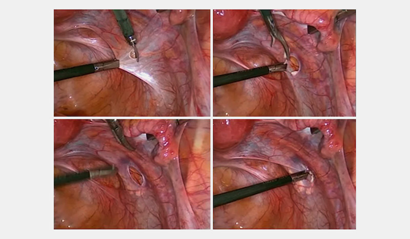
Orthotopic transplantation of ovarian tissue in a peritoneal pocket of the pelvic peritoneum of the ovarian fossa.
Fig. 6.
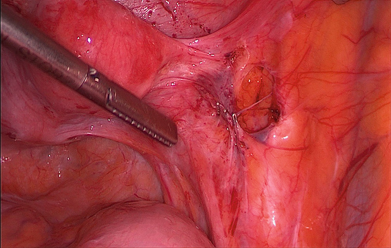
Marking the transplantation site with titanium clips.
Please refer to the corresponding literature in the case of transplantation in/on the ovary 2 , 27 .
In general, it is not recommended to transplant all of the cryopreserved material at once but rather to implant only approximately ⅓ to ½ of the available tissue in order to have tissue in reserve in the event of repeat loss of ovarian function 1 . However, the patientʼs age in the case of an ovarian transplantation should also be taken into account.
A natural pregnancy is possible in principle in the case of an orthotopic transplantation under the pelvic peritoneum. Therefore in the case of an ovarian tissue transplantation, a fertility assessment (tubal, uterine and extrauterine factors) and correction, if necessary, should be performed 1 ; that is, in the case of an ovarian tissue transplantation, it is recommended to check the patency of the Fallopian tubes by means of “chromo”-pertubation (with NaCl) and also to consider a hysteroscopy to rule out polyps, myomas, adhesions or chronic endometritis. The transplantation is performed on the side on which the tube is patent and the anatomical conditions are the most favourable ( Fig. 7 ).
Fig. 7.

Algorithm for selecting the transplantation side.
The risks of surgery are comparable to the risks in the case of other laparoscopic procedures. Possible risks are secondary haemorrhage or a wound infection which occur in < 1% of cases 27 , 28 , 29 .
Follow-up Care of Patients Who Have Undergone Transplantation
What follow-up care should be provided?
The ovarian function can be restored in 80 – 85% of cases through transplantations of ovarian tissue 15 . It takes between 2 to 6 months for the transplanted ovarian tissue to show signs of resumption of its function (drop in FSH, rise in oestradiol) 48 . A spontaneous pregnancy can be attempted if the tubes are patent and there are no other relevant infertility factors. To increase the likelihood of conception, the patient can be offered cycle monitoring with induction of ovulation through HCG within the framework of intercourse at the optimal time. In the case of occluded tubes or other infertility factors, assisted reproductive medicine techniques (IVF; ICSI) must be used. If there is a high follicle density (antral follicle count and AMH), gonadotropin stimulation can be considered, depending on the patientʼs age and the underlying oncological disease, in order to accelerate conception, if applicable. Alternatively, egg cells obtained in several spontaneous cycles or stimulated cycles can also be vitrified so that an egg cell pool is created for later use in an assisted reproductive medicine technique 52 . To increase the rate of implantation, particularly in the case of whole-body radiation and radiation of the lesser pelvis, other diagnostic measures, such as diagnostic endometrial procedures to analyse the endometrial receptivity profile, can be offered. After one year, approximately ⅔ of the transplants are still active. The tissue transplants which initially showed good activity are generally active for several years. The mean length of ovarian function after transplantation is about 4 to 7 years on average, however this depends on the number of follicles in the ovarian tissue and the age of the woman at the time of cryopreservation 11 . If there are no signs of activity seen after approximately ½ year after transplantation, another transplantation of cryopreserved ovarian tissue should be considered.
Conflict of Interest/Interessenkonflikt The authors declare that they have no conflict of interest./Die Autoren geben an, dass kein Interessenkonflikt besteht.
Equal authorship
gleichberechtigte Autorenschaft
Joint Decision.
The cryopreservation of ovarian tissue should be performed prior to the start of gonadotoxic therapy in girls and women only up to the age of 40 years. Ovarian cryopreservation can also be considered on a case-by-case basis in prepubertal girls, taking the gonadotoxic therapy and the risks of the surgical procedure into account.
Joint Decision.
The cryopreservation of ovarian tissue should be offered when there is a high or intermediate risk and – on a case-by-case basis – also when there is a low risk of premature ovarian insufficiency due to gonadotoxic therapy.
Joint Decision.
The gold standard for the removal of ovarian tissue is laparoscopy.
A laparotomy should only be performed if this is necessary within the scope of the underlying oncological disease.
The ovarian tissue should be detached using scissors and sources of bleeding should only be coagulated in a targeted manner afterwards (punctate coagulation).
Histology to rule out malignant cells in the ovarian tissue (at least 2 × 2 mm) must be performed by the centre at which the ovarian tissue was removed.
Joint Decision.
At least ½ to ⅔ of an ovary (preferably without ovarian pathologies) should be removed. Bilateral removal should be performed only in exceptional cases. Biopsies are not recommended.
Joint Decision.
Prior to cryopreservation, ovarian tissue should be transported chilled (4 – 8 °C) in special transport containers.
Joint Decision.
The transport of ovarian tissue up to 24 hours prior to cryopreservation is possible.
Joint Decision.
Custodiol ® or a comparable medium should be used under chilled conditions (4 – 8 °C) as a transport medium.
Joint Decision.
Based on the better pool of data available, ovarian tissue should at present be cryopreserved in routine practice with the established slow-freezing method.
Joint decision.
The ovarian tissue should be processed in a suitable room under a sterile class II laminar air flow.
The ovarian tissue pieces should measure between 3 × 2 × 1 mm up to a maximum of 4 × 8 × 1 mm. The size of the ovarian tissue pieces should be listed in the transplantation record.
Joint Decision.
DMSO or, alternatively, ethylene glycol can be used as a cryoprotective.
Joint Decision.
Vitality testing with a suitable assay should be performed before freezing and before transplantation.
Joint Decision.
Transplantation of ovarian tissue should be performed in women with a manifest desire for a child and failure of ovarian function (amenorrhoea/oligomenorrhoea), up to the age of 45 years.
A basic clarification of causes of infertility should be performed prior to transplantation.
Joint Decision.
The transplantation of ovarian tissue is performed laparoscopically.
In the case of a transplantation, a “chromo”-pertubation (for example, with NaCl) should be performed.
Blunt preparation of a pocket is performed in the pelvic peritoneum for this purpose and the ovarian tissue is introduced into this pocket.
If the ovarian tissue preparation is to be taken again later, it is recommended to mark the transplantation site (e.g. titanium clips).
Joint Decision.
Patients with amenorrhoea should be offered a monthly blood test (FSH, oestradiol, progesterone) 8 – 10 weeks before the transplantation until signs of activity are detected, and every two months, an ultrasound examination should be offered.
If there are signs of activity of the transplanted ovarian tissue, it is recommended to perform cycle monitoring to increase the chances of pregnancy, after other causes of infertility have been clarified.
In patients with causes of infertility (such as tubal infertility, reduced male fertility) ART measures must be taken.
Patients who still had an active cycle prior to transplantation should be offered monthly cycle monitoring 8 – 10 weeks after the transplantation.
Stimulation can be considered depending on the individual situation (follicular density, number of attempts to conceive, tumour entity).
References/Literatur
- 1.Fertility preservation for patients with malignant disease. Guideline of the DGGG, DGU and DGRM (S2k-Level, AWMF Registry No. 015/082, November 2017)Online:http://www.awmf.org/leitlinien/detail/ll/015-082.htmllast access: 13.06.2018 [DOI] [PMC free article] [PubMed]
- 2.von Wolff M, Germeyer A, Liebenthron J. Practical recommendations for fertility preservation in women by the FertiPROTEKT network. Part II: fertility preservation techniques. Arch Gynecol Obstet. 2018;297:257–267. doi: 10.1007/s00404-017-4595-2. [DOI] [PMC free article] [PubMed] [Google Scholar]
- 3.Winkler-Crepaz K, Bottcher B, Toth B. What is new in 2017? Update on fertility preservation in cancer patients. Minerva Endocrinol. 2017;42:331–339. doi: 10.23736/S0391-1977.17.02633-5. [DOI] [PubMed] [Google Scholar]
- 4.International Society for Fertility Preservation–ESHRE–ASRM Expert Working Group . Martinez F. Update on fertility preservation from the Barcelona International Society for Fertility Preservation-ESHRE-ASRM 2015 expert meeting: indications, results and future perspectives. Fertil Steril. 2017;108:407–4.15E13. doi: 10.1016/j.fertnstert.2017.05.024. [DOI] [PubMed] [Google Scholar]
- 5.Practice Committee of American Society for Reproductive Medicine . Ovarian tissue cryopreservation: a committee opinion. Fertil Steril. 2014;101:1237–1243. doi: 10.1016/j.fertnstert.2014.02.052. [DOI] [PubMed] [Google Scholar]
- 6.Donnez J, Dolmans M M, Demylle D. Livebirth after orthotopic transplantation of cryopreserved ovarian tissue. Lancet. 2004;364:1405–1410. doi: 10.1016/S0140-6736(04)17222-X. [DOI] [PubMed] [Google Scholar]
- 7.Muller A, Keller K, Wacker J. Retransplantation of cryopreserved ovarian tissue: the first live birth in Germany. Dtsch Arztebl Int. 2012;109:8–13. doi: 10.3238/arztebl.2012.0008. [DOI] [PMC free article] [PubMed] [Google Scholar]
- 8.Van der Ven H, Liebenthron J, Beckmann M. Ninety-five orthotopic transplantations in 74 women of ovarian tissue after cytotoxic treatment in a fertility preservation network: tissue activity, pregnancy and delivery rates. Hum Reprod. 2016;31:2031–2041. doi: 10.1093/humrep/dew165. [DOI] [PubMed] [Google Scholar]
- 9.Jensen A K, Macklon K T, Fedder J. 86 successful births and 9 ongoing pregnancies worldwide in women transplanted with frozen-thawed ovarian tissue: focus on birth and perinatal outcome in 40 of these children. J Assist Reprod Genet. 2017;34:325–336. doi: 10.1007/s10815-016-0843-9. [DOI] [PMC free article] [PubMed] [Google Scholar]
- 10.Pacheco F, Oktay K. Current Success and Efficiency of Autologous Ovarian Transplantation: A Meta-Analysis. Reprod Sci. 2017;24:1111–1120. doi: 10.1177/1933719117702251. [DOI] [PubMed] [Google Scholar]
- 11.Donnez J, Dolmans M M. Ovarian cortex transplantation: 60 reported live births brings the success and worldwide expansion of the technique towards routine clinical practice. J Assist Reprod Genet. 2015;32:1167–1170. doi: 10.1007/s10815-015-0544-9. [DOI] [PMC free article] [PubMed] [Google Scholar]
- 12.Dittrich R, Hackl J, Lotz L. Pregnancies and live births after 20 transplantations of cryopreserved ovarian tissue in a single center. Fertil Steril. 2015;103:462–468. doi: 10.1016/j.fertnstert.2014.10.045. [DOI] [PubMed] [Google Scholar]
- 13.Jadoul P, Guilmain A, Squifflet J. Efficacy of ovarian tissue cryopreservation for fertility preservation: lessons learned from 545 cases. Hum Reprod. 2017;32:1046–1054. doi: 10.1093/humrep/dex040. [DOI] [PubMed] [Google Scholar]
- 14.Lotz L, Maktabi A, Hoffmann I. Ovarian tissue cryopreservation and retransplantation – what do patients think about it? Reprod Biomed Online. 2016 doi: 10.1016/j.rbmo.2015.12.012. [DOI] [PubMed] [Google Scholar]
- 15.Gellert S E, Pors S E, Kristensen S G. Transplantation of frozen-thawed ovarian tissue: an update on worldwide activity published in peer-reviewed papers and on the Danish cohort. J Assist Reprod Genet. 2018 doi: 10.1007/s10815-018-1144-2. [DOI] [PMC free article] [PubMed] [Google Scholar]
- 16.Pacheco F, Oktay K. Current Success and Efficiency of Autologous Ovarian Transplantation: A Meta-Analysis. Reprod Sci. 2017;24:1111–1120. doi: 10.1177/1933719117702251. [DOI] [PubMed] [Google Scholar]
- 17.von Wolff M, Montag M, Dittrich R. Fertility preservation in women–a practical guide to preservation techniques and therapeutic strategies in breast cancer, Hodgkinʼs lymphoma and borderline ovarian tumours by the fertility preservation network FertiPROTEKT. Arch Gynecol Obstet. 2011;284:427–435. doi: 10.1007/s00404-011-1874-1. [DOI] [PMC free article] [PubMed] [Google Scholar]
- 18.Dittrich R, Mueller A, Binder H. First retransplantation of cryopreserved ovarian tissue following cancer therapy in Germany. Dtsch Arztebl Int. 2008;105:274–278. doi: 10.3238/arztebl.2008.0274. [DOI] [PMC free article] [PubMed] [Google Scholar]
- 19.Wallace W H, Smith A G, Kelsey T W. Fertility preservation for girls and young women with cancer: population-based validation of criteria for ovarian tissue cryopreservation. Lancet Oncol. 2014;15:1129–1136. doi: 10.1016/S1470-2045(14)70334-1. [DOI] [PMC free article] [PubMed] [Google Scholar]
- 20.Schuring A N, Fehm T, Behringer K. Practical recommendations for fertility preservation in women by the FertiPROTEKT network. Part I: Indications for fertility preservation. Arch Gynecol Obstet. 2018;297:241–255. doi: 10.1007/s00404-017-4594-3. [DOI] [PMC free article] [PubMed] [Google Scholar]
- 21.Jensen A K, Rechnitzer C, Macklon K T. Cryopreservation of ovarian tissue for fertility preservation in a large cohort of young girls: focus on pubertal development. Hum Reprod. 2017;32:154–164. doi: 10.1093/humrep/dew273. [DOI] [PubMed] [Google Scholar]
- 22.Anderson R A, Hindmarsh P C, Wallace W H. Induction of puberty by autograft of cryopreserved ovarian tissue in a patient previously treated for Ewing sarcoma. Eur J Cancer. 2013;49:2960–2961. doi: 10.1016/j.ejca.2013.04.031. [DOI] [PubMed] [Google Scholar]
- 23.Poirot C, Abirached F, Prades M. Induction of puberty by autograft of cryopreserved ovarian tissue. Lancet. 2012;379:588. doi: 10.1016/S0140-6736(11)61781-9. [DOI] [PubMed] [Google Scholar]
- 24.Demeestere I, Simon P, Dedeken L. Live birth after autograft of ovarian tissue cryopreserved during childhood. Hum Reprod. 2015;30:2107–2109. doi: 10.1093/humrep/dev128. [DOI] [PubMed] [Google Scholar]
- 25.Senthilingam M.Women is first to have baby with ovaries frozen in childhood. CNN-Meldung vom 16. Dezember 2016Online:http://daebl.de/UX32last access: 01.05.2018
- 26.Wallace W H, Thomson A B, Saran F. Predicting age of ovarian failure after radiation to a field that includes the ovaries. Int J Radiat Oncol Biol Phys. 2005;62:738–744. doi: 10.1016/j.ijrobp.2004.11.038. [DOI] [PubMed] [Google Scholar]
- 27.Beckmann M W, Dittrich R, Lotz L. Fertility protection: complications of surgery and results of removal and transplantation of ovarian tissue. Reprod Biomed Online. 2018;36:188–196. doi: 10.1016/j.rbmo.2017.10.109. [DOI] [PubMed] [Google Scholar]
- 28.Beckmann M W, Dittrich R, Lotz L. Operative techniques and complications of extraction and transplantation of ovarian tissue: the Erlangen experience. Arch Gynecol Obstet. 2017;295:1033–1039. doi: 10.1007/s00404-017-4311-2. [DOI] [PubMed] [Google Scholar]
- 29.Beckmann M W, Dittrich R, Findeklee S. Surgical Aspects of Ovarian Tissue Removal and Ovarian Tissue Transplantation for Fertility Preservation. Geburtsh Frauenheilk. 2016;76:1057–1064. doi: 10.1055/s-0042-115017. [DOI] [PMC free article] [PubMed] [Google Scholar]
- 30.von Wolff M, Dittrich R, Liebenthron J. Fertility-preservation counselling and treatment for medical reasons: data from a multinational network of over 5000 women. Reprod Biomed Online. 2015;31:605–612. doi: 10.1016/j.rbmo.2015.07.013. [DOI] [PubMed] [Google Scholar]
- 31.Rosendahl M, Simonsen M K, Kjer J J. The influence of unilateral oophorectomy on the age of menopause. Climacteric. 2017;20:540–544. doi: 10.1080/13697137.2017.1369512. [DOI] [PubMed] [Google Scholar]
- 32.Liebenthron J, Montag M. Chapter 15 Development of a Nationwide Network for Ovarian Tissue Cryopreservation. Methods Mol Biol. 2017;1568:205–220. doi: 10.1007/978-1-4939-6828-2_15. [DOI] [PubMed] [Google Scholar]
- 33.Schmidt K L, Ernst E, Byskov A G. Survival of primordial follicles following prolonged transportation of ovarian tissue prior to cryopreservation. Hum Reprod. 2003;18:2654–2659. doi: 10.1093/humrep/deg500. [DOI] [PubMed] [Google Scholar]
- 34.Dittrich R, Lotz L, Keck G. Live birth after ovarian tissue autotransplantation following overnight transportation before cryopreservation. Fertil Steril. 2012;97:387–390. doi: 10.1016/j.fertnstert.2011.11.047. [DOI] [PubMed] [Google Scholar]
- 35.Klocke S, Tappehorn C, Griesinger G. Effects of supra-zero storage on human ovarian cortex prior to vitrification-warming. Reprod Biomed Online. 2014;29:251–258. doi: 10.1016/j.rbmo.2014.03.025. [DOI] [PubMed] [Google Scholar]
- 36.Rosendahl M, Timmermans Wielenga V, Nedergaard L. Cryopreservation of ovarian tissue for fertility preservation: no evidence of malignant cell contamination in ovarian tissue from patients with breast cancer. Fertil Steril. 2011;95:2158–2161. doi: 10.1016/j.fertnstert.2010.12.019. [DOI] [PubMed] [Google Scholar]
- 37.Gastal G D, Alves B G, Alves K A. Ovarian fragment sizes affect viability and morphology of preantral follicles during storage at 4 degrees C. Reproduction. 2017;153:577–587. doi: 10.1530/REP-16-0621. [DOI] [PubMed] [Google Scholar]
- 38.Dittrich R, Lotz L, Keck G. Live birth after ovarian tissue autotransplantation following overnight transportation before cryopreservation. Fertil Steril. 2012;97:387–390. doi: 10.1016/j.fertnstert.2011.11.047. [DOI] [PubMed] [Google Scholar]
- 39.Bastings L, Liebenthron J, Westphal J R. Efficacy of ovarian tissue cryopreservation in a major European center. J Assist Reprod Genet. 2014;31:1003–1012. doi: 10.1007/s10815-014-0239-7. [DOI] [PMC free article] [PubMed] [Google Scholar]
- 40.Rodriguez-Wallberg K A, Tanbo T, Tinkanen H. Ovarian tissue cryopreservation and transplantation among alternatives for fertility preservation in the Nordic countries – compilation of 20 years of multicenter experience. Acta Obstet Gynecol Scand. 2016;95:1015–1026. doi: 10.1111/aogs.12934. [DOI] [PMC free article] [PubMed] [Google Scholar]
- 41.Meirow D, Roness H, Kristensen S G. Optimizing outcomes from ovarian tissue cryopreservation and transplantation; activation versus preservation. Hum Reprod. 2015;30:2453–2456. doi: 10.1093/humrep/dev210. [DOI] [PubMed] [Google Scholar]
- 42.Shi Q, Xie Y, Wang Y. Vitrification versus slow freezing for human ovarian tissue cryopreservation: a systematic review and meta-anlaysis. Sci Rep. 2017;7:8538. doi: 10.1038/s41598-017-09005-7. [DOI] [PMC free article] [PubMed] [Google Scholar]
- 43.Donnez J, Dolmans M M. Ovarian tissue freezing: current status. Curr Opin Obstet Gynecol. 2015;27:222–230. doi: 10.1097/GCO.0000000000000171. [DOI] [PubMed] [Google Scholar]
- 44.Gosden R G, Baird D T, Wade J C. Restoration of fertility to oophorectomized sheep by ovarian autografts stored at − 196 degrees C. Hum Reprod. 1994;9:597–603. doi: 10.1093/oxfordjournals.humrep.a138556. [DOI] [PubMed] [Google Scholar]
- 45.Newton H, Aubard Y, Rutherford A. Low temperature storage and grafting of human ovarian tissue. Hum Reprod. 1996;11:1487–1491. doi: 10.1093/oxfordjournals.humrep.a019423. [DOI] [PubMed] [Google Scholar]
- 46.Bastings L, Westphal J R, Beerendonk C C. Clinically applied procedures for human ovarian tissue cryopreservation result in different levels of efficacy and efficiency. J Assist Reprod Genet. 2016;33:1605–1614. doi: 10.1007/s10815-016-0816-z. [DOI] [PMC free article] [PubMed] [Google Scholar]
- 47.Dolmans M M, Jadoul P, Gilliaux S. A review of 15 years of ovarian tissue bank activities. J Assist Reprod Genet. 2013;30:305–314. doi: 10.1007/s10815-013-9952-x. [DOI] [PMC free article] [PubMed] [Google Scholar]
- 48.Donnez J, Dolmans M M, Pellicer A. Restoration of ovarian activity and pregnancy after transplantation of cryopreserved ovarian tissue: a review of 60 cases of reimplantation. Fertil Steril. 2013;99:1503–1513. doi: 10.1016/j.fertnstert.2013.03.030. [DOI] [PubMed] [Google Scholar]
- 49.Donnez J, Dolmans M M. Transplantation of ovarian tissue. Best Pract Res Clin Obstet Gynaecol. 2014;28:1188–1197. doi: 10.1016/j.bpobgyn.2014.09.003. [DOI] [PubMed] [Google Scholar]
- 50.von Wolff M, Dittrich R, Stute P. Transplantation of ovarian tissue to postpone menopause – is it really more advantageous for womenʼs health than menopause hormone therapy? Reprod Biomed Online. 2015;31:827. doi: 10.1016/j.rbmo.2015.08.015. [DOI] [PubMed] [Google Scholar]
- 51.Rodriguez-Wallberg K A, Tanbo T, Tinkanen H. Ovarian tissue cryopreservation and transplantation among alternatives for fertility preservation in the Nordic countries – compilation of 20 years of multicenter experience. Acta Obstet Gynecol Scand. 2016;95:1015–1026. doi: 10.1111/aogs.12934. [DOI] [PMC free article] [PubMed] [Google Scholar]
- 52.Cobo A, Garrido N, Crespo J. Accumulation of oocytes: a new strategy for managing low-responder patients. Reprod Biomed Online. 2012;24:424–432. doi: 10.1016/j.rbmo.2011.12.012. [DOI] [PubMed] [Google Scholar]


