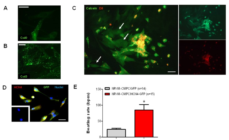
Figure 2.
Functional coupling of CMPCs to cardiomyocytes. (A,B) Immunolabeling of connexin proteins Cx40 and Cx43. Scale bars represent 30 and 25 µm, respectively. (C) Fluorescent microscopy of dye transfer from CMPCs labeled with DiI (red) and Calcein (green) to unlabeled cardiomyocytes. Cardiomyocytes that have imported Calcein are indicated by white arrows. Scale bar represents 45 µm. (D) Immunolabeling of GFP (green) and HCN4 (red) in CMPCs transduced with LV-HCN4-GFP. Yellow indicates co-staining. Nuclei were counterstained blue with DAPI. Non-transduced CMPCs, shown in the inset, stained negative for GFP and HCN4. Scale bar represents 45 µm. (E) Average beating rates of neonatal rat ventricular myocyte (NRVM) monolayers co-cultured with CMPCs expressing GFP alone (grey) and HCN4 and GFP (red). * indicates P < 0.05.

