. 2019 Jan 18;8:e43821. doi: 10.7554/eLife.43821

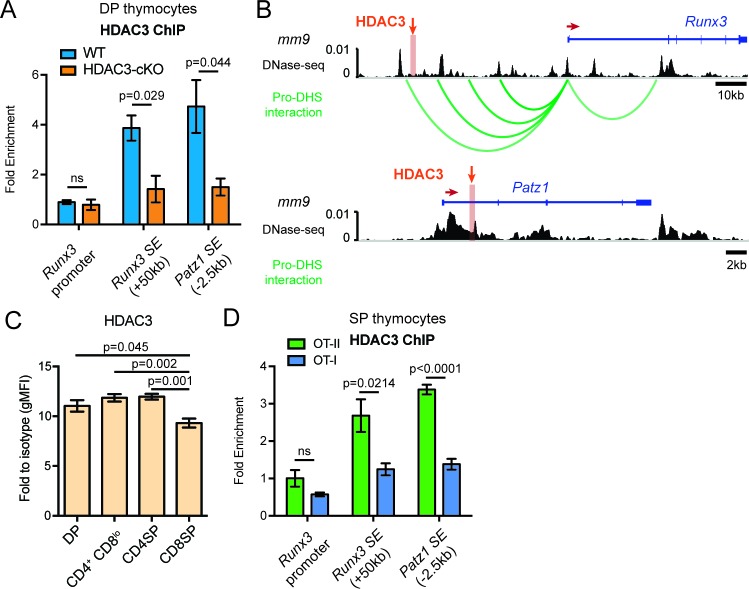
Figure 7. HDAC3 binds to Runx3 and Patz1 regulatory regions in DP and CD4SP thymocytes.

(A) HDAC3 qChIP in DP thymocytes from WT and HDAC3-cKO mice. Plots show mean ± SEM of fold enrichment over Rpl30 (n = 3 mice/group from three independent experiments). (B) DNase-seq and Hi-C arc plots at the Runx3 and Patz1 gene loci in DP thymocytes and pooled DN3-to-DP thymocytes, respectively. Shaded region highlights where HDAC3 binds, as shown in (A). (DHS, DNA hypersensitivity sites). (C) HDAC3 protein expression measured by flow cytometry in DP, CD4+CD8lo, CD4SP, and CD8SP thymocytes from WT mice. Plots show mean ± SEM of fold enrichment over isotype (n = 4 mice/group from three independent experiments). (D) HDAC3 qChIP in SP thymocytes from OT-II and OT-I mice. Plots show mean ± SEM of fold enrichment over Rpl30 (n = 4 mice/group from two independent experiments). See also Figure 7—figure supplements 12.

Figure 7.

Figure 7—figure supplement 1. HDAC3 ChIP-seq in pro-B cells and human CD4 T cells.

Figure 7—figure supplement 1.

ChIP-seq snapshots at Runx3 and Patz1 of publicly available HDAC3 ChIP-seq datasets in pro-B cells (GEO: GSM2096648, genome alignment mm10) and human CD4 T cells (GEO: GSM393952, genome alignment hg18). Alongside the HDAC3 ChIP-seq, DNase-seq (GEO: GSM2195840, genome alignment mm10) and H3K27ac ChIP seq from Immature OT-II and OT-II HDAC3-cKO thymocytes (as shown in Figure 4) were provided to visualize where HDAC3 binds in relation to regulatory elements and changes in histone acetylation between WT and HDAC3-cKO mice, respectively. Orange shaded regions demarcate super enhancers (SE) and blue shaded regions highlight where primers were made for qPCR. In pro-B cells, HDAC3 binds 50 kb upstream of Runx3, at the Runx3 promoter, and approximately 2 kb downstream of the Patz1 promoter. Likewise, HDAC3 binds approximately 57 kb upstream of Runx3 and 2 kb downstream of the Patz1 promoter in human CD4 T cells. These three regions were used to make primers for HDAC3 qChIP performed in Figure 7.
Figure 7—figure supplement 2. Model.

Figure 7—figure supplement 2.

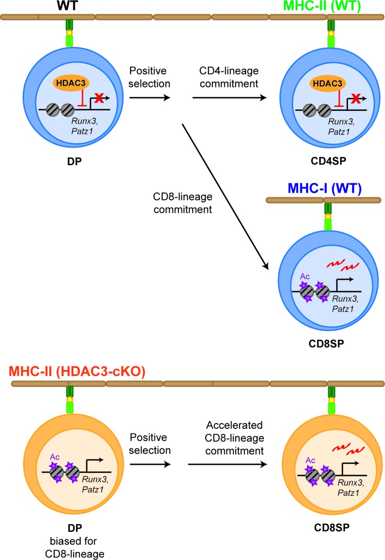
HDAC3 is required to restrain CD8-lineage genes for CD4-lineage choice. In WT thymocytes (upper panel), HDAC3 associates with Runx3 and Patz1 in DP thymocytes to restrain CD8-lineage gene expression. After positive selection, HDAC3 stays bound to these regions in CD4SP thymocytes for CD4-lineage commitment, while in CD8SP thymocytes HDAC3 no longer binds to these regions for expression of CD8-lineage genes and CD8-lineage commitment. However, deletion of HDAC3 (lower panel) results in an increase in histone acetylation at CD8-lineage genes (Runx3, Patz1) and priming DP thymocytes for the CD8-lineage. As a result, Runx3 is pre-maturely expressing during CD4/CD8-lineage choice and cells commit to the CD8-lineage, which is accelerated.