Abstract
Purpose
Axial loading of the spine while supine, simulating upright posture, decreases intervertebral disc (IVD) height and lumbar length and increases lumbar lordosis. The purpose of this study is to measure the adult lumbar spine’s response to upright posture and a backpack load using upright magnetic resonance imaging (MRI). We hypothesize that higher spinal loads, while upright and with a backpack, will compress lumbar length and IVD height as well as decrease lumbar lordosis.
Methods
Six volunteers (45 ± 6 years) underwent 0.6 T MRI scans of the lumbar spine while supine, upright, and upright with a 10 % body weight (BW) backpack. Main outcomes were IVD height, lumbar spinal length (distance between anterior–superior corners of L1 and S1), and lumbar lordosis (Cobb angle between the superior end-plates of L1 and S1).
Results
The 10 % BW load significantly compressed the L4–L5 and L5–S1 IVDs relative to supine (p < 0.05). The upright and upright plus 10 % BW backpack conditions significantly compressed the anterior height of L5–S1 relative to supine (p < 0.05), but did not significantly change the lumbar length or lumbar lordosis.
Conclusions
The L4–L5 and L5–S1 IVDs compress, particularly anteriorly, when transitioning from supine to upright position with a 10 % BW backpack. This study is the first radiographic analysis to describe the adult lumbar spine wearing common backpack loads. The novel upright MRI protocol described allows for functional, in vivo, loaded measurements of the spine that enables the study of spinal biomechanics and therapeutic interventions.
Keywords: Disc compression, Backpack, MRI, Upright MRI, Intervertebral disc
Introduction
With an upright magnetic resonance imaging (MRI) machine, investigators can measure the functional weight-bearing positions of the spine in vivo. Upright MRI allows patients to recreate positions that bring about their symptoms and may uncover occult findings not seen with traditional supine imaging [1, 2]. Upright MRI has facilitated the study of upright posture’s effect on herniated discs, spinal canal stenosis, and lumbar disc heights [3–8].
Previous studies report a decrease in intervertebral (IVD) height and lumbar spinal length, as well as an increase in lumbar lordosis, when transitioning from supine to an upright or a simulated upright position [5, 9, 10]. In addition, investigators record an increase in anterior disc height and a decrease in posterior disc height when moving from supine to kneeling [6]. In active-duty US Marines, with prolonged standing and marching, a 51 kg load of body armor and a backpack compresses all the lumbar IVD heights and decreases lumbar lordosis [11]. In children, a 10 % BW load, via a 4 kg backpack, has been shown to compress each lumbar IVD; however, these results cannot be applied to an adult population [7]. To date, there is no published study performing standing measurements in adults wearing common backpack loads.
In an adult population with upright MRI, we imaged the spine consecutively while supine, standing, and standing with a 10 % body weight (BW) backpack load to determine changes in IVD height, lumbar lordosis, and lumbar spine length with increasing spinal load. This novel upright MRI protocol allows for functional, in vivo, loaded measurements of the spine that enables the study of spinal bio-mechanics and therapeutic interventions. We hypothesize that increased spinal load will compress IVD height, posterior disc height, and lumbar spinal length as well as increase anterior disc height and decrease lumbar lordosis.
Materials and methods
An upright MRI scanner (FONAR Upright MRI, Melville, NY; 0.6 T) imaged the lumbar spine [12]. Six subjects (five male and one female) were recruited per Institutional Review Board guidelines. The subjects were non-smokers with no history of back pain and no previous medical and/or surgical treatments for back pain. The subjects’ weights were 72.1 ± 3.5 kg (mean ± SEM) and heights were 171 ± 1 cm (BMI 24.8 ± 1.0 kg/m2). Average age was 45 ± 6 years.
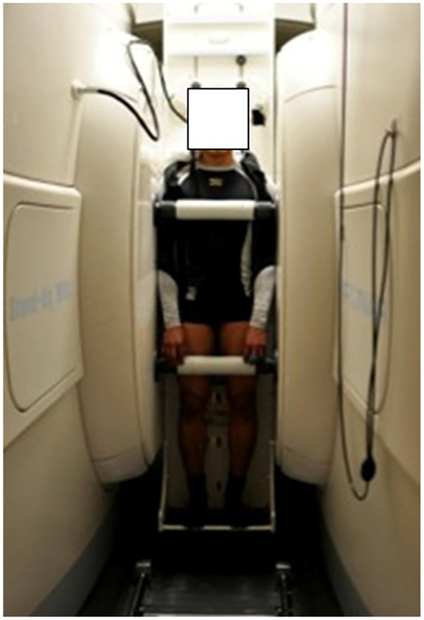
Subjects were allowed ad lib activity prior to being imaged in the mid-morning or early afternoon, but no exercising occurred within 24 h of the testing. Subjects were initially weighed (to within 0.1 kg) with an EatSmart scale (Wyckoff, NJ). As done in a previous upright MRI study, subjects were required to initially lie supine for 30 min to establish an equilibrated baseline supine measurement prior to the onset of the protocol [7]. Subjects then underwent sagittal T2 scans of the lumbar spine while supine and then “upright” at 84° from the horizontal plane, where bolsters and close verbal/visual communication were used to minimize subject motion during scans (Fig. 1). The subjects spent about 8 min standing prior to imaging, which is the amount of time needed to change the subject and the MR machine position to the upright condition. The 8 min of standing served as a preload equilibration time for the IVDs, which was considered an adequate amount of time as supported by in vitro IVD biomechanical studies [13].
Fig. 1.
Subject in the standing position within the upright MRI scanner
After the upright scan, the subject stepped out of the scanner and a Quadrature planar coil (lumbar) was attached to the small of the subject’s back, to enhance image resolution, via Velcro® straps placed horizontally around the center of the MRI coil, waist, and stomach of the subject. Next, a backpack (Dana design, Hoodoo Spine, Rohnert Park, CA; 1.2 kg) was filled with ceramic tiles and worn in the 2-strap condition without any waist or chest straps and adjusted so that the weight was mainly on the back and evenly distributed across both shoulders. The subject had a total load of 10 % BW made up by the MRI coil (2.3 kg), backpack (1.2 kg), and a certain amount of ceramic tiles. Sagittal T2 scans were then repeated. The subjects wore the backpack for 10 min during imaging.
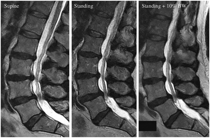
Midsagittal images measured the lumbar spinal length, lumbar IVD heights, and lumbar lordosis (Fig. 2). The midsagittal plane was determined using the spinous processes as a confirmatory landmark. Lumbar spinal length was defined as the distance between the horizontal line drawn at the anterosuperior corner of the L1 vertebral body and the horizontal line drawn at the anterosuperior corner of the S1 vertebral body [14]. For anterior disc height, the distance from the most anterior–inferior corner of the upper vertebrae to the most anterior–superior corner of the lower vertebrae was measured. For posterior disc height, the distance from the most posterior–inferior corner of the upper vertebrae to the most posterior–superior corner of the lower vertebrae was measured. Disc height was obtained from the average of anterior and posterior disc heights according to the Dabbs’ method [15]. Disc compression (mm) was the difference in IVD height between the control supine condition and either the standing or the standing plus 10 % BW condition. Lumbar lordosis was defined as the Cobb angle between the upper surfaces of L1 and S1 vertebral bodies [16]. Distances and angles were measured by one independent observer. All measurements were carried out blinded to any clinical information or subject data. Measurements were made with Osirix software, free version 3.6.1 (University Hospital of Geneva, Switzerland), with a screen resolution of 1,024 × 768 pixels.
Fig. 2.
Midsagittal T2 MRI images of the lumbar spine while supine, standing, and standing bearing 10 % of the subjects’ body weight (BW) in a backpack
To investigate the difference in anterior disc height and posterior disc height over the three conditions (supine, standing, and standing + 10 % BW), we conducted a two-way analysis of variance (ANOVA) with repeated measures for each disc level. To determine the effect of load on lumbar disc height, anterior disc height, and posterior disc height, one-way repeated measures ANOVAs were performed. To measure the effect of loading on lumbar lordosis and lumbar spinal length, one-way repeated measures ANOVAs were performed. All ANOVAs and post hoc analyses were conducted using SPSS software (SPSS, Chicago, IL). A priori power analyses with G*Power (version 3.0.10) indicated a sample size of six subjects to be sufficient (power = 0.80) [17]. An α level of p < 0.05 indicated a statistically significant difference. Data are reported as Mean ± SEM.
To examine inter-observer reproducibility, we compared the measurements of L4–L5 IVD height, lumbar lordosis, and lumbar spinal length for one subject in the standing condition between four independent observers. We investigated intra-observer reproducibility by having one observer record the same measurements for separate instances. Uncertainty was calculated as the standard deviation divided by the mean of the numbers used and then multiplied by 100. Intra-observer analysis had a mean of 11.4 ± 0.3 mm (Mean ± SD) and an uncertainty value of 1.8 %, indicating that 68 % (one standard deviation) of repeated measurements between different observations falls within a 0.6 mm range (Table 1).
Table 1.
Means, Standard Deviations, and Uncertainties of Intra-observer and Inter-observer Measurements of L4–L5 Disc Height, Lumbar Lordosis, and Lumbar Spine Length for one subject in the standing condition
| Inter-observer | Intra-observer | |||
|---|---|---|---|---|
| Mean ± SDb | Uncertainty (%)c | Mean ± SDb | Uncertainty (%)c | |
| L4–L5 Disc height (n = 4)a | 11.6 ± 0.2 mm | 1.8 | 11.4 ± 0.3 mm | 2.9 |
| Lumbar lordosis (n = 4)a | 62.2 ± 2.3° | 3.8 | 59.7 ± 2.9° | 4.8 |
| Lumbar spine length (n = 4)a | 186.4 ± 1.4 mm | 0.7 | 186.6 ± 1.2 mm | 0.6 |
n is defined as the number of measurements done either by one independent observer on four separate occasions (Intra-observer) or by four independent observers (Inter-observer)
SD stands for standard deviation from the mean
Uncertainty (%) was calculated as the standard deviation divided by the mean and then multiplied by 100
Results
The images were obtained expeditiously in a protocol that was well tolerated by the subjects. Inspection of MR images revealed no significant motion artifact.
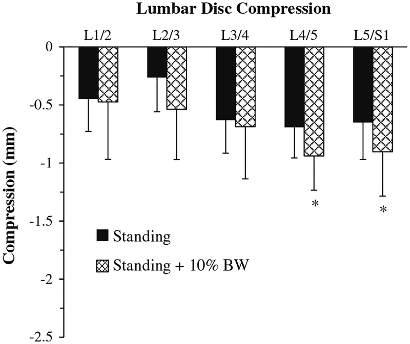
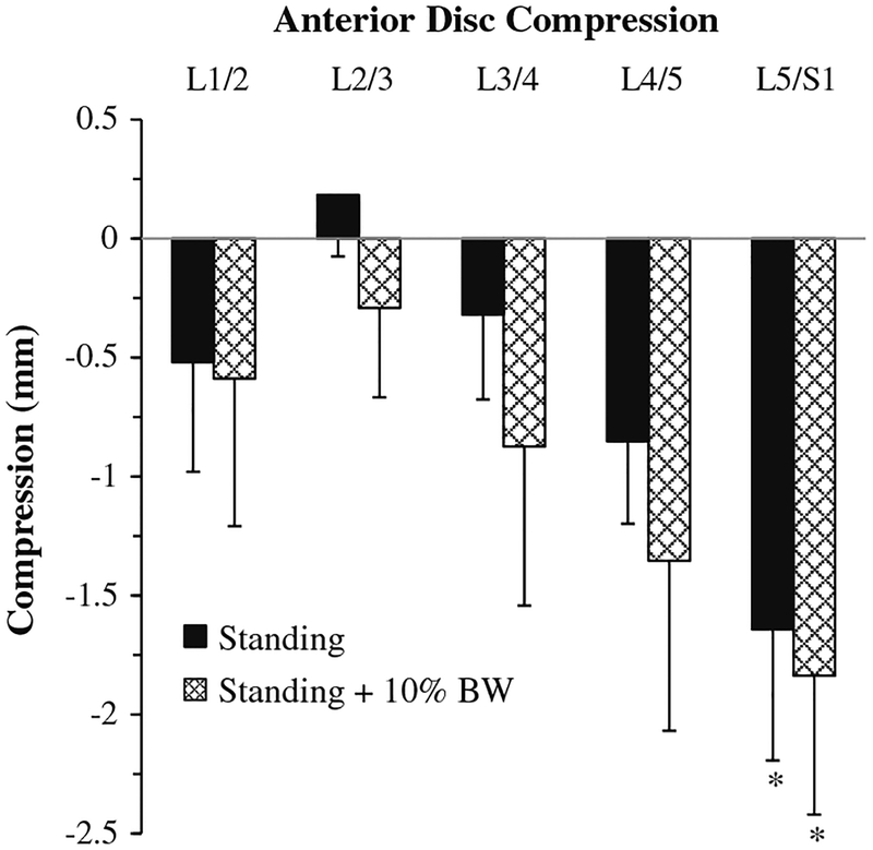
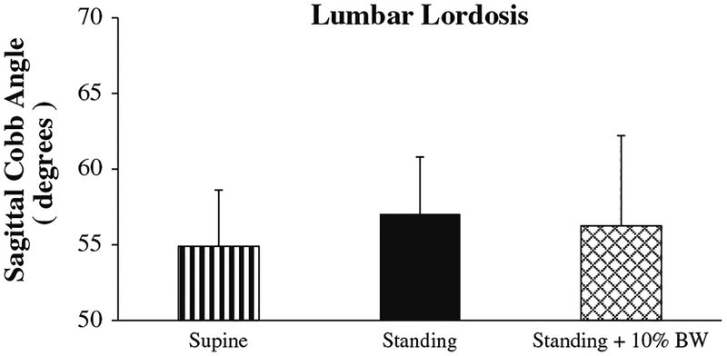
The 10 % BW load significantly compressed the L4–L5 disc by 0.94 ± 0.30 mm (~10 %) and the L5–S1 disc by 0.65 ± 0.32 mm (~10 %) in relation to supine (Fig. 3; p < 0.05). However, lumbar disc heights did not significantly change at other levels with added load (Fig. 3). In addition, as spinal load increased, the anterior height of L5–S1 disc was significantly compressed in relation to supine by 1.64 ± 0.55 mm (14 %) and by 1.84 ± 0.58 mm (16 %) in the standing (p < 0.05) and standing +10 % BW conditions (p < 0.01), respectively (Fig. 4). However, the anterior and posterior disc heights did not significantly change at the other levels with added load (Fig. 4). The magnitude of the anterior disc height compression for L5–S1 in the standing and standing + 10 % BW conditions in relation to supine was significantly greater than the posterior disc height change (p < 0.05 for anterior/posterior and disc height; p > 0.05 loading condition and disc height as well as loading condition and anterior/posterior). In addition, increasing spinal load did not significantly change lumbar spine length or lumbar lordosis (Fig. 5).
Fig. 3.
Lumbar disc compression was significantly greater in the standing plus 10 % body weight (BW) condition compared to the supine control at L4–L5 and L5–S1 (*p < 0.05)
Fig. 4.
Anterior lumbar disc compression was significantly greater in the standing and standing plus 10 % body weight (10 % BW) conditions compared to the supine control at L5–S1 (*p < 0.05)
Fig. 5.
Lumbar lordosis did not significantly change in the standing, and standing plus 10 % body weight (10 % BW) condition compared to the supine control
Discussion
To our knowledge, this is the first adult, upright MRI study to document decreases in lumbar disc heights in comparison to supine posture due to common backpack loads. The similarity of our results to previous studies demonstrates the accuracy of our upright MRI protocol for studying the spine while bearing a load in the functional, upright position. Our results for lumbar disc compression when transitioning from supine to upright are supported by previous studies done only with simulated upright and true upright postures (Table 2) [5, 10]. In addition, the observed lumbar disc height compression due to a 10 % BW load is comparable to that measured in a pediatric population (Table 2) [7].
Table 2.
Comparison of studies on lumbar disc compression
| Current study | Neuschwander et al. [7] | Kimura et al. [5]a | Lee et al. [6]b | Tarantino et al. [10] | |
|---|---|---|---|---|---|
| n (number of subjects) | 6 | 8 | 8 | 15 | 57 |
| Disc compression (standing-supine) (mm) | |||||
| L1/2 | 0.45 ± 0.28 | 0.64 ± 0.18 | 0.1 | ||
| L2/L3 | 0.26 ± 0.29 | 1.21 ± 0.20 | 0.2 | 1.7 | |
| L3/L4 | 0.63 ± 0.29 | 0.86 ± 0.24 | 0.8 | 1.4 | 1.7 |
| L4/L5 | 0.69 ± 0.26 | 0.93 ± 0.30 | 0.8 | 1.0 | |
| L5/S1 | 0.65 ± 0.32 | 1.00 ± 0.30 | 0.5 | 0.9 | |
| Disc compression (standing ± 10 % BW-supine) (mm) | |||||
| L1/L2 | 0.47 ± 0.49 | 1.10 ± 0.14 | |||
| L2/L3 | 0.54 ± 0.43 | 1.63 ± 0.14 | |||
| L3/L4 | 0.69 ± 0.45 | 1.16 ± 0.34 | |||
| L4/L5 | 0.94 ± 0.30 | 1.59 ± 0.27 | |||
| L5/S1 | 0.90 ± 0.38 | 1.76 ± 0.30 | |||
When transitioning from supine to upright with and without a backpack, we measured a compression of the anterior L5–S1 IVD height, while previous research documents an increase in the L5–S1 anterior disc height (1.2 ± 0.9; Mean ± SD) when transitioning from supine to kneeling [6]. In addition, the posterior IVD heights did not significantly compress with increasing spinal load as had been previously observed [6]. Nevertheless, the nonsignificant reductions in posterior disc height for L2–3, L3–4, and L4–5 in our study are similar in magnitude (0.5–1.0 mm) to those previously recorded [6]. In our study, the subjects were standing, while the previous study had their subjects kneeling [6]. The loading state of the spine significantly changes between the standing and kneeling position secondary to the changes in flexion and extension [18]. Therefore, the reason for this contrast in results may be due to the differences in spinal posture when kneeling and standing.
The compression of the L4–L5 and L5–S1 IVDs when transitioning from supine to upright posture with a 10 % BW load suggests that these discs are either bearing more of the load or are more compliant than the other IVDs. Physiologically, the L5–S1 disc has the lowest proteoglycan content and therefore has the lowest swelling pressure, which is the force that counteracts axial loads [19]. Thus, a disc with the lowest swelling pressure would inherently have the least resistance to spinal loading. On the other hand, the highest incidence of lumbar disc disease occurs at L4–5 and L5–S1 and is attributed to the wider range of motion and higher loads experienced by these discs with lumbar flexion or extension [20, 21]. The compression of the L4–L5 and the L5–S1 disc heights in the loaded upright position may be related to the higher risk of disc disease in this region. Further investigation of lumbar discs under upright loaded conditions in the flexed and extended positions may help to understand this proposed relationship between disc compression, movement, and disease.
Previous research documents a decrease in lumbar spinal length while supine with a 50 % BW axial load, which simulates upright posture [5]. This previous study measured lumbar spinal length from T12, instead of L1 as was done in previous research and this study [5, 14]. Taking this into consideration, our lumbar spinal length measurements of 180.6 ± 3.8 and 181.7 ± 4.6 mm for supine and standing, respectively, are similar to their measurements of 191 and 194 mm before and after compression with a 50 % BW axial load, respectively [5]. The lumbar spine shortens over the first 8 h of upright posture, with 26 % of the loss occurring in the first hour [22]. Lengthening the time spent in the loaded conditions might help elucidate the behavior of the lumbar spinal length under spinal load. However, a longer duration of spinal loading will increase the subjects’ discomfort and fatigue potentially contributing to motion artifact in the imaging. The subjects spent about 10 min in each of the spinal loading conditions, which was considered a reasonable preload equilibration time for the IVDs [13].
Increased spinal loading with a backpack did not change lumbar lordosis, which is similar to a pediatric study (Table 3) [7]. In their study, Neuschwander and associates utilized upright MRI to study the lumbar spines of children (ages 11 ± 2 years) wearing 4 kg (~10 % BW), 8 kg (~20 % BW), and 12 kg (~30 % BW) backpacks. However, Chow and associates, using a five-camera motion system, reported a decrease in lumbar lordosis in teenage males wearing similar backpack loads of 10 % BW as well as 15 and 20 % BW. The camera motion system utilized skin markers, which is a less accurate measurement tool than upright MRI and may be influenced by body fat distribution and skin/soft tissue deformation. Alternatively, in an adult population, backpacks heavier than 10 % BW load may be necessary to observe a decrease in lumbar lordosis.
Table 3.
Comparison of studies on lumbar lordosis
This novel upright MRI protocol for studying the loaded spine, in vivo, has many clinical and research applications. The measurements of IVD height change give us a functional measure of mechanical compressibility, which can be correlated to structural and compositional changes [23]. In addition, the measurement uncertainty of IVD compression (0.6 mm) establishes the lower limit of resolution for the clinical application of this MRI protocol. Clinically this protocol could be utilized to study the effectiveness of therapeutic interventions to improve spine health [23]. Given recent research documenting limitations in the diagnostic accuracy of non-functional supine MRI for detecting IVD herniation and spinal stenosis, we believe that our functional, upright MRI protocol could improve both the sensitivity and specificity of this imaging modality [24]. Current research is underway, using this protocol, to study the effects of microgravity on the lumbar spine of astronaut crewmembers after prolonged microgravity exposure on the International Space Station, because low back pain is a serious concern among the astronaut corps [25–27].
Our study has some limitations. We did not rigorously control for the time of day when the subjects were tested. This was because of logistical reasons related to availability of the subjects and the MRI scanner. The subjects generally were tested a few hours before or after noon. On the day of testing, the subjects did not exercise or participate in an unusual level of activity prior to testing. We acknowledge that throughout the day the spine shortens, but the most height decrease occurs during the first hour after rising [28, 29]. Conversely, the majority of that IVD height loss is regained within the first hour after lying supine [28]. As has been done in previous upright MRI research, we required 30 min of supine rest prior to imaging to establish an equilibrated baseline supine measurement prior to the onset of the protocol [7]. In addition, due to the weight of the backpack and the time spent standing, there is the possibility of motion artifact in the images. Fortunately, the images did not show any significant evidence of motion. We minimized the time spent standing with and without the backpack, and provided the subjects with adequate rest breaks between imaging cycles. The subjects also had bolsters within the MR scanner for stabilization (Fig. 1). In addition, in the MRIs of one subject, who was a flight-ready NASA crewmember, revealed asymptomatic, multilevel spinal pathology (Fig. 2). The presence of these spinal abnormalities in MRI is common in asymptomatic adults [30].
Conclusions
The L4–L5 and L5–S1 IVD heights compress, particularly anteriorly, when transitioning from supine to upright position with a 10 % BW backpack. This IVD height compression may be due to higher forces applied at these levels or due to different material properties that result in a higher compliance. This study is the first radiographic analysis to describe lumbar spine deformation in an adult population wearing common backpack loads. The novel, upright MRI protocol described will allow for future comparison of spinal therapeutic interventions in a more functional, weight-bearing environment.
Acknowledgments
The authors would like to gratefully acknowledge the participation of our six subjects. We thank JR Bachman for technical support and help with revisions. We thank Dr. Brandon Macias for help with manuscript editing, Dr. Stephen Chiang for his assistance with MR imaging and analysis, Clifford Mao for his help with the repeatability measurements, Dr. Lin Liu for her help with the statistical analysis, and Dr. Steven Garfin for support. This study was supported by the National Aeronautics and Space Administration Grant NNX10AM18G and the UCSD Clinical Translational Research Institute fellowship award.
Footnotes
Conflict of interest None.
Contributor Information
Stephen Shymon, Department of Orthopaedic Surgery, University of California, San Diego, 350 Dickinson Street, Suite 121, San Diego, CA 92103-8894, USA.
Alan R. Hargens, Department of Orthopaedic Surgery, University of California, San Diego, 350 Dickinson Street, Suite 121, San Diego, CA 92103-8894, USA
Lawrence A. Minkoff, Fonar Corporation, Melville, NY, USA
Douglas G. Chang, Email: dchang@ucsd.edu, Department of Orthopaedic Surgery, University of California, San Diego, 350 Dickinson Street, Suite 121, San Diego, CA 92103-8894, USA.
References
- 1.Alyas F, Connell D, Saifuddin A (2008) Upright positional MRI of the lumbar spine. Clin Radiol 63:1035–1048 [DOI] [PubMed] [Google Scholar]
- 2.Saifuddin A, Blease S, MacSweeney E (2003) Axial loaded MRI of the lumbar spine. Clin Radiol 58:661–671 [DOI] [PubMed] [Google Scholar]
- 3.Alexander LA, Hancock E, Agouris I et al. (2007) The response of the nucleus pulposus of the lumbar intervertebral discs to functionally loaded positions. Spine (Phila Pa 1976) 32:1508–1512 [DOI] [PubMed] [Google Scholar]
- 4.Madsen R, Jensen TS, Pope M et al. (2008) The effect of body position and axial load on spinal canal morphology: an MRI study of central spinal stenosis. Spine (Phila Pa 1976) 33:61–67 [DOI] [PubMed] [Google Scholar]
- 5.Kimura S, Steinbach GC, Watenpaugh DE, Hargens AR (2001) Lumbar spine disc height and curvature responses to an axial load generated by a compression device compatible with magnetic resonance imaging. Spine (Phila Pa 1976) 26:2596–2600 [DOI] [PubMed] [Google Scholar]
- 6.Lee SH, Hargens AR, Fredericson M, Lang P (2003) Lumbar spine disc heights and curvature: upright posture vs. supine compression harness. Aviat Sp Env Med 74:512–516 [PubMed] [Google Scholar]
- 7.Neuschwander TB, Cutrone J, Macias BR et al. (2010) The effect of backpacks on the lumbar spine in children: a standing magnetic resonance imaging study. Spine (Phila Pa 1976) 35:83–88 [DOI] [PubMed] [Google Scholar]
- 8.Dimitriadis A, Papagelopoulos P, Smith F et al. (2011) Intervertebral disc changes after 1 h of running: a study on athletes. J Int Med Res 39:569–579 [DOI] [PubMed] [Google Scholar]
- 9.Macias BR, Cao P, Watenpaugh DE, Hargens AR (2007) LBNP treadmill exercise maintains spine function and muscle strength in identical twins during 28-day simulated microgravity. J Appl Physiol 102:2274–2278 [DOI] [PubMed] [Google Scholar]
- 10.Tarantino U, Fanucci E, Iundusi R et al. (2012) Lumbar spine MRI in upright position for diagnosing acute and chronic low back pain: statistical analysis of morphological changes. J Orthop Traumatol 14:15–22 [DOI] [PMC free article] [PubMed] [Google Scholar]
- 11.Rodríguez-Soto AE, Jaworski R, Jensen A et al. (2013) Effect of load carriage on lumbar spine kinematics. Spine (Phila Pa 1976) 38:783–791 [DOI] [PubMed] [Google Scholar]
- 12.Shymon S, Hargens AR, Minkoff LA, Chang DG (2013) Body position and backpack loading: an upright magnetic resonance imaging study of the adult lumbar spine. Poster session presented at: Federation of American Societies for Experimental Biology Conference, Boston [Google Scholar]
- 13.Recuerda M, Périé D, Gilbert G, Beaudoin G (2012) Assessment of mechanical properties of isolated bovine intervertebral discs from multi-parametric magnetic resonance imaging. BMC Musculoskelet Disord 13:195. [DOI] [PMC free article] [PubMed] [Google Scholar]
- 14.Cao P, Kimura S, Macias BR, Ueno T, Watenpaugh DE, Hargens AR (2005) Exercise within lower body negative pressure partially counteracts lumbar spine deconditioning associated with 28-day bed rest. J Appl Physiol 99:39–44 [DOI] [PubMed] [Google Scholar]
- 15.Dabbs VM, Dabbs LG (1990) Correlation between disc height narrowing and low-back pain. Spine (Phila Pa 1976) 15:1366–1369 [DOI] [PubMed] [Google Scholar]
- 16.Andersson GBJ, Murphy RW, Ortengren R, Nachemson AL (1979) The influence of backrest inclination and lumbar support on lumbar lordosis. Spine (Phila Pa 1976) 4:52–58 [DOI] [PubMed] [Google Scholar]
- 17.Faul F, Erdfelder E, Lang AG, Buchner A (2007) G*Power 3: a flexible statistical power analysis program for the social, behavioral, and biomedical sciences. Behav Res Methods 39:175–191 [DOI] [PubMed] [Google Scholar]
- 18.Schmid MR, Dueweii S, Romanowski B, Hodler J (1999) Changes in cross-sectional measurements of the spinal canal and intervertebral foramina as a function of body position: in vivo studies on an open-configuration MR system. Am J Roentgenol 1095–1102 [DOI] [PubMed] [Google Scholar]
- 19.Urban JP, McMullin JF (1988) Swelling pressure of the lumbar intervertebral discs: influence of age, spinal level, composition, and degeneration. Spine (Phila Pa 1976) 13:179–187 [DOI] [PubMed] [Google Scholar]
- 20.Wisneski RJ, Garfin SR, Rothman RH (1999) Lumbar disc disease In: Herkowitz HN, Garfin SR, Balderston RA (eds) Spine, vol 1 WB Saunders, Philadelphia, pp 613–679 [Google Scholar]
- 21.White AA, Panjabi MM (1990) Clinical biomechanics of the spine, 2nd edn JB Lippincott, Philadelphia [Google Scholar]
- 22.Krag M, Cohen M, Haugh L, Pope M (1990) Body height change during upright and recumbent posture. Spine (Phila Pa 1976) 15:202–207 [DOI] [PubMed] [Google Scholar]
- 23.Lotz JC, Haughton V, Boden SD et al. (2012) New treatments and imaging strategies in degenerative disease of the intervertebral disks. Radiology 264:6–19 [DOI] [PubMed] [Google Scholar]
- 24.Rijn RM, Tulder MW, Verhagen AP et al. (2012) Magnetic resonance imaging for diagnosing lumbar spinal pathology in adult patients with low back pain or sciatica: a diagnostic systematic review. Eur Spine J 21:220–227 [DOI] [PMC free article] [PubMed] [Google Scholar]
- 25.Hargens AR, Bhattacharya R, Schneider SM (2013) Space physiology VI: exercise, artificial gravity, and countermeasure development for prolonged space flight. Eur J Appl Physiol 113:2183–2192 [DOI] [PubMed] [Google Scholar]
- 26.Kerstman EL, Scheuring RA, Barnes MG, DeKorse TB, Saile LG (2012) Space adaptation back pain: a retrospective study. Aviat Sp Env Med 83:2–7 [DOI] [PubMed] [Google Scholar]
- 27.Sayson JV, Hargens AR (2008) Pathophysiology of low back pain during exposure to microgravity. Aviat Sp Env Med 79:364–373 [DOI] [PubMed] [Google Scholar]
- 28.Styf JR, Ballard RE, Fechner K, Watenpaugh DE, Kahan NJ, Hargens AR (1997) Height increase, neuromuscular function, and back pain during 6 degrees head-down tilt with traction. Aviat Sp Env Med 68:24–29 [PubMed] [Google Scholar]
- 29.Tyrrell AR, Reilly T, Troup JD (1985) Circadian variation in stature and the effects of spinal loading. Spine (Phila Pa 1976) 10:161–164 [DOI] [PubMed] [Google Scholar]
- 30.Jensen MC, Brant-Zawadzki MN, Obuchowski N et al. (1994) Magnetic resonance imaging of the lumbar spine in people without back pain. N Engl J Med 331:69–73 [DOI] [PubMed] [Google Scholar]


