Abstract
With the ever-increasing laser power and repetition rate, thermal control of laser media is becoming increasingly important. Except for widely used air cooling or a bonded heat sink, water cooling of a laser medium is more effective in removing waste heat. However, how to protect deliquescent laser media from water erosion is a challenging issue. Here, novel waterproof coatings were proposed to shield Nd:Glass from water erosion. After clarifying the dependence of the waterproof property of single layers on their microstructures and pore characteristics, nanocomposites that dope SiO2 in HfO2 were synthesized using an ion-assisted co-evaporation process to solve the issue of a lack of a high-index material that simultaneously has a dense amorphous microstructure and wide bandgap. Hf0.7Si0.3O2/SiO2 multifunctional coatings were finally shown to possess an excellent waterproof property, high laser-induced damage threshold (LIDT) and good spectral performance, which can be used as the enabling components for thermal control in high-power laser cavities.
Subject terms: Applied optics, Optical materials and structures
Although hydrophobic coatings have been well studied regarding their water repellency or moisture proof property1–3, they are not applicable under cooling water conditions4–9, where coatings are immersed in water over long periods of time. For waterproof coatings, Murahara has proposed a hard-water-resistant coating that can protect KDP from dissolving10. However, his approach was limited to the use of SiO2 single layers and failed to meet the requirement of multilayers in which both high- and low-index materials are needed11,12. Although the microstructures of widely used optical coatings prepared using physical vapor deposition techniques have been extensively studied regarding stress, the refractive index, humidity-induced spectral shift and so on13–15, the correlation between the coating microstructure and waterproof property has not been specifically addressed. To meet the challenging requirement of waterproof cavity mirrors, the dependence of water resistance on the microstructure of single- and multilayers was studied first. On the basis of these results, microstructure and bandgap engineering were performed to synthesize nanocomposite-based cavity mirrors with exceptional multifunctionality, which has been previously unattainable. The proposed multifunctional coating can be used as an enabling technology to realize high-repetition-rate laser inertial confinement fusion, which can then be used as a fusion power plant.
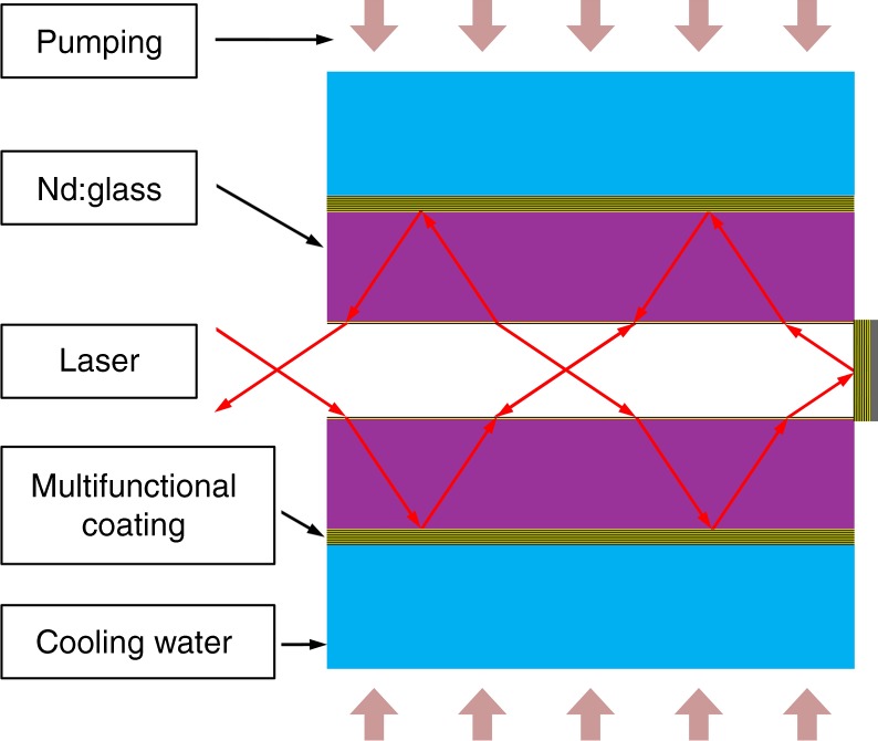
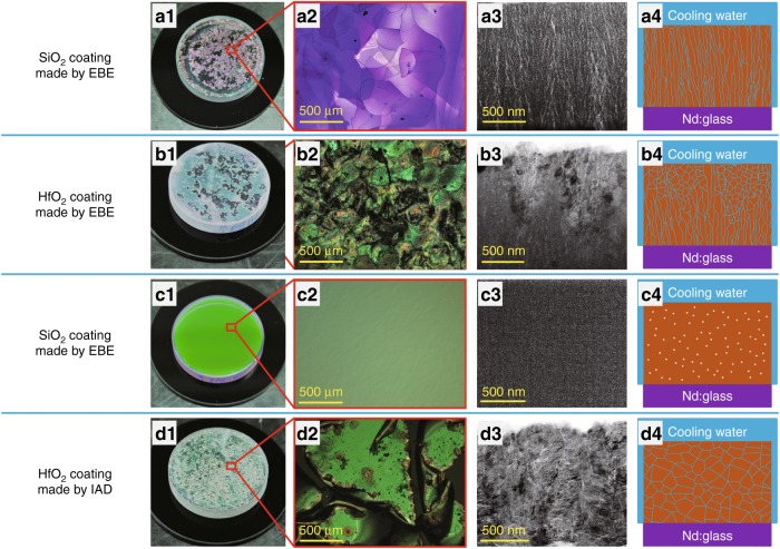
The schematic of a water-cooled Nd:Glass laser cavity is shown in Fig. 1. SiO2 and HfO2 are the dominant low and high index materials for the near-infrared region16,17. The water resistance of single layers was investigated first. SiO2 and HfO2 single layers were prepared using electron beam evaporation (EBE) and ion assisted deposition (IAD) processes, respectively. The coatings prepared by the EBE process had a porous microstructure and failed to protect Nd:Glass from water erosion. The SiO2 layers peeled off of Nd:Glass substrates with heating in a water bath over a period of several hours. The eroded morphologies of the SiO2 layer were recorded using a camera and an optical microscope, as shown in Fig. 2a1, a2. A cross-sectional transmission electron microscopy (TEM) image of a Ta2O5 coating was used instead to reveal the microstructure of the SiO2 coating, where the brighter areas represent pores. It can be seen that an abundance of pores exist between the columns, which are open, elongated, and oriented perpendicular to the coating surface, as shown in Fig. 2a3. Water can quickly penetrate into the Nd:Glass surface through these open microscopic channels by capillarity, which is schematically illustrated in Fig. 2a4. Erosion of the Nd:Glass surface led to the delamination of SiO2 coatings. For porous HfO2 layers on Nd:Glass substrates, visible damage was observed after testing the samples in a hot water bath for several days, as shown in Fig. 2b1, b2. The relatively longer survival time of these layers is attributed to their different microstructure and pore characteristics. Figure 2b3 shows that there is a transition from an amorphous microstructure to a polycrystalline microstructure as the coating grows thicker. Compared to the elongated pores in the amorphous coatings, the polycrystalline microstructure in the upper part of the HfO2 coating results in a substantially lower number of long and open pores. Water diffusion through the HfO2 layer is delayed due to these crisscrossing channels, which are illustrated in Fig. 2b4. This comparison shows that for porous coatings, the polycrystalline microstructure offers advantages with respect to water resistant compared to an amorphous microstructure.
Fig. 1.

Schematic of a water-cooled Nd:Glass laser cavity
Fig. 2. SiO2 and HfO2 coatings prepared by EBE and IAD processes.
a1–d1 Optical photographs, a2–d2 microscopic images, a3–d3 cross-sectional TEM images, a4–d4 schematics of water diffusion along the nanopores in the coatings. It is worth noting that the TEM images of the SiO2 coatings are replaced using TEM images of Ta2O5 coatings to better reveal the microstructure. TEM images and the electron diffraction pattern of the SiO2 coatings are shown in Figure S2 and S3 in the Supplementary Information
IAD can significantly reduce the number of pores in coatings via energetic ion bombardment during film growth. The densified SiO2 layers did not peel off of Nd:Glass substrates with water bath heating for several months, as shown in Fig. 2c1, c2. Although no visible pores can be identified in the cross-sectional TEM image in Fig. 2c3, some nanometer-sized closed pores are assumed to be embedded in the SiO2 coatings18. These closed nanopores are isolated from each other, as schematically presented in Fig. 2c4. There are no channels for water diffusion in dense amorphous SiO2 coatings, so Nd:Glass substrates can be protected from corrosion. HfO2 coatings prepared by the IAD process exhibited a polycrystalline microstructure. Although these coatings were also very dense, they failed to protect Nd:Glass from water erosion. Figure 2d1, d2 shows that HfO2 coatings were severely damaged after immersion in a hot water bath for several weeks. The cross-sectional TEM image shown in Fig. 2d3 shows an abundance of complex grain boundaries with nanopores. These nanopores are connected with each other, resulting in a network of zigzag channels from which water can find ways to reach and erode the Nd:Glass, as shown in Fig. 2d4. This comparison reveals that for dense single layers, an amorphous microstructure rather than a crystallized microstructure exhibits a good waterproof property.
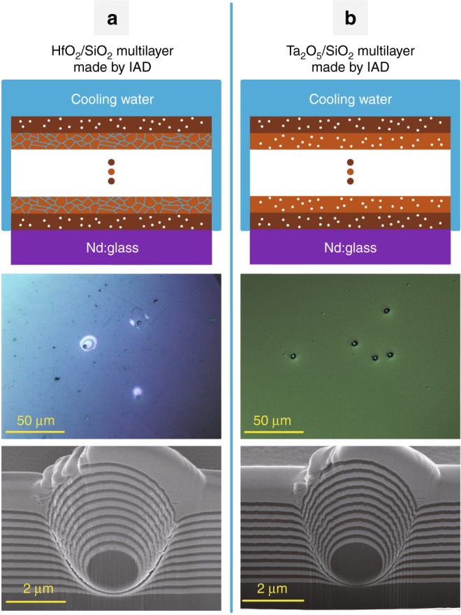
The water resistance of multilayers was further studied. HfO2/SiO2 multilayers were prepared using the IAD process. After immersing samples in a hot water bath for several months, severe delamination was observed. The erosion morphologies of HfO2/SiO2 multilayers are shown in Figure S1 in the Supplementary Information. This result means that a multilayer does not possess a good waterproof property when one coating material is water resistant while another is not. Although water penetration perpendicular to the surface might be effectively blocked by dense amorphous SiO2 layers, water can diffuse along zigzag channels in HfO2 layers that are parallel to the surface, as illustrated in Fig. 3a1. Since there are always extrinsic defects in real multilayer coatings19, water molecules in HfO2 layers could reach and corrode the Nd:Glass substrate along defect-induced channels, which could be linked to the Nd:Glass surface.
Fig. 3.

Schematic of water diffusion in nanopores; optical photographs; cross-sectional SEM images of HfO2/SiO2 and Ta2O5/SiO2 multilayers prepared by an IAD process
To verify the influence of defects, artificial nodules created from 2μm silica microspheres on Nd:Glass were tested in a hot water bath20. Figure 3a2 shows that blisters were observed at artificial nodules after serval hours of water bath heating. The cross-sectional scanning electron microscopy (SEM) image shown in Fig. 3a3 shows that large pores at nodular boundaries are open to the Nd:Glass surface, through which water can reach and erode the Nd:Glass. For comparison, artificial nodules in dense amorphous Ta2O5/SiO2 multilayers were also prepared using the IAD process. Because both Ta2O5 and SiO2 have a dense amorphous microstructure with closed nanopores, there are no channels for water diffusion along defect-induced channels, as shown in Fig. 3b. The Ta2O5/SiO2 multilayers did not peel off from the Nd:Glass substrate for the same water bath time. However, the LIDT for the Ta2O5/SiO2 multilayers is much lower than that for the HfO2/SiO2 multilayers because the bandgap of Ta2O5 is ~30% less than that of HfO221. It is desirable to find novel coating materials to achieve an excellent waterproof property without sacrificing the LIDT.
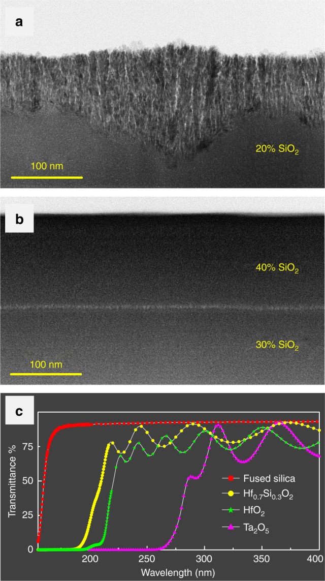
Nanocomposites that dope SiO2 into HfO2 were synthesized using an ion-assisted co-evaporation process to obtain a dense amorphous microstructure, wider bandgap and higher LIDT. The higher the SiO2 concentration in HfxSi1−xO2 nanocomposites, the better the resistance to crystallization. SiO2 (20%) in HfxSi1−xO2 nanocomposites was not enough to suppress crystallization. Figure 4a shows that severe crystallization occurred after an initial amorphous growth phase. HfxSi1−xO2 nanocomposites with a 30% SiO2 concentration or higher maintained a dense amorphous microstructure, as shown in Fig. 4b. From the aspects of coating design and electric-field control, it is optimal to use an amorphous HfxSi1−xO2 nanocomposite, which has the highest refractive index. Therefore, an Hf0.7Si0.3O2 nanocomposite layer was used. The ultraviolet transmission spectra of the Hf0.7Si0.3O2, SiO2, HfO2 and Ta2O5 layers are compared in Fig. 4c. Together with the reflection spectrum, the bandgap of the Hf0.7Si0.3O2 nanocomposite layer was derived to be 6.4 eV using the Tauc algorithm22. The bandgap of this is much wider than that Ta2O5 and its LIDT should also be much higher.
Fig. 4.

Comparisons among HfxSi1−xO2 nanocomposite films and oxide films. Cross-sectional TEM images of HfxSi1−xO2 nanocomposite layers with a 20% SiO2 concentration, b 30 and 40% SiO2 concentration. c Transmission spectra of single layers of Hf0.7Si0.3O2, SiO2, HfO2 and Ta2O5
Hf0.7Si0.3O2/SiO2 cavity mirrors were then prepared with a high reflectance at 1053 nm for s-polarization at the Brewster angle and a high transmittance at 802 nm for normal incidence23. Their LIDT was 34 ± 4 J/cm2 according to the method described by Borden et al.24, which is almost three times higher than the LIDT of Ta2O5/SiO2 cavity mirrors. This value is close to the LIDT value of ~42 ± 4 J/cm2 for a bare Nd:Glass substrate. By carefully controlling defects during the preparation of Nd:Glass substrates and coatings, Hf0.7Si0.3O2/SiO2 cavity mirrors can protect Nd:Glass from corrosion for over 1 year, even in a hot water bath.
Here, the influence of the pore characteristics on the waterproof property of single layers was determined. Dense amorphous coatings are water resistant because no channels exist for water diffusion. If one material is water resistant while another is not, the multilayer does not possess a waterproof property when extrinsic defects are present. HfxSi1−xO2 nanocomposites were then synthesized to generate Hf0.3Si0.7O2/SiO2 cavity mirrors with the multifunctionality of an excellent waterproof property, high LIDT and good spectral performance.
Materials and methods
Preparation of laser coatings
SiO2, HfO2 and Ta2O5 single layers with a thickness of ~500 nm were deposited on Nd:Glass substrates using EBE and IAD processes. The deposition temperature was approximately 393 K, and the chamber was pumped down to a base pressure of 2.3 × 10⁻⁴ Pa. The IAD process employed was based on an RF-type ion source with a working condition of 600 V and 600 mA. Further details of the deposition process can be found in our previous paper25. HfxSi1−xO2 nanocomposites were prepared using an ion-assisted co-evaporation process including annealing at 600 °C to reduce their absorption. A schematic of the ion-assisted co-evaporation process is shown in Figure S4 in the Supplementary Information. Further details of the process can be found in a previous paper26.
Characterization of laser coatings
The water resistance of the prepared coatings was evaluated by immersing samples in a temperature-controlled water bath. Compared to a practical water-cooling situation, the temperature of the water bath was set to 90 °C to accelerate the failure process.
The microstructure of SiO2, HfO2 and Ta2O5 single layers was characterized using TEM. However, due to the low contrast of electron scattering between pores and the SiO2 matrix, it is difficult to clearly observe its microstructure. Based on the fact that the SiO2 coating has the same microstructure as a Ta2O5 coating27, a cross-sectional TEM image of a Ta2O5 coating was used instead to determine the microstructure of the SiO2 coating. For comparison, TEM images and electron diffraction patterns of SiO2 coatings are shown in Figures S2 and S3 in the Supplementary Information. The electron diffraction pattern of the crystallized part of the HfO2 coating is also shown in Figure S2 in the Supplementary Information. The microstructure of the artificial nodules in the HfO2/SiO2 and Ta2O5/SiO2 multilayers was determined by cutting them through the middle using focused ion beam technology. Then, the cross-sectional images shown in Fig. 3 were taken using SEM.
The concentration of SiO2 in HfxSi1−xO2 nanocomposites was determined by fitting the refractive indices of the HfxSi1−xO2 single layers using a Lorentz–Lorentz model28.
Supplementary information
Acknowledgements
This work was supported by the National Natural Science Foundation of China (Nos. 61522506, 51475335, 61621001 and 91536111), Joint Sino-German Research Project (No. GZ1275), National Program on Key Research Project (No. 2016YFA0200900), Major projects of Science and Technology Commission of Shanghai (No. 17JC1400800), “Shu Guang” project supported by Shanghai Municipal Education Commission and Shanghai Education (No. 17SG22), Development Foundation National Key Scientific Instrument and Equipment Development Project (No. 2014YQ090709).
Conflict of interest
The authors declare that they have no conflict of interest.
Supplementary material
Supplementary information is available for this paper at 10.1038/s41377-018-0118-6.
References
- 1.Voïtchovsky K, Giofrè D, Segura JJ, Stellacci F, Ceriotti M. Thermally-nucleated self-assembly of water and alcohol into stable structures at hydrophobic interfaces. Nat. Commun. 2016;7:13064. doi: 10.1038/ncomms13064. [DOI] [PMC free article] [PubMed] [Google Scholar]
- 2.Liu KC, et al. A flexible and superhydrophobic upconversion- luminescence membrane as an ultrasensitive fluorescence sensor for single droplet detection. Light Sci. Appl. 2016;5:e16136. doi: 10.1038/lsa.2016.136. [DOI] [PMC free article] [PubMed] [Google Scholar]
- 3.Lee H, Alcaraz ML, Rubner MF, Cohen RE. Zwitter- wettability and antifogging coatings with frost- resisting capabilities. ACS Nano. 2013;7:2172–2185. doi: 10.1021/nn3057966. [DOI] [PubMed] [Google Scholar]
- 4.Jauregui C, Limpert J, Tünnermann A. High-power fibre lasers. Nat. Photonics. 2013;7:861–867. doi: 10.1038/nphoton.2013.273. [DOI] [Google Scholar]
- 5.Fan ZW, et al. High beam quality 5 J, 200 Hz Nd:YAG laser system. Light Sci. Appl. 2017;6:e17004. doi: 10.1038/lsa.2017.4. [DOI] [PMC free article] [PubMed] [Google Scholar]
- 6.Tauer J, Kofler H, Wintner E. Laser-initiated ignition. Laser Photonics Rev. 2010;4:99–122. doi: 10.1002/lpor.200810070. [DOI] [Google Scholar]
- 7.Weber R, Neuenschwander B, Mac Donald M, Roos MB, Weber HP. Cooling schemes for longitudinally diode laser-pumped Nd:YAG rods. IEEE J. Quantum Electron. 1998;34:1046–1053. doi: 10.1109/3.678602. [DOI] [Google Scholar]
- 8.Waldburger D, et al. High-power 100 fs semiconductor disk lasers. Optica. 2016;3:844–852. doi: 10.1364/OPTICA.3.000844. [DOI] [Google Scholar]
- 9.Tokita S, Murakami M, Shimizu S, Hashida M, Sakabe S. Liquid-cooled 24 W mid-infrared Er:ZBLAN fiber laser. Opt. Lett. 2009;34:3062–3064. doi: 10.1364/OL.34.003062. [DOI] [PubMed] [Google Scholar]
- 10.Murahara M, Sato N, Ikadai A. Hard protective waterproof coating for high-power laser optical elements. Opt. Lett. 2005;30:3416–3418. doi: 10.1364/OL.30.003416. [DOI] [PubMed] [Google Scholar]
- 11.Jung BH, Lee DK, Sohn SH, Kim HS. Thermal, dielectric, and optical properties of neodymium borosilicate glasses for thick films. J. Am. Ceram. Soc. 2003;86:1202–1204. doi: 10.1111/j.1151-2916.2003.tb03448.x. [DOI] [Google Scholar]
- 12.Šuminas R, Tamošauskas G, Valiulis G, Dubietis A. Spatiotemporal light bullets and supercontinuum generation in β-BBO crystal with competing quadratic and cubic nonlinearities. Opt. Lett. 2016;41:2097–2100. doi: 10.1364/OL.41.002097. [DOI] [PubMed] [Google Scholar]
- 13.Vinnichenko M, et al. Highly dense amorphous Nb2O5 films with closed nanosized pores. Appl. Phys. Lett. 2009;95:081904. doi: 10.1063/1.3212731. [DOI] [Google Scholar]
- 14.Tolenis T, et al. Next generation highly resistant mirrors featuring all-silica layers. Sci. Rep. 2017;7:10898. doi: 10.1038/s41598-017-11275-0. [DOI] [PMC free article] [PubMed] [Google Scholar]
- 15.Lupoi R, et al. Hardfacing steel with nanostructured coatings of Stellite-6 by supersonic laser deposition. Light Sci. Appl. 2012;1:e10. doi: 10.1038/lsa.2012.10. [DOI] [Google Scholar]
- 16.Wang AQ, et al. HfO2/SiO2 multilayer enhanced aluminum alloy-based dual-wavelength high reflective optics. Thin. Solid. Films. 2015;592:232–236. doi: 10.1016/j.tsf.2015.04.032. [DOI] [Google Scholar]
- 17.Cheng XB, et al. The effect of an electric field on the thermomechanical damage of nodular defects in dielectric multilayer coatings irradiated by nanosecond laser pulses. Light Sci. Appl. 2013;2:e80. doi: 10.1038/lsa.2013.36. [DOI] [Google Scholar]
- 18.Stenzel O. A model for calculating the effect of nanosized pores on refractive index, thermal shift and mechanical stress in optical coatings. J. Phys. D Appl. Phys. 2009;42:055312. doi: 10.1088/0022-3727/42/5/055312. [DOI] [Google Scholar]
- 19.Stolz CJ, et al. Substrate and coating defect planarization strategies for high-laser-fluence multilayer mirrors. Thin. Solid. Films. 2015;592:216–220. doi: 10.1016/j.tsf.2015.04.047. [DOI] [Google Scholar]
- 20.Cheng XB, et al. Contribution of angle-dependent light penetration to electric-field enhancement at nodules in optical coatings. Opt. Lett. 2017;42:2086–2089. doi: 10.1364/OL.42.002086. [DOI] [PubMed] [Google Scholar]
- 21.Jensen L, Mende M, Schrameyer S, Jupé M, Ristau D. Role of two-photon absorption in Ta2O5 thin films in nanosecond laser-induced damage. Opt. Lett. 2012;37:4329–4331. doi: 10.1364/OL.37.004329. [DOI] [PubMed] [Google Scholar]
- 22.Cody GD, Tiedje T, Abeles B, Brooks B, Goldstein Y. Disorder and the optical-absorption edge of hydrogenated amorphous silicon. Phys. Rev. Lett. 1981;47:1480–1483. doi: 10.1103/PhysRevLett.47.1480. [DOI] [Google Scholar]
- 23.Cheng XB, et al. Optimal coating solution for a compact resonating cavity working at Brewster angle. Opt. Express. 2016;24:24313–24320. doi: 10.1364/OE.24.024313. [DOI] [PubMed] [Google Scholar]
- 24.Borden, M. R., et al. Improved method for laser damage testing coated optics. In Proceedings of SPIE 5991, Laser-Induced Damage in Optical Materials. p59912A (SPIE, Boulder, 2006).
- 25.Wang ZS, et al. Interfacial damage in a Ta2O5/SiO2 double cavity filter irradiated by 1064 nm nanosecond laser pulses. Opt. Express. 2013;21:30623–30632. doi: 10.1364/OE.21.030623. [DOI] [PubMed] [Google Scholar]
- 26.Liu F, et al. Interface and material engineering for zigzag slab lasers. Sci. Rep. 2017;7:16699. doi: 10.1038/s41598-017-16968-0. [DOI] [PMC free article] [PubMed] [Google Scholar]
- 27.Kaiser N. Review of the fundamentals of thin-film growth. Appl. Opt. 2002;41:3053–3060. doi: 10.1364/AO.41.003053. [DOI] [PubMed] [Google Scholar]
- 28.Cheng, X. B., Fan, B., Haruo, T. & Wang Z. S. Optical and structural properties of NbxSiyO composite films prepared by metallic co-sputtering process. In Proceedings Volume 7101, Advances in Optical Thin Films III. p71011G (SPIE, Glasgow, 2008).
Associated Data
This section collects any data citations, data availability statements, or supplementary materials included in this article.


