
Figure 4.
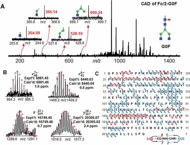
Mass spectrum, fragment ions, and sequence map of Fc/2-G0F glycoform fragmentation. A Mass spectrum of CAD fragmentation of Fc/2-G0F. Inset, representative fragment ions from labile glycan fragmentation. B Representative fragment ions from ECD and CAD fragmentations localizing the glycosylation site at Asn61. C Fc/2 subunit sequence map showing bond cleavages and glycosylation site. The glycosylation site at Asn61 is highlighted in red. The amino acid number here is based on the Fc/2 subunit sequence.