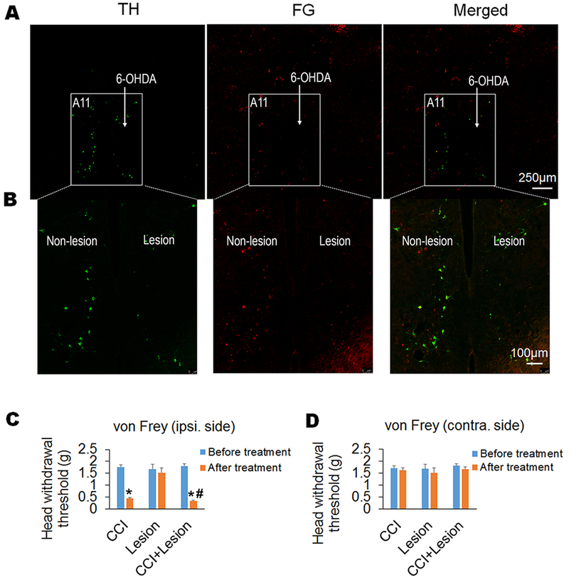
Figure 5. Chemical lesion-produced specific ablation of A11 dopaminergic neurons exacerbates trigeminal neuropathic pain.

(A and B) Chemical lesion of A11 nucleus was conducted by injecting 6-OHDA into unilateral A11 nucleus of mice after systemic injection of desipramine (i.p.) to protect noradrenergic neurons. Double staining of harvested A11-containing brain slices with antibodies against TH and the retrograde tracer FG (injected into Sp5C) showed that the chemical lesion specifically ablated A11 dopaminergic neurons, which projected to Sp5C. The expression of TH and/or FG in bilateral A11 was showed at low magnification in (A). And the expression of TH and/or FG in the non-lesion and lesion sides of A11 within the respective boxed areas in (A) was showed at high magnification in (B). (C and D) The chemical lesion-produced specific ablation of A11 dopaminergic neurons further reduced CCI-decreased head withdrawal threshold in the ipsilateral side compared to the CCI-ION alone group (C), but the A11 lesion had no effect on the head withdrawal threshold in the contralateral side of the mice with CCI-ION (D). As a control, the unilateral A11 lesion alone did not affect the head withdrawal thresholds in both ipsilateral and contralateral sides of the mice without CCI (C and D). n = 7–10 mice per group. *P < 0.05 vs. the corresponding Baseline values; #P < 0.05 vs. the CCI alone group.
