

Abstract
Forensic odontology is the application of dental principles to legal issues. It is an investigative aspect of dentistry that analyzes dental evidence for human identification. Sex determination is a subdivision of forensic odontology, and it is important especially when information relating to the deceased is unavailable. Sex determination becomes the first priority in the process of identification of a person by a forensic investigator in the case of mishaps, chemical and nuclear bomb explosions, natural disasters, crime investigations, and ethnic studies. Determination of sex/gender using skeletal remains presents a great problem to forensic experts, especially when only fragments of body are recovered. Forensic odontologist can assist other experts to determine the sex of the remains using teeth and skull traits. Various features of teeth such as morphology, crown size, and root length are characteristics for male and female sexes. There are also differences in the skull pattern and skull traits of two sexes. These will help forensic odontologists to identify the sex of the remains. The library dissertation contents and several articles and books were electronically searched in Google using the keywords “sex determination,” “forensic dentistry,” “sex determination in forensic dentistry.” The contents were screened between 1950 and 2015 by going through the title and abstracts and full-text reading. The purpose of this article is to familiarize the different methods of sex determination.
Keywords: Amelogenin, canine dimorphism, cheiloscopy, forensic odontology, polymerase chain reaction, rugoscopy, sex determination
Introduction
Forensic odontology may play an important role in establishing the sex of victims with bodies mutilated beyond recognition due to major mass disaster.
Determination of sex using skeletal remains presents a great problem to forensic experts, especially when only fragments of the body are recovered.[1] Forensic dentists can assist other experts to determine sex of the remains using teeth and skull.
Various features of the teeth, such as morphology, crown size, and root lengths, are characteristic for male and female sexes. There are also differences in the skull patterns. These will help a forensic odontologist to identify the sex. New developments such as polymerase chain reaction (PCR) amplification will assist in accurately determining the sex of the remains.[2]
The contents of library dissertation and several articles and books were electronically searched in Google using the keywords “sex determination,” “forensic dentistry,” “sex determination in forensic dentistry.” The contents were screened between 1950 and 2015 by going through the title and abstracts and full-text reading. The purpose of this article is to familiarize the different methods of sex determination.
Classification of Methods Used for Sex Determination
Visual method or clinical method
Microscopic methods
Advanced methods.
Visual/clinical methods
Sex differences in tooth size
Teeth may be used for differentiating sex by measuring their mesiodistal (m-d) and buccolingual dimensions. This is of special importance in young individuals where skeletal secondary sexual characters have not yet developed. Studies show significant differences between male and female permanent and deciduous tooth crown dimensions.
Among teeth, mandibular canines show the greatest dimensional difference with larger teeth in males than in females. Premolars, first and second molars, as well as maxillary incisors, are also known to have significant differences.[3,4]
Root length and crown diameter
Using an optical scanner and radiogrammetric measurements on mandibular permanent teeth, sex determination can be performed with 80% accuracy by measuring root length and crown diameters.[2]
Sex determination using canine dimorphism
In the field of forensic odontology, permanent canine teeth and their arch width (distance between the canine tip) contribute to sex identification through dimorphism. The dimensions of canine teeth have been studied by several methods, including Fourier analysis (Mizuno, 1990), Moire topography (Suzuki et al., 1984), and the measurement of linear dimensions such as m-d width, buccolingual width, and incisocervical height.[5,6,7,8]
A study by Anderson and Thompson[5] showed that mandibular canine width and intercanine distance were greater in males than in females and permitted a 74% correct classification of sex.
Garn et al.[6] studied sexual dimorphism by measuring the m-d width of canine teeth in different ethnic groups. They concluded that the magnitude of canine teeth sexual dimorphism varies among different ethnic groups. Furthermore, the mandibular canine showed a greater degree of sexual dimorphism than the maxillary canine.
However, other investigators (Kuwana, 1983; Mizuno, 1990) reported that, in a Japanese population, the maxillary canine showed a higher degree of sexual dimorphism compared to the mandibular canine. Thus, controversy exists related to the degree of sexual dimorphism between maxillary and mandibular canines in different ethnic groups.
Rao et al.[7] reported that the m-d width of mandibular canines was significantly greater in males than in females.
In another study by Rao et al.,[7] 88% accuracy of sex identification was reported. The crown length was less significant in establishing sex identity.
Sherfudhin et al.[4] investigated the occurrence of canine tooth dimorphism in Indian subjects and the use of two statistical methods of evaluation compared. The results indicated significant dimorphism of the maxillary and mandibular canine teeth. In another study, Işcan and Kedici[9] could accurately establish sex in 77% of the cases using maxillary and mandibular canines and mandibular second molar. The role of the maxillary canine arch width in establishing sex identity has not been reported in the literature.
Dental index
In addition to absolute tooth size, tooth proportions have been suggested for differentiating the sexes. Aitchison presented the “incisor index” (Ii), which is calculated by the formula Ii = (MDI2/MDI1) ×100, where MDI2 is the maximum m-d diameter of the maxillary lateral incisor and MDI1 is the maximum m-d diameter of the central incisor. This index is higher in males, confirming the suggestion of Schrantz and Bartha[10] that the lateral incisor is distinctly smaller than the central incisor in females.
Another index the “mandibular canine index” proposed by Rao et al.[8] has given an accurate indication of sex in an Indian population. Using the m-d dimension of the mandibular canines, these researchers obtained the formula:
([Mean m-d canine dimension + [mean m-d canine dimension in female + standard deviation (SD)] in males – SD])/2
The value obtained using this formula was 7.1, i.e., 7.1 mm is the maximum possible m-d dimension of mandibular canines in females. The same dimension is greater in males. The success rate of determining sex using the above formula was close to 89%. However, relative to the near 100% accuracy using pelvis and skull, sexing by odontometrics is relatively poor.[3,5]
Odontometric differences
The odontometric difference between males and females is generally explained as a result of greater genetic expression in males.[9]
Işcan and Kedici[9] caution that an overlap exists between male and female tooth dimensions, and this makes accurate diagnosis of sex challenging, even for experienced dentists. They emphasize that success is greater when all available teeth are used.
Tooth morphology and sexing
Distal accessory ridge, a nonmetric feature on the canine, is the most sexually dimorphic crown trait in the human dentition, with males showing significantly higher frequencies and more pronounced expression than females.[11]
Microscopic methods
Sex determination using Barr bodies
Sex can also be determined by the study of X and Y chromosomes in the cells which are not undergoing active division. The presence or absence of X chromosome can be studied from buccal smears, skin biopsy, blood, cartilage, hair root sheath, and tooth pulp. After death, it persists for variable periods depending on the humidity and temperature of the ambient atmosphere. X chromatin and intranuclear structure are also known as Barr body as they were first discovered by Barr et al. (1950).[12] It is present as a mass usually lying against the nuclear membrane in the females.[12]
In a study done by Das et al.,[13] it has been shown that up to 4 weeks after death, we can determine the sex accurately from the study of X and Y chromosomes keeping in view the variation of temperature and humidity.
Whittaker et al. determined sex from necrotic pulp tissue stained by quinacrine mustard using fluorescent Y chromosome test for maleness and claimed that up to 5 weeks after death, sex determination can be done with high degree of accuracy.[14]
Duffy et al.[15] have shown that Barr bodies and F bodies Y chromosomes are preserved in dehydrated pulp tissues up to 1 year and pulp tissues retain sex diagnostic characteristics when heated up to 100°C for 1 h.
Advanced methods
Sex determination using polymerase chain reaction
PCR is a method of amplifying small quantities of relatively short target sequences of DNA using sequence-specific oligonucleotide primers and thermostable Taq DNA polymerase.[16]
The teeth can withstand high temperature and are used for personal identification in forensic medicine. In the case of few teeth or missing dental records, there is not enough information to identify the person. The dental pulp enclosed by the hard tissue is not influenced by temperature unlike the buccal mucous membrane, saliva, and calculus.[17]
In a study by Tsuchimochi et al., they used Chelex method to extract DNA from the dental pulp and amplified it with PCR and typing at Y chromosomal loci to determine the effects of temperature on the sex determination of the teeth.[16]
Hanaoka and Minaguchi conducted a study to determine sex from blood and teeth by PCR amplification of the alphoid satellite family using amplification of X-specific (131 bp) and Y-specific (172 bp) sequences in males and Y-specific sequences in females. It was shown to be a useful method in determining the sex of an individual.[18]
Sivagami et al. prepared DNA from teeth by ultrasonication and subsequent PCR amplification and obtained 100% success in determining the sex of the individual.[19]
Sex determination from the enamel protein
Amelogenin or AMEL is a major matrix protein found in the human enamel. It has a different signature (or size and pattern of the nucleotide sequence) in male and females.
The AMEL gene that encodes for female amelogenin is located on the X chromosome and AMEL gene that encodes for male amelogenin is located on the Y chromosome. The female has two identical AMEL genes or alleles, whereas the male has two different AMEL genes. This can be used to determine the sex of the remains with very small samples of DNA.[2]
Miscellaneous methods of sex determination
Sex determination from craniofacial morphology and dimensions
It involves morphology of the skull and mandible with a constellation of six traits and frontal sinus dimensions. The use of morphological features of the skull and mandible is a common approach used by anthropologists in sexing (Sweet, 2001). Williams and Rogers found that sex could be predicted correctly in 96% of cases using different features of skull and mandible.[20]
Morphology of skull and mandible
Constellation of six traits are mastoid, supraorbital ridge, size and architecture of skull, zygomatic extensions, nasal aperture, and mandible gonial angle, and it was said that the determination of sex using only these six traits shows accuracy of 94% [Table 1].
Table 1.
The difference in skull morphology among male and female

Frontal sinus dimensions
Sinuses are mucosa-lined air spaces within the bones of the face and skull. Frontal sinuses are situated between the internal and external laminae of the frontal bone. Frontal sinuses are absent at birth and fully developed around 8 years and reach full size after puberty. Frontal sinuses are important parameters in the determination of sex as it presents a distinctive difference in shape, measurements, and symmetry.
Uthman et al.[21] in their study of evolution of frontal sinuses and frontal measurements using spiral computed tomography scanning of 90 patients concluded that frontal sinus measurements are valuable aid in differentiating sex and stated that including skull measurements along with frontal sinus measurements improved the accuracy.
Belaldavar et al. showed a greater mean value of frontal sinus height, width, and area in male compared to female.[22]
Supplementary Method of Sex Determination
Stenberg and Borrman in 1998 stated that dental prostheses labeled with at least the patient's name and further unique identifiers such as sex, phone number, address, job and national identity number may play an important role in forensic casework's. This labeled prosthesis can be used as antemortem record for forensic identification. Denture labeling can be classified as inclusion system and marking system. Inclusion system uses metal, nonmetal, microlabel, and chips. Marking system uses spirit-based pen or pencil. Borrman et al. in 1999 suggested that due to the biologically inert nature, durability, and ability to withstand even elevated temperature, lead paper as the best-suited denture markers that help in forensic identification.[23,24]
Soft Tissue Method of Sex Determination
Rugoscopy
Palatal rugae have been shown to be highly individual and consistent in shape throughout life. The anatomical position of the rugae inside the oral cavity (surrounded by cheek, lips, tongue, and the buccal pad of fat) also gives some protection in cases of trauma or incineration. When the identification of an individual by other methods is difficult, palatal rugae may thus be considered as an alternative source of information (usually if comparative material is available) enabling the search field to be narrowed.
Palatal rugoscopy or rugoscopy is the study of the pattern on the palatal rugae to identify a person. Trobo Hermosa, a Spanish Investigator in 1932, first proposed on palatal rugoscopy. Due to its internal position, stability, perennity, that is, it persists throughout life, it is selected in forensic for human identification.[25]
According to the Glossary of Prosthodontic Terms-8, rugae are anatomical folds or wrinkles (usually used in the plural sense), the irregular fibrous connective tissue located on the anterior third of the palate. They are also called “plica palatine” or “rugae palatine.”
Despite being protected by their internal position within the head, some events can contribute to changes in rugae pattern, including trauma, extreme finger sucking in infancy, and persistent pressure with orthodontic treatment and dentures. In one study, it has been reported that no two palates are alike in their configuration and that the palatal print did not change with time or age. Even between twins, the studies indicated that the patterns are similar but not identical.[26] Once formed, they do not undergo any changes except in length, due to normal growth, remaining in the same position throughout an entire person's life.
Stereoscopy is a technique in which three-dimensional image of palatal rugae anatomy is obtained, based on the analysis of pictures taken with the same camera, from two different points, using special equipment. Stereophotogrammetry, using a special device called Traster Marker, allows for an accurate determination of the length and position of every single palatal ruga. However, the overlay print of palatal rugae in a maxillary cast is termed as calcorrugoscopy.[27]
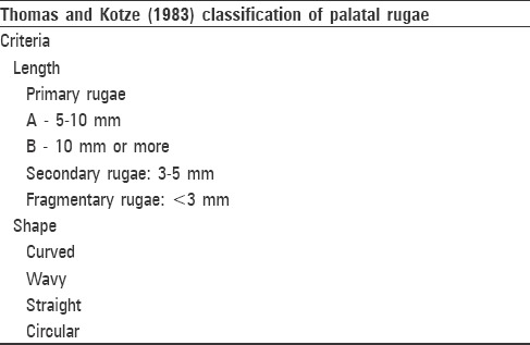
The rugae pattern is classified based on their length, shape, direction, and unification, proposed by Lysell and later modified by Thomas and Kotze[28,29] [Table 2].
Table 2.
Classification of rugae pattern
Caldes et al. have sited in a study that there is no bilateral symmetry in the number of primary rugae or in their distribution from the midline. It has been observed that there are slightly more rugae in males and on the left side in both genders.
Various studies have compared the rugae pattern in male and female. A study on the Japanese population concluded that female had fewer rugae than male. Shetty et al. compared the rugae pattern between Indian and Tibetan population. The result of their study showed that Indian male possessed more primary palatal rugae on the left side when compared to female. Furthermore, more curved rugae were observed in Indian male compared to Tibetan male, and more wavy rugae were observed in Tibetan female when compared to Indian females. The study results of Bharat et al. among males and females showed a specific pattern of palatal rugae pattern among both sexes of coastal Andhra population.[30]
Cheiloscopy
The word Chelios comes from the Greek word meaning lip. The study of lip prints is called cheiloscopy. Lip prints can be identified even at 6th week of intrauterine life. These prints then do not change. Therefore, lip prints are unique patterns on lip which help in the identification of a person.[25,31]
Lip print studies are unique to one person, except in monozygotic twins. Like fingerprints, lip grooves are permanent and unchangeable. The 10 mm wide area in the middle part of lower lip is used as the best-suited area of the study.
It has been verified that lip prints recover after undergoing alterations such as minor trauma, inflammation, and diseases such as herpes. The form of furrows does not vary with environmental factors. However, major trauma to the lips may lead to scarring and pathosis, and the surgical treatment rendered to correct the pathosis may affect the size and shape of the lip, thereby altering the pattern and morphology of grooves. The variations in patterns among males and females could help in sex determination.[26,27]
Lip anatomy is also analyzed, i.e. their thickness and position. Caldas et al.[32] analyzing the anatomical aspects, i.e., thickness and position of lips, have stated that lips can be horizontal, elevated, or depressed. Lip thickness also varies, e.g., thin lips in European Caucasian, medium, thick lips seen in Africans-Americans.
Lips prints are classified using the classification given by Tsuchihashi (1974).[26]
Type I: Clear-cut vertical grooves that run across the entire lips
Type I: Similar to Type I but do not cover the entire lip
Type II: Branched grooves (branching Y- shaped pattern)
Type III: Intersected grooves
Type IV: Criss-cross pattern, reticular grooves
Type V: Other patterns.
For classification, the middle part of the lower lip (10 mm wide) was taken as study area, as proposed by Sivapathasundharam et al.[33]
The sex of the individual is determined as per the descriptions given by Vahanwala et al.[34]
Type I, I’: Pattern dominant - female
Type II: Pattern is dominant - female
Type III: Pattern present - male
Type IV: Male
Type V: Varied pattern - male
Same patterns in all quadrants – female.
Thus, lip prints and palatal rugae hold potential as a supplementary tool, along with the dentition, to establish the identity of an individual.
Conclusion
Sex determination becomes one of the first priorities in the process of identification of a person by a forensic investigator in the case of mishaps, chemical and nuclear bomb explosions, natural disasters crime investigations, and ethnic studies. Forensic odontologist may help in determination of gender where skeletal remains create difficulty to forensic experts, especially when only fragments of body are recovered. Thus, forensic odontologist may play a key role in identifying the gender.
Financial support and sponsorship
Nil.
Conflicts of interest
There are no conflicts of interest.
References
- 1.Sopher IM. Forensic Dentistry. Indian Journal of Forensic Medicine and Pathology. Vol. 4. Bannerstone House, Illinois, USA: Charles Thompson Co; 2011. p. 156. [Google Scholar]
- 2.Dayal PK. 1st ed. Hyderabad Paras Medical Publishers; 1998. Textbook of Forensic Odontology. [Google Scholar]
- 3.Rajendran R, Sivaparthasundharam B. 6th ed. New Delhi: Elsevier; 2009. Shafer's Text Book of Oral Pathology. [Google Scholar]
- 4.Sherfudhin H, Abdullah MA, Khan N. A cross-sectional study of canine dimorphism in establishing sex identity: Comparison of two statistical methods. J Oral Rehabil. 1996;23:627–31. doi: 10.1046/j.1365-2842.1996.00406.x. [DOI] [PubMed] [Google Scholar]
- 5.Anderson DL, Thompson GW. Interrelationships and sex differences of dental and skeletal measurements. J Dent Res. 1973;52:431–8. doi: 10.1177/00220345730520030701. [DOI] [PubMed] [Google Scholar]
- 6.Garn SM, Lewis AB, Swindler DR, Kerewsky RS. Genetic control of sexual dimorphism in tooth size. J Dent Res. 1967;46:963–72. doi: 10.1177/00220345670460055801. [DOI] [PubMed] [Google Scholar]
- 7.Rao NG, Rao NN, Pai ML, Kotian MS. Mandibular canine index – A clue for establishing sex identity. Forensic Sci Int. 1989;42:249–54. doi: 10.1016/0379-0738(89)90092-3. [DOI] [PubMed] [Google Scholar]
- 8.Rao NG, Pai ML, Rao NN, Rao KT. Mandibular canine in establishing sex identity. J Indian Forensic Med. 1988;10:5–12. [Google Scholar]
- 9.Işcan MY, Kedici PS. Sexual variation in bucco-lingual dimensions in Turkish dentition. Forensic Sci Int. 2003;137:160–4. doi: 10.1016/s0379-0738(03)00349-9. [DOI] [PubMed] [Google Scholar]
- 10.Schrantz D, Bartha M. Gender determination from teething. German Journal of All Judicial Medicine. 1963;54:10–5. [Google Scholar]
- 11.Scott GR, Turner-II CG. The Anthropology of Modem Human Teeth: Dental Morphology and its Variation in Recent Human Populations. Cambridge: Cambridge University Press; 1997. [Google Scholar]
- 12.Barr ML, Bertram LF, Lindsay HA. The morphology of the nerve cell nucleus, according to sex. Anat Rec. 1950;107:283–97. doi: 10.1002/ar.1091070307. [DOI] [PubMed] [Google Scholar]
- 13.Das N, Gorea RK, Gargi J, Singh JR. Sex determination from pulpal tissue. J Indian Acad Forensic Med. 2004;26:122–5. [Google Scholar]
- 14.Whittaker DK, Llewelyn DR, Jones RW. Sex determination from necrotic pulpal tissue. Br Dent J. 1975;139:403–5. doi: 10.1038/sj.bdj.4803645. [DOI] [PubMed] [Google Scholar]
- 15.Duffy JB, Waterfield JD, Skinner MF. Isolation of tooth pulp cells for sex chromatin studies in experimental dehydrated and cremated remains. Forensic Sci Int. 1991;49:127–41. doi: 10.1016/0379-0738(91)90073-r. [DOI] [PubMed] [Google Scholar]
- 16.Tsuchimochi T, Iwasa M, Maeno Y, Koyama H, Inoue H, Isobe I, et al. Chelating resin-based extraction of DNA from dental pulp and sex determination from incinerated teeth with Y-chromosomal alphoid repeat and short tandem repeats. Am J Forensic Med Pathol. 2002;23:268–71. doi: 10.1097/00000433-200209000-00013. [DOI] [PubMed] [Google Scholar]
- 17.Hemanth M, Vidya M, Karkera BV. Sex determination using dental tissue. Med Leg Update. 2008;12:2. [Google Scholar]
- 18.Hanaoka Y, Minaguchi K. Sex determination from blood and teeth by PCR amplification of the alphoid satellite family. J Forensic Sci. 1996;41:855–8. [PubMed] [Google Scholar]
- 19.Sivagami AV, Rao AR, Varshney U. A simple and cost-effective method for preparing DNA from the hard tooth tissue, and its use in polymerase chain reaction amplification of amelogenin gene segment for sex determination in an Indian population. Forensic Sci Int. 2000;110:107–15. doi: 10.1016/s0379-0738(00)00155-9. [DOI] [PubMed] [Google Scholar]
- 20.Williams BA, Rogers TL. Evaluatiing the accuracy and precision of cranial morphological traits for sex determination. Journal of forensic sciences. 2006;4:729–35. doi: 10.1111/j.1556-4029.2006.00177.x. [DOI] [PubMed] [Google Scholar]
- 21.Uthman AT, Al-Rawi NH, Al-Naaimi AS, Tawfeeq AS, Suhail EH. Evaluation of frontal sinus and skull measurements using spiral CT scanning: An aid in unknown person identification. Forensic Sci Int. 2010;197:124.e1–7. doi: 10.1016/j.forsciint.2009.12.064. [DOI] [PubMed] [Google Scholar]
- 22.Belaldavar C, Kotrashetti VS, Hallikerimath SR, Kale AD. Assessment of frontal sinus dimensions to determine sexual dimorphism among Indian adults. J Forensic Dent Sci. 2014;6:25–30. doi: 10.4103/0975-1475.127766. [DOI] [PMC free article] [PubMed] [Google Scholar]
- 23.Stenberg I, Borrman HI. Dental condition and identification marking of dentures in homes for the elderly in Göteborg, Sweden. J Forensic Odontostomatol. 1998;16:35–7. [PubMed] [Google Scholar]
- 24.Borrman HI, DiZinno JA, Wasen J, Rene N. On denture marking. J Forensic Odontostomatol. 1999;17:20–6. [PubMed] [Google Scholar]
- 25.Ramakrishnan K, Sharma S, Sreeja C, Pratima DB, Aesha I, Vijayabanu B. Sex determination in forensic odontology: A review. J Pharm Bioallied Sci. 2015;7:398–402. doi: 10.4103/0975-7406.163469. [DOI] [PMC free article] [PubMed] [Google Scholar]
- 26.Saraf A, Bedia S, Indurkar A, Degwekar S, Bhowate R. Rugae patterns as an adjunct to sex differentiation in forensic identification. J Forensic Odontostomatol. 2011;29:14–9. [PMC free article] [PubMed] [Google Scholar]
- 27.Saxena S, Sharma P, Gupta N. Experimental studies of forensic odontology to aid in the identification process. J Forensic Dent Sci. 2010;2:69–76. doi: 10.4103/0975-1475.81285. [DOI] [PMC free article] [PubMed] [Google Scholar]
- 28.Lysell L. Plicae palatinae transversae and papilla incisiva in man. A morphologic and genetic study. Acta Odontol Scand. 1955;13:5–137. [PubMed] [Google Scholar]
- 29.Thomas CJ, Kotze TJ, Nash JM. Papillarity of the palatal mucosa. Papilla. J Oral Rehabil. 1985;12:491–7. doi: 10.1111/j.1365-2842.1985.tb01296.x. PubMed. [DOI] [PubMed] [Google Scholar]
- 30.Bharath ST, Kumar GR, Dhanapal R, Saraswathi T. Sex determination by discriminant function analysis of palatal rugae from a population of coastal Andhra. J Forensic Dent Sci. 2011;3:58–62. doi: 10.4103/0975-1475.92144. [DOI] [PMC free article] [PubMed] [Google Scholar]
- 31.Tsuchihashi Y. Studies on personal identification by means of lip print. Forensic Sci Int. 1974;3:233–48. doi: 10.1016/0300-9432(74)90034-x. [DOI] [PubMed] [Google Scholar]
- 32.Caldas IM, Magalhães T, Afonso A. Establishing identity using cheiloscopy and palatoscopy. Forensic Sci Int. 2007;165:1–9. doi: 10.1016/j.forsciint.2006.04.010. [DOI] [PubMed] [Google Scholar]
- 33.Sivapathasundharam B, Prakash PA, Sivakumar G. Lip prints (cheiloscopy) Indian J Dent Res. 2001;12:234–7. [PubMed] [Google Scholar]
- 34.Vahanwala S, Nayak CD, Pagare SS. Study of lip-prints as aid for sex determination. Med Leg Update. 2005;5:93–8. [Google Scholar]