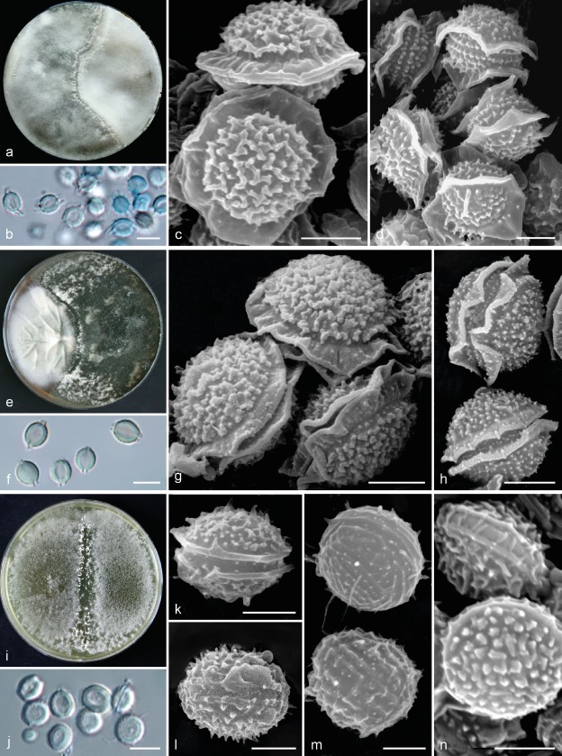
Fig. 7.
Comparison of morphology of sexual morphs of A. felis, A. udagawae and A. wyomingensis. a. Fertile cleistothecia of A. felis as a result of crossing of isolates IFM 60053 × FRR 5680; b. ascospores in light microscopy; c–d. ascospores in scanning electron microscopy: CBS 130245T × CCF 5627 (c), CBS 130245T × IFM 60053 (d); e. fertile cleistothecia of A. udagawae as a result of crossing of isolates IFM 46972T × IFM 46973; f. ascospores in light microscopy; g–h. ascospores in scanning electron microscopy; i. fertile cleistothecia of A. wyomingensis as a result of crossing of isolates CCF 4416 × CCF 4417T; j. ascospores in light microscopy (CCF 4416 × CCF 4169); k–n. ascospores in scanning electron microscopy: CCF 4416 × CCF 4417T (k–l), CCF 4417T × CCF 4419 (m–n). — Scale bars: b, f, j = 5 μm; c–d, g–h, k–n = 2 μm.

