Abstract
Out of body organ perfusion is a concept that has been around for a long time. As technology has evolved, so have the systems available for out of body perfusion making whole organ preservation for extended evaluation, resuscitation and discovery routine. Clinical use of ex vivo lung perfusion (EVLP) systems has continued to expand as evidence has accumulated to suggest EVLP transplants experience similar mortality, ICU length of stay, length of mechanical ventilation, hospital length of stay and rates of primary graft dysfunction as conventional lung transplants. In 2017, more lung transplants were performed than any previous year in the US history. Early success of EVLP has motivated groups to evaluate additional donor types and methods for expanding the donor pool. The ability to keep a lung alive in a physiologically neutral environment opens the ability to better understand organ quality, define pathophysiology in certain disease conditions and provides a platform for interventions to prevent or repair injury. The next several years will usher in significant changes in understanding and interventions focused on lung injury. This manuscript highlights applications of ex vivo lung perfusion (EVLP) to clarify how this system can be used for basic and translational research.
Keywords: Lung Transplantation, Ex Vivo Lung Perfusion
Lung transplantation is an effective therapy for patients with end-stage lung disease. However, a major challenge facing this therapeutic option is the limited availability of suitable organs for transplantation.[1, 2] As a consequence of this limitation, strategies have been employed to increase the number of lung transplants by expanding the donor pool through improved donor management strategies, use of extended criteria donors and ex vivo lung perfusion (EVLP) technologies.[3, 4] As experience in lung transplantation with EVLP has continued to expand, further interest in use of this technology for use in other contexts has become increasingly common. Our group has utilized this technology to create a high-fidelity human lung model for basic and translational work in drug discovery pipelines, toxicant research and molecular characterization of acute lung injury. For the purposes of this manuscript, we will describe the closed atrium acellular method of EVLP to highlight clinical and research applications of out-of-body perfusion. Our intent is to provide a basic understanding of out of body perfusion in order to facilitate basic and translational research efforts that capitalize from clinical experience and expand the multidisciplinary potential of this technology.
History of out of body lung perfusion
The first experimental lung transplant was performed by Vladamir Demikhov in 1947. Almost 20 years passed before the first human technical success was reported by James Hardy and almost 40 years before the first clinical success reported by Joel Cooper in 1983. Significant progress had been made in lung transplant, but there are still too few organs for transplant. This reinvigorated interest in out of body perfusion techniques proposed by Lindbergh and Carrel in the 1930s.[5] The concept of out of body whole organ perfusion for organ evaluation had been previously described by the French physiologist Le Gallois in 1812 but was not successfully realized until Lindbergh and Carrel in 1935. Their success was in part due to surgical innovation to allow for organ procurement without organ damage, improved aseptic techniques and the development of an apparatus that could be sterilized and perfuse an organ indefinitely. [5] Though successful, this technique for whole organ perfusion was not used clinically and remained a relatively infrequently used research technique. With increasing numbers of patients waiting for transplant and limited numbers of suitable donor available for transplant, efforts were made to identify new methods for expanding the donor pool. A promising solution for this organ shortage was to use organs from non-brain dead donors whom had elected to have a natural death and donate their organs to help other patients in need. Donation after cardiac death donors (DCDs) present unique challenges since the assessment of such donors can be significantly limited. To overcome these limitations, Stig Steen, used a low potassium dextran augmented blood-based perfusate and a modified perfusions system to successfully perfuse and transplant the first patient to use this technology in 2000 (Figure 1).[6] This report encouraged other groups to investigate similar methods but with slight modifications to the specific procedures. These innovations led to the current era of EVLP for use for evaluation, preservation and rehabilitation of organs for transplant. The conceptual framework of EVLP in most cases is to allow for risk assessment for extended criteria organs. This is important because improper organ selection can increase the risk of primary graft dysfunction (PGD) which is the most common cause of death in the first 90 days and impacts short, long and functional outcomes after transplant.[7–13] For this reason, safe donor pool expansion through use of EVLP has stimulated significant enthusiasm and innovation in clinical lung transplantation.
Figure 1. Timeline of events in lung transplantation leading to out of body organ perfusion.
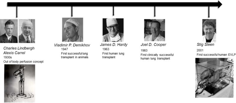
Images of Charles Lindbergh and Alexis Carrel courtesy of Wikipedia. Images of James Hardy and the Lindbergh and Carrel perfusion pump courtesy of Pinterest. Image of Joel Cooper courtesy of the University of Pennsylvania. Images of Stig Steen and his ex vivo perfusion chamber courtesy of Stig Steen.
Technical Aspects of EVLP
The equipment used for EVLP is no different from standard cardiopulmonary bypass systems which include a pump, reservoir, oxygenator, leukocyte filter and heat exchanger.[3] Any typical ICU ventilator system and circuit can be used for ventilation. The perfusate is composed of a low potassium dextran solution that is balanced with recombinant human albumin. This is commercially available as Steen solution (XVIVO, Inc).
For straight forward cannulation that does not require any surgical repair or augmentation, there are several anatomic requirements which can be employed during standard organ procurement procedures.[3, 14] First, the trachea must be long enough to cannulate which is typically >5 cm. Second, the left atrium must contain all 4 veins as a single cuff with sufficient length for transplant. Third, the pulmonary arteries must also be contiguous as a single cuff, ideally with sufficient main pulmonary artery to place a suture circumferentially to secure a cannula without obstruction. Given these requirements, specially designed cannulas can be sewn to the left atrium and fixated to the pulmonary artery (Figure 2). Modifications can be made to perfuse a single lung.
Figure 2. Standard cannulation for EVLP.
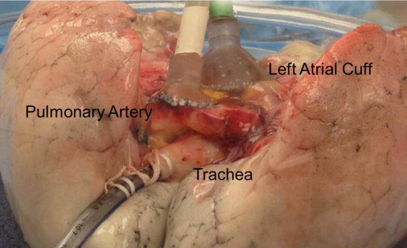
Funnel shaped cannulas are sutured to the left atrium and pulmonary artery using a monofilament polypropylene suture. Care is taken to avoid inadvertent obstruction of the pulmonary artery or veins by placing cannulas on tension as illustrated. An endotracheal tube is secured using umbilical tapes.
Following organ cannulation, the circuit is de-aired and perfusion established. The organs are re-warmed to 37 °C over the course of an hour with graded increases in target perfusion to 40% total cardiac output taking care to ensure pulmonary artery pressure remains <12 mmHg. At 32 °C, lung protective ventilation is initiated with a tidal volume of 6 cc/kg, 7 breaths/minute, at a peep of 5 cm H2O on a fractional inspired concentration of oxygen at 21%. Continuous monitoring of pulmonary artery and left atrial pressure, peak airway pressure, pulmonary artery and left atrial oxygenation, pulmonary vascular resistance and lung compliance are monitored. Chest radiographs, bronchoscopy and other adjuncts are employed as needed.[3, 14, 15]
Conceptually, EVLP can be considered as 2 oxygenators in series, the first is the lung (oxygenates the perfusate) and the second is a standard oxygenator (de-oxygenates the perfusate). This setup allows for isolated evaluation of the organ (Figure 3).
Figure 3. Circuit diagram of EVLP.
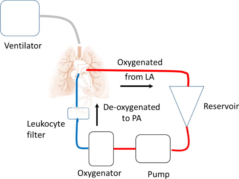
Blue depicts deoxygenated and red oxygenated perfusate. Starting from the left atrium, oxygenated perfusate is returned to the hard-shell reservoir then pumped through an oxygenator and leukocyte filter before reentering the pulmonary artery. In this set up, the oxygenator de-oxygenates the perfusate to facilitate evaluation of lung oxygenation potential. The ventilator maintains respiration and controls the concentration of oxygen delivery. Typically, the lungs are ventilated with room air (21% oxygen) and only delivered 100% when an oxygen challenge is performed to gauge maximal potential of the organ to oxygenate the perfusate.
Organ Assessment
Standard criteria for assessment include a stable or improving chest radiograph without evidence of significant edema or consolidation; stable (no more than a 15% decrease) or improving pulmonary vascular resistance, pulmonary compliance, or airway pressures; an absolute partial pressure of oxygen in the left atrium >400 mmHg or a difference from the left atrium and pulmonary artery >350 mmHg.[14] Though an organ meeting these criteria is considered acceptable, the ultimate decision is based on surgical judgement which includes standard and other criteria such as organ weight and/or feel, bronchoscopy assessment and additional ad hoc assessments judged necessary (e.g. computer tomography or endobronchial ultrasound).
Clinical Results
Reports using a closed atrium acellular perfusion EVLP technique (Toronto method) have confirmed safety and efficacy of clinical EVLP in lung transplantation. The HELP trial, a nonrandomized cohort study of extended criteria organs evaluated with EVLP for transplant, demonstrated no significant difference between standard and EVLP lung transplant recipients with respect to PGD grade 2-3 at 72 hours. Secondary endpoints (length of mechanical ventilation, ICU and hospital length of stay and 30-day mortality) similarly demonstrated no statistically significant differences.[14] Updated reports from the same group in 50 EVLP transplants continue to demonstrate equivalent length of mechanical ventilation, ICU and hospital length of stay, PGD incidence and 30-day mortality.[16] Similar results have been reported by other groups.[17–19] Given these reports, there is increasing enthusiasm and utilization of EVLP across the US with more centers establishing new programs.
Current practice at our center is to use EVLP as a method to safely expand donor use (Figure 4). Whether it is a new donor type, a logistical issue or a donor related factor that would otherwise make use prohibitively risky, EVLP has become a tool to make more transplants possible.[4] As a consequence of traveling to evaluate more donors, we have also found that we are transplanting more donors without EVLP. How EVLP ultimately is used in clinical lung transplantation still remains to be seen since the technology is still fairly new and early in the technology adoption cycle.
Figure 4. Conceptual framework of EVLP.
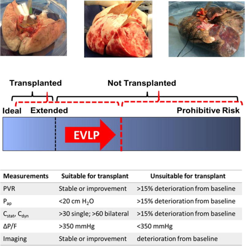
Images at the top of the figure demonstrate the variation in organ injury from minimal on the far left to severe at the far right. The goal of EVLP is to make more organs available for transplant by extending the evaluation of less ideal organs and demonstrating adequate function for transplantation (arrow depicts the level of risk being shifted to the right by EVLP). The means of determining adequate function for transplantation is demonstrated in the bottom table. Abbreviations: PVR, pulmonary vascular resistance; Pap, Peak airway pressure; Cstat, static compliance; Cdyn, dynamic compliance; ∆P/F; difference in partial pressure of oxygen in the perfusate between the left atrium (oxygenated) and pulmonary artery (deoxygenated).
Applications for research
The human lung model developed by our group offers the potential for study designs that capitalize on highly phenotyped organs and modifiable EVLP strategies suited to the research question asked (Figure 5). The typical EVLP utilizes a common perfusate (simultaneous left and right lung perfusion) and common ventilation (simultaneous left and right lung ventilation) strategy to assess both lungs. However, scenarios which would require different permutations of perfusate and ventilation strategies may be necessary. To highlight the potential of out of body organ perfusion, we will describe two previously unpublished groups of experiments conducted within our drug discovery pipeline.
Figure 5. Potential experimental EVLP configurations.
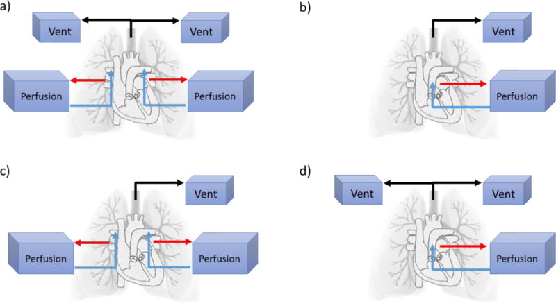
There are multiple EVLP configurations possible that can be individualized to the questions to be addressed. With respect to a lung block, a left and right lung from the same donor, there are 4 potential configurations: (a) independently perfused and ventilated lungs; (b) commonly perfused and ventilated lungs; (c) commonly ventilated but independently perfused lungs; and (d) independently ventilated but commonly perfused lungs. Note: in single perfusion systems perfusate leaves the left atrium and returns via the main pulmonary artery while in independently perfused systems perfusate leaves the pulmonary vein confluence and returns via the branch pulmonary artery.
Experimental design 1: Understanding kinetics of agent delivery method
Because of the extreme organ shortage in the US, we and other groups have been interested in potential therapeutics that may reduce lung injury. When considering how and when a therapeutic will be delivered to a potential organ donor, questions arise as to how to deliver an agent that can avoid or minimize bystander effects to other organs. These questions are important because they have practical implications as to how the agent is administered and who needs to be consented for this treatment. For example, an agent that is delivered in situ via a route that only exposes the target organ may only need recipient consent, but an agent that has systemic distribution and significant bystander effects would require all organ recipients’ consent which may be difficult if not impossible to obtain. For this reason, our group designed a series of experiments to answer basic questions regarding kinetics of an aerosolized agent in lungs with acute lung injury. We were particularly interested in this question because we have identified several agents that may reduce lung injury and potentially expand the donor pool.
The first series of experiments utilized a common perfusate and separate ventilation strategy (Figure 5a). This was achieved by modifying standard EVLP to include a double lumen endotracheal tube and 2 separate ventilators. There are two unique advantages for this study design. First, a genetically identical similarly injured organ can serve as the control for a treated organ. Second, the untreated lung simulates a bystander organ with exposure being only possible by perfusion.
A therapeutic agent (LGM2605)[20] was delivered to one lung and a saline vehicle control to the other using two aeroneb nebulizer systems attached to the endotracheal tubes. The agent was resuspended in saline (vehicle) by a study coordinator. Investigators were blinded to allocation of the agent and vehicle. Perfusate and tissue sampling were performed at 30 minute intervals. Perfusate was obtained from the reservoir manifold and tissue biopsies were obtained directly from peripheral lung using a mechanical stapler. Bronchoalveolar lavage (BAL) was performed before administration, at 3 hours and at 6 hours post administration of the agent or vehicle control. Liquid chromatography tandem-mass spectrometry (LC-MS/MS) of tissue, BAL and perfusate samples was performed to determine agent concentrations.[21]
Figure 6 summarizes the kinetics of this agent delivered to the left lung via aerosol. From these experiments, a few conclusions can be made. There is rapid transfer across the epithelium and endothelium to the vascular space of this agent. There is accumulation of this agent in bystander organs (red line). The agent is detectable in perfusate (black line) and tissue (blue line) for at least 4 hours, but probably longer given the detection of the agent in BAL (grey line) to 6 hours.
Figure 6. Kinetics of LGM2605 delivered by aerosol.
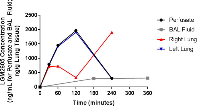
LGM2605 concentrations from perfusate, BAL fluid and tissue biopsies are depicted serially over time in a common perfusate independent ventilation configuration. Aerosolized LGM2605 delivered to the left lung (blue line) quickly traverses the epithelial and endothelial membranes to be found in the circulating perfusate (black line). BAL fluid from the left lung demonstrates persistence of LGM2605 in the airway up to 6 hours (grey line). Off target delivery is seen in the right lung tissue (red line) with delayed kinetics.
Transcriptomic evaluation of proinflammatory genes demonstrated statistically significant reductions in IL-1β, IL-6 and COX-2 as early as 30 minutes after administration with persistence to at least 120 minutes between vehicle and LGM2605 (Figure 7). Additionally, IL-6 and COX-2 down regulation was seen in the bystander organ at later time points coinciding with detection of the agent in that organ.
Figure 7. Transcriptomic response of lung tissue to LGM2605.
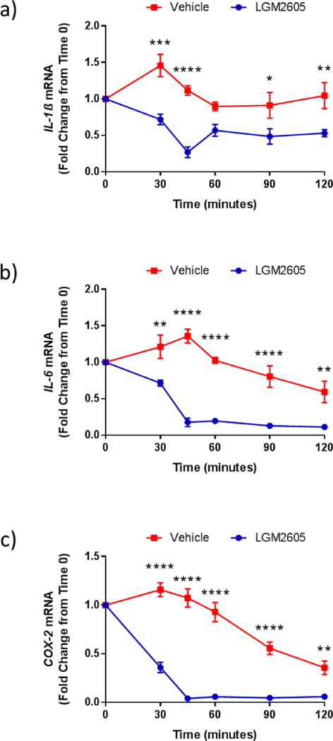
Effects of LGM2605 (blue line) were compared to vehicle (red line) in a common perfusate independent ventilation configuration. Decreased proinflammatory gene expression was demonstrated in IL-1β (a); IL-6 (b); and COX-2 (c) transcripts as early as 30 minutes after delivery of LGM2605 in the treated lung and subsequently in the bystander lung after a short delay. Levels of target gene mRNA are reported as the mean fold change from baseline (time 0). Statistically significant differences were determined by two-way analysis of variance (ANOVA) followed by Tukey’s multiple comparisons tests (vehicle versus LGM2605) using GraphPad Prism version 6.00 for Windows, GraphPad Software, La Jolla California USA, www.graphpad.com. Results are reported as the mean ± the standard error of the mean (SEM). Asterisks shown in figures indicate statistically significant differences between vehicle and LGM2605 (* = p<0.05, ** = p<0.01, *** = p<0.001 and **** = p<0.0001).
Experimental design 2: Defining efficacy for an inhalational agent
Having demonstrated that the dose and route of our therapeutic was adequate to demonstrate targeting and significant proinflammatory gene transcript reduction, attention was next focused on evaluating efficacy of this agent as part of an EVLP strategy. The goal of this strategy is to increase the number of EVLP organs that are suitable for transplant thereby further expanding the donor pool.
This series of experiments utilized a standard EVLP configuration with common ventilation and perfusion for both lungs (Figure 2). LGM2605 was delivered via aerosol once normothermia was reached in organs not suitable for transplant. Continuous monitoring of pulmonary artery and left atrial pressure, peak airway pressure, pulmonary artery and left atrial oxygenation, pulmonary vascular resistance and lung compliance were collected.
Figure 8 demonstrates a representative case summarizing the effects of the agent on two parameters of lung function. After the administration of aerosolized LGM2605, there is a significant increase in oxygenation and compliance that persists for at least 4 hours. This is contrary to what is seen in control experiments where oxygenation and compliance continue to deteriorate. Given these preliminary efficacy studies, further work is now underway to bring this agent forward for human clinical trials.
Figure 8. Physiologic response to aerosolized LGM2605.
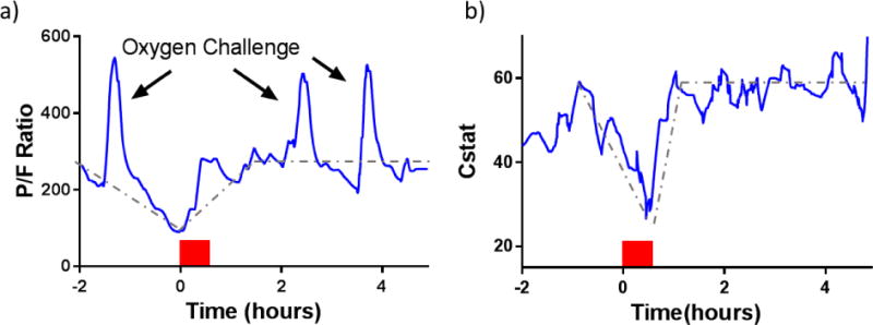
(a) Partial pressure of arterial oxygen to the fractional inspired oxygen ratio (P/F) and (b) static compliance are graphed in the treated lung. The arrows highlight the effect of an oxygen challenge with 100% oxygen. The red blocks demonstrate the delivery interval of aerosolized LGM2605. Dotted lines highlight trends in oxygenation and compliance. Note: when the oxygenation and compliance deterioratethey do not later improve.
As these two studies illustrate, human EVLP studies can accelerate drug discovery pipelines without increasing risk to patients. Optimization of dose, route and efficacy is possible in humans in out of body systems and future work may also facilitate discovery of new diagnostics, non-invasive imaging systems, and high-fidelity toxicant exposure models.
Future
As the highlighted studies illustrate, human EVLP studies can accelerate drug discovery pipelines without increasing risk to patients. Optimization of dose, route and efficacy is possible in humans in out of body systems and future work may also facilitate discovery of new diagnostics, non-invasive imaging systems, and high-fidelity toxicant exposure models.
The modular design and controlled conditions of EVLP allow for flexibility of exposure and measurement of parameters of interest. This can facilitate real-time ex vivo imaging studies (Figure 9). For example, our group has used a combination of fluorescent dyes added to the perfusate to label the endothelial layer and/or to label reactive oxygen species (ROS) and other oxidants. Imaging using confocal microscopy can provide real-time ROS production and catalogue oxidative damage in the lungs. Additionally, removal of the leukocyte filter and addition of pre-labeled polymorphonuclear neutrophils (PMNs) to this preparation facilitates the visualization of early interaction and recruitment in lungs with acute injury (Figure 9E).
Figure 9. Real-time confocal microscopy of ex vivo lungs for reactive oxygen species (ROS) during reperfusion.
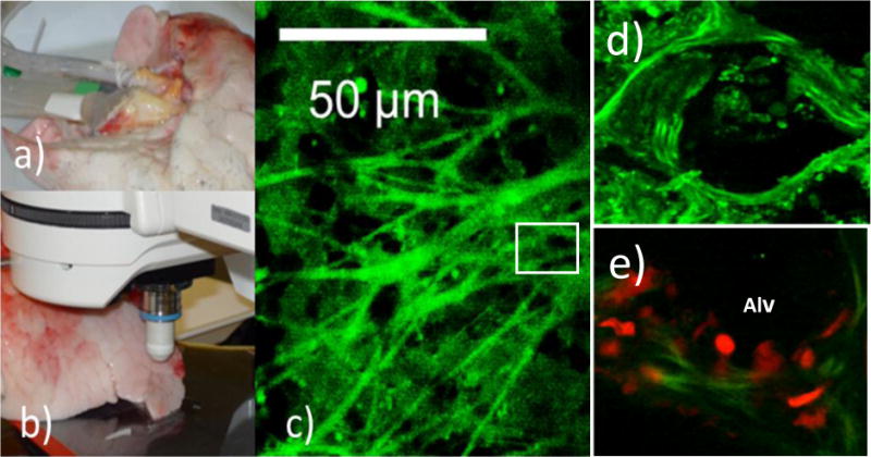
The ROS sensitive dye dihydrodifluorofluorescein (H2DFFDA, 10 μM) (Invitrogen, Molecular Probes, OR, USA) was added to the perfusate for 5-10 min prior to reperfusion. Images were acquired using a 10X lens on a Zeiss LSM 510 microscope. (a) Standard cannulation for continuous perfusion. (b) Stage preparation on the movable stage of Zeiss LSM 510 microscope for image acquisition. (c) Real-time imaging of ROS as detected by the green fluorescence of the oxidized H2DFFDAreactive oxygen species (d) A magnification of the box in c. (e). Polymorphonuclear Neutrophils (PMN) were labeled with red dye (CellTracker™ Red CMTPX, ThermoFisher Scientific, Waltham, MA, USA) and injected into the perfusate. After reperfusion, the lungs were washed and the PMN as detected by red fluorescence were observed sticking to the alveolar (Alv) and perivascular regions of the lungs.
We have also used this model to simplify complex systems to better characterize lung specific responses. Because the perfusate is acellular and a large volume perfusate exchange takes place, the only source of cytokines, metabolites, or extracellular vesicles is the lung. We have capitalized on this to begin to explore lung specific paracellular communication and lung injury status through characterization of extracellular vesicle release.[22] Further applications are not only possible but can begin to dissect the interaction of added cell populations to organ injury and repair. Furthermore, the application of this technology in drug and therapeutic discovery pipelines may soon result in new lung injury diagnostics and therapeutics.[23, 24]
In summary, the advantage of human EVLP research is the potential for rapid expansion of knowledge and understanding of lung physiological responses to injuries sustained during death and aggravated by ischemia reperfusion. As potential therapies and donor management strategies are improved through human EVLP studies, further donor expansion will be possible. Future treatment strategies may include gene or cell therapies, inhaled agents, targeted drugs and perfusion directed therapies.[6] Additional work is under way to allow for engineered organs and immunomodulation strategies to improve long-term outcomes and reduce the need for immunosuppression.[25–27] Taken together the knowledge gained using EVLP will undoubtedly improve outcomes in lung transplantation and may also facilitate new treatments for other non-transplant related lung injury.
Acknowledgments
The authors want to thank Dr. Thais Sielecki, Lignamed LLC, for helpful discussions.
Funding Sources: This work was supported by the National Institutes of Health R21AT008291 (MCS), P42ES023720 (MCS), P30 ES013508 (MCS), HL116656 (EC), HL135227 (EC) and the Robert Wood Johnson Foundation AMFDP11642 (EC).
Abbreviations
- BAL
Bronchoalveolar Lavage
- DCD
Donation after Cardiac Death
- EVLP
Ex Vivo Lung Perfusion
- ICU
Intensive Care Unit
- PGD
Primary Graft Dysfunction
- LC-MS/MS
Liquid Chromatography Tandem-Mass Spectrometry
Footnotes
Publisher's Disclaimer: This is a PDF file of an unedited manuscript that has been accepted for publication. As a service to our customers we are providing this early version of the manuscript. The manuscript will undergo copyediting, typesetting, and review of the resulting proof before it is published in its final citable form. Please note that during the production process errors may be discovered which could affect the content, and all legal disclaimers that apply to the journal pertain.
The author affirms no potential conflicts of interest.
References
- 1.Naik PM, Angel LF. Special issues in the management and selection of the donor for lung transplantation. Semin Immunopathol. 2011;33(2):201–10. doi: 10.1007/s00281-011-0256-x. [DOI] [PubMed] [Google Scholar]
- 2.Suzuki Y, Cantu E, Christie JD. Primary graft dysfunction. Semin Respir Crit Care Med. 2013;34(3):305–19. doi: 10.1055/s-0033-1348474. [DOI] [PMC free article] [PubMed] [Google Scholar]
- 3.Machuca TN, Cypel M. Ex vivo lung perfusion. J Thorac Dis. 2014;6(8):1054–62. doi: 10.3978/j.issn.2072-1439.2014.07.12. [DOI] [PMC free article] [PubMed] [Google Scholar]
- 4.Suzuki Y, et al. Should we reconsider lung transplantation through uncontrolled donation after circulatory death? Am J Transplant. 2014;14(4):966–71. doi: 10.1111/ajt.12633. [DOI] [PMC free article] [PubMed] [Google Scholar]
- 5.Carrel A, Lindbergh CA. The Culture of Whole Organs. Science. 1935;81(2112):621–3. doi: 10.1126/science.81.2112.621. [DOI] [PubMed] [Google Scholar]
- 6.Cypel M, Keshavjee S. Extending the donor pool: rehabilitation of poor organs. Thorac Surg Clin. 2015;25(1):27–33. doi: 10.1016/j.thorsurg.2014.09.002. [DOI] [PubMed] [Google Scholar]
- 7.Christie JD, et al. The effect of primary graft dysfunction on survival after lung transplantation. Am J Respir Crit Care Med. 2005;171(11):1312–6. doi: 10.1164/rccm.200409-1243OC. [DOI] [PMC free article] [PubMed] [Google Scholar]
- 8.Christie JD, et al. Clinical risk factors for primary graft failure following lung transplantation. Chest. 2003;124(4):1232–41. doi: 10.1378/chest.124.4.1232. [DOI] [PubMed] [Google Scholar]
- 9.Christie JD, et al. Impact of primary graft failure on outcomes following lung transplantation. Chest. 2005;127(1):161–5. doi: 10.1378/chest.127.1.161. [DOI] [PubMed] [Google Scholar]
- 10.King RC, et al. Reperfusion injury significantly impacts clinical outcome after pulmonary transplantation. Ann Thorac Surg. 2000;69(6):1681–5. doi: 10.1016/s0003-4975(00)01425-9. [DOI] [PubMed] [Google Scholar]
- 11.Lee JC, Christie JD, Keshavjee S. Primary graft dysfunction: definition, risk factors, short- and long-term outcomes. Semin Respir Crit Care Med. 2010;31(2):161–71. doi: 10.1055/s-0030-1249111. [DOI] [PubMed] [Google Scholar]
- 12.Whitson BA, et al. Primary graft dysfunction and long-term pulmonary function after lung transplantation. J Heart Lung Transplant. 2007;26(10):1004–11. doi: 10.1016/j.healun.2007.07.018. [DOI] [PubMed] [Google Scholar]
- 13.Yusen RD, et al. The registry of the International Society for Heart and Lung Transplantation: thirty-first adult lung and heart-lung transplant report–2014; focus theme: retransplantation. J Heart Lung Transplant. 2014;33(10):1009–24. doi: 10.1016/j.healun.2014.08.004. [DOI] [PubMed] [Google Scholar]
- 14.Cypel M, et al. Normothermic ex vivo lung perfusion in clinical lung transplantation. N Engl J Med. 2011;364(15):1431–40. doi: 10.1056/NEJMoa1014597. [DOI] [PubMed] [Google Scholar]
- 15.Reeb J, Keshavjee S, Cypel M. Expanding the lung donor pool: advancements and emerging pathways. Curr Opin Organ Transplant. 2015;20(5):498–505. doi: 10.1097/MOT.0000000000000233. [DOI] [PubMed] [Google Scholar]
- 16.Cypel M, et al. Experience with the first 50 ex vivo lung perfusions in clinical transplantation. J Thorac Cardiovasc Surg. 2012;144(5):1200–6. doi: 10.1016/j.jtcvs.2012.08.009. [DOI] [PubMed] [Google Scholar]
- 17.Andreasson A, et al. The effect of ex vivo lung perfusion on microbial load in human donor lungs. J Heart Lung Transplant. 2014;33(9):910–6. doi: 10.1016/j.healun.2013.12.023. [DOI] [PubMed] [Google Scholar]
- 18.Boffini M, Bonato R, Rinaldi M. The potential role of ex vivo lung perfusion for the diagnosis of infection before lung transplantation. Transpl Int. 2014;27(2):e5–7. doi: 10.1111/tri.12232. [DOI] [PubMed] [Google Scholar]
- 19.Zych B, et al. Early outcomes of bilateral sequential single lung transplantation after ex-vivo lung evaluation and reconditioning. J Heart Lung Transplant. 2012;31(3):274–81. doi: 10.1016/j.healun.2011.10.008. [DOI] [PubMed] [Google Scholar]
- 20.Mishra OP, et al. Synthesis and antioxidant evaluation of (S, S)- and (R, R)-secoisolariciresinol diglucosides (SDGs) Bioorg Med Chem Lett. 2013;23(19):5325–8. doi: 10.1016/j.bmcl.2013.07.062. [DOI] [PMC free article] [PubMed] [Google Scholar]
- 21.Chikara S, et al. Flaxseed Consumption Inhibits Chemically Induced Lung Tumorigenesis and Modulates Expression of Phase II Enzymes and Inflammatory Cytokines in A/J Mice. Cancer Prev Res (Phila) 2018;11(1):27–37. doi: 10.1158/1940-6207.CAPR-17-0119. [DOI] [PMC free article] [PubMed] [Google Scholar]
- 22.Vallabhajosyula P, et al. Ex Vivo Lung Perfusion Model to Study Pulmonary Tissue Extracellular Microvesicle Profiles. Ann Thorac Surg. 2017;103(6):1758–1766. doi: 10.1016/j.athoracsur.2016.11.074. [DOI] [PubMed] [Google Scholar]
- 23.Mordant P, et al. Mesenchymal stem cell treatment is associated with decreased perfusate concentration of interleukin-8 during ex vivo perfusion of donor lungs after 18-hour preservation. J Heart Lung Transplant. 2016;35(10):1245–1254. doi: 10.1016/j.healun.2016.04.017. [DOI] [PubMed] [Google Scholar]
- 24.Machuca TN, et al. Protein expression profiling predicts graft performance in clinical ex vivo lung perfusion. Ann Surg. 2015;261(3):591–7. doi: 10.1097/SLA.0000000000000974. [DOI] [PubMed] [Google Scholar]
- 25.Ott HC, et al. Regeneration and orthotopic transplantation of a bioartificial lung. Nat Med. 2010;16(8):927–33. doi: 10.1038/nm.2193. [DOI] [PubMed] [Google Scholar]
- 26.Petersen TH, et al. Tissue-engineered lungs for in vivo implantation. Science. 2010;329(5991):538–41. doi: 10.1126/science.1189345. [DOI] [PMC free article] [PubMed] [Google Scholar]
- 27.Martens A, et al. Immunoregulatory effects of multipotent adult progenitor cells in a porcine ex vivo lung perfusion model. Stem Cell Res Ther. 2017;8(1):159. doi: 10.1186/s13287-017-0603-5. [DOI] [PMC free article] [PubMed] [Google Scholar]


