Abstract
Gfi1 supports neutrophil development at the expense of monopoiesis, but the underlying molecular mechanism is incompletely understood. We recently showed that the G-CSFR Y729F mutant, in which tyrosine 729 was mutated to phenylalanine, promoted monocyte rather than neutrophil development in myeloid precursors, which was associated with prolonged activation of Erk1/2 and enhanced activation of c-Fos and Egr-1. We show here that Gfi1 inhibited the expression of c-Fos, Egr-1 and Egr-2, and rescued neutrophil development in cells expressing G-CSFR Y729F. Gfi1 directly bound to and repressed c-Fos and Egr-1, as has been shown for Egr-2, all of which are the immediate early genes (IEGs) of the Erk1/2 pathway. Interestingly, G-CSF- and M-CSF-stimulated activation of Erk1/2 was augmented in lineage-negative (Lin−) bone marrow (BM) cells from Gfi1−/− mice. Suppression of Erk1/2 signaling resulted in diminished expression of c-Fos, Egr-1 and Egr-2, and partially rescued the neutrophil development of Gfi1−/− BM cells, which are intrinsically defective for neutrophil development. Together, our data indicate that Gfi1 inhibits the expression of c-Fos, Egr-1 and Egr-2 through direct transcriptional repression and indirect inhibition of Erk1/2 signaling, and that Gfi1-mediated downregulation of c-Fos, Egr-1 and Egr-2 may contribute to the role of Gfi1 in granulopoiesis.
Introduction
Gfi1 plays a critical role in hematopoiesis1–3. Gfi1 ablation in mice impaired the self-renewal ability of hematopoietic stem cells (HSCs)4–6, indicating that Gfi1 is required for the functional integrity of HSCs. Gfi1−/− mice also show defects in B and T-cell development. The most significant phenotype of Gfi1−/− mice is a lack of mature neutrophils accompanied by an expansion of granulocyte-monocyte progenitors (GMPs) and immature monocyte-like myeloid cells7–10. Myeloid precursors from Gfi1−/− mice are unable to differentiate into mature neutrophils in vitro, but instead give rise to atypical monocytes. In contrast, overexpression of Gfi1 accelerates neutrophilic differentiation at the expense of monocyte formation11,12. Thus, Gfi1 promotes the neutrophil development and antagonize the alternative monocyte/macrophage fate. Consistent with its role in granulopoiesis, mutations in GFI1 have been identified in patients with severe congenital neutropenia (SCN)3,13,14, a condition characterized by a profound absolute neutropenia occurring in early life and a maturation arrest of myeloid precursors in the bone marrow. Expression of an SCN-derived dominant negative (DN) Gfi1 mutant, N382S, in mouse Lin− BM cells led to monocyte development in response to G-CSF at the expense of granulopoiesis11.
The molecular mechanism by which Gfi1 favors neutrophil over monocyte development is incompletely understood.Gfi1 has been shown to repress PU.1, Egr-2 and CSF1 encoding M-CSF11,15,16, which promotes monocyte development. Notably, targeted deletion of Csf1 rescued granulopoiesis that was blocked by the Gfi1 N382S mutant in mouse Lin− BM cells11, suggesting that repression of Csf1 represents an important mechanism by which Gfi1 supports granulopoiesis. In addition, Gfi1 has been shown to repress miR21 and miR-196b, which may also contribute to its role in resolving neutrophil versus monocyte fate17.
We recently demonstrated that G-CSF and M-CSF instruct neutrophil and monocyte fates, respectively, in part through differential activation of Erk1/2 and the downstream targets c-Fos and Egr-118. Specifically, M-CSF stimulates strong and prolonged activation of Erk1/2-c-Fos/Egr1 signaling pathway in comparison with the weak and transient activation of the pathway in response to G-CSF. Here we show that Gfi1 directly represses c-Fos, Egr-1 and Egr-2, and negatively regulates Erk1/2 activation. We further demonstrate that inhibition of Erk1/2 signaling led to diminished expression of c-Fos, Egr-1 and Egr-2, and partially rescued the neutrophil development in Lin− BM cells from Gfi1−/− mice.
Results
Gfi1 rescues neutrophil development in 32D/Y729F and FDCP/Y729F cells independent of M-CSF signaling
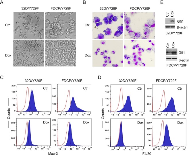
We recently showed that murine myeloid 32D and multipotent FDCP-mix A4 cells expressing the human G-CSFR Y729F (32D/Y729F and FDCP/Y729F), in which tyrosine 729 in the cytoplasmic domain was mutated to phenylalanine, underwent monocyte rather than neutrophil development in response to G-CSF18. Parental 32D and FDCP-mix A4 cells expressed no detectable M-CSF receptor and showed no response to M-CSF (data not shown). To address whether Gfi1 suppressed monopoiesis independent of M-CSF signaling, we transduced 32D/Y729F and FDCP/Y729F with the lentiviral pPMPrtTA-Gfi1-IRES-GFP construct in which Gfi1 expression was driven by the tetracycline-response element (TRE)19. The transduced cells (32D/Y729F/Gfi1 and FDCP/Y729F/Gfi1) were cultured in G-CSF-containing medium without or with doxycycline (Dox). In the absence of Dox, 32D/Y729F/Gfi1 and FDCP/Y729F/Gfi1 cells displayed features typical of monocyte development, including increased cell sizes, adherence to culture dishes, monocyte/macrophage morphology, and increased expression of macrophage surface marker F4/80 (Fig. 1). Dox induction of Gfi1 expression completely restored G-CSF-induced neutrophil development in both cell types, which was associated with upregulation of neutrophil differentiation markers neutrophil elastase (NE) and lactoferrin (LF), and downregulation of monocyte/macrophage differentiation markers M-CSF and MMP-12 (Suppl. Fig. 1). Induction of Gfi1 expression alone did not lead to neutrophil development in the two cell lines cultured in IL-3. Thus, Gfi1 may promote neutrophil fate independent of its inhibitory effect on M-CSF expression.
Figure 1.
Gfi1 overexpression rescues neutrophil development. 32D/Y729F/Gfi1 and FDCP/Y729F/Gfi1 cells were cultured in G-CSF in the absence (Ctr) or presence of Dox. (A) Cell growth behaviors were examined on day 6 for 32D/Y729F/Gfi1 cells and day 2 for FDCP/Y729F/Gfi1 cells. (B) Cell morphology was evaluated on day 6 for 32DY729F/Gfi1 cells, and on day 3 and day 5 for FDCP/Y729F/Gfi1 cells in the absence and presence of Dox, respectively (May Grünwald Giemsa staining). The expression of Mac-3 (C) and F4/80 (D) was examined on day 3. (E) Gfi1 expression following Dox induction for 24 hours was examined by Western blot analysis.
Gfi1 represses c-Fos and Egr-1
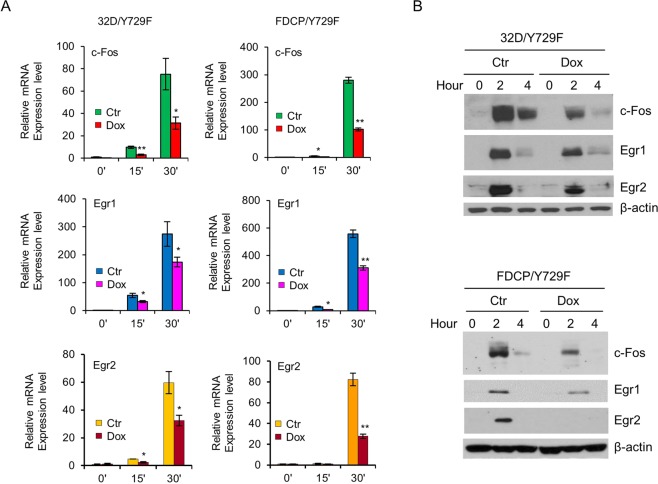
Monocyte development mediated by G-CSFR Y729F was associated with prolonged activation of Erk1/2 and subsequently augmented activation of c-Fos and Egr-1, and knockdown of c-Fos or Egr-1 in 32D/Y729 and FDCP/Y729F cells rescued neutrophil development18. Notably, Gfi1 has been shown to repress Egr-2 and downregulate Egr-1 expression20. We examined whether Gfi1 had an effect on the expression of c-Fos and Egr-1 in response to G-CSF. The mRNAs and protein levels of c-Fos and Egr-1 were rapidly induced following G-CSF stimulation, but their induction was markedly attenuated in Dox-treated 32D/Y729F/Gfi1 and FDCP/Y729F/Gfi1 cells (Fig. 2). Consistent with previous report20, Gfi1 repressed Egr-2 expression. We further investigated whether Gfi1 regulated the expression of c-Fos and Egr-1 in primary BM cells. As shown in Fig. 3, the mRNA levels of c-Fos and Egr-1 were considerably higher in Lin− BM cells from Gfi1−/− mice than in cells from Gfi1+/+ mice. As expected, Egr-2 expression was also increased in Gfi1−/− BM cells.
Figure 2.
Gfi1 suppresses the expression of c-Fos, Egr-1 and Egr-2. (A) 32D/Y729F/Gfi1 and FDCP/Y729F/Gfi1 cells were cultured in the absence (Ctr) or presence of Dox for 12 hours. Cells were then starved for 2 hours and stimulated with G-CSF for the indicated times prior to evaluation of c-Fos, Egr-1 and Egr-2 mRNA (A) and protein levels (B).
Figure 3.
The expression of c-Fos, Egr-1 and Egr-2 mRNAs is markedly increased in Lin− BM cells from Gfi1−/− mice. Cells were isolated from 6–8 week old Gfi1+/+ and Gfi1−/− mice. The mRNA levels of c-Fos, Egr-1 and Egr-2 were assessed by qRT-PCR.
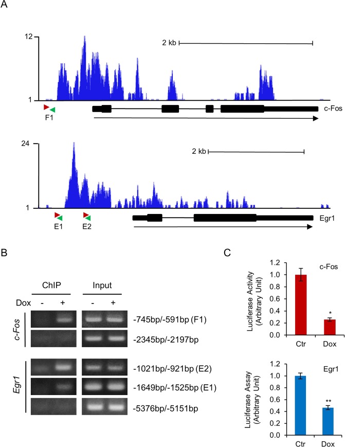
Gfi1 has been shown to bind to Egr-220. Analysis of the ChIP-seq data (GSE31657) submitted by Möröy’s research group21 indicated that Gfi1 bound to the promoters of c-Fos and Egr-1 in murine hematopoietic progenitor cells (Fig. 4A). Examination of c-Fos and Egr-1 promoters using the online transcription factor prediction tool TFBIND (http://tfbind.hgc.jp/) revealed potential Gfi1 binding sites at approximate nucleotide positions −786 and −661 of c-Fos, and at −1614 and −997 of Egr-1. ChIP assays demonstrate that Gfi1 indeed bound to these sites, but not the upstream promoter regions of c-Fos and Egr1 in 32D/Y729F/Gfi1 cells (Fig. 4B). We further performed luciferase reporter assays to determine whether Gfi1 repressed the c-Fos and Egr-1 promoters. As shown in Fig. 4C, Gfi1 repressed the activities of murine c-Fos promoter fragment spanning from −1070 to + 30 bp and Egr-1 promoter fragment spanning from −1780 to + 50 bp in 32DGR/Y729F/Gfi1 cells. As reported in the previous study20, Gfi1 also repressed the Egr-2 promoter (data not shown). Together, these data revealed that c-Fos and Egr-1 are the novel target genes of Gfi1.
Figure 4.
Gfi1 represses c-Fos and Egr-1 through direct binding to their promoters. (A) Gfi1 binding patterns at the c-Fos (upper panel) and Egr-1 (lower panel) based on the ChIP-seq data submitted by Möröy’s research group21. The pink and green arrows denote the forward and reverse primers, respectively, used to amplify the promoter regions at c-Fos (F1) and Egr-1 (E1 and E2) loci. (B) 32D/Y729F/Gfi1 cells were cultured with or without Dox for 24 hours. ChIP assays were carried out using the anti-mouse Gfi1 antibody. The indicated regions of c-Fos and Egr-1 promoters were amplified by PCR. (C) 32D/Y729F/Gfi1 cells were transfected with pGL3-basic vector containing c-Fos or Egr-1 promoter fragment and cultured in G-CSF with or without Dox. Luciferase activities were measured 24 hours later.
Enhanced activation of Erk1/2 contributes to increased expression of c-Fos, Egr-1 and Egr-2 in Lin− BM cells from Gfi1−/− mice
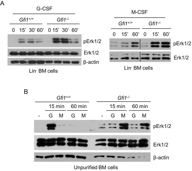
It has been shown that c-Fos, Egr-1 and Egr-2 are the IEGs of the Erk1/2 signaling pathway22,23. A previous study showed that G-CSF-stimulated activation of Erk1/2 was significantly reduced in unpurified BM cells from Gfi1−/− mice12. However, Gfi1−/− mice lack mature neutrophils accompanied by an expansion of atypical monocytes in BM and peripheral blood. We therefore examined Erk1/2 activation in Lin− BM cells. Unexpectedly, Erk1/2 activation in response to G-CSF and M-CSF was stronger in Gfi1−/− cells than in Gfi1+/+ cells (Fig. 5A). Notably, when unpurified BM cells were used, G-CSF-stimulated activation of Erk1/2 was strong in Gfi1+/+ cells, but extremely weak or barely activated in Gfi1−/− cells (Fig. 5B), in line with the previous study12. In contrast, M-CSF stimulation led to strong Erk1/2 activation in Gfi1−/− cells, but not in Gfi1+/+ cells. Flow cytometric analyses revealed that unpurified BM cells from Gfi1+/+ mice abundantly expressed G-CSFR, but only a small percentage of these cells weakly expressed M-CSFR whereas unpurified Gfi1−/− cells expressed high levels of M-CSFR with minimal expression of G-CSFR (Suppl. Fig. 2). The expression levels of G-CSFR and M-CSFR in the Lin− cells from Gfi1+/+ and Gfi1−/− mice were relatively comparable. Together, these results indicate that Gfi1 negatively regulates Erk1/2 activation in Lin− BM cells whereas the differential activation of Erk1/2 in response to G-CSF and M-CSF in the unpurified BM cells from Gfi1+/+ and Gfi1−/− mice may largely result from the differential expression of G-CSFR and M-CSFR.
Figure 5.
G-CSF- and M-CSF-induced activation of Erk1/2 in different populations of BM cells from Gfi1+/+ and Gfi1−/− mice. Lin− (A) and unpurified (B) BM cells were isolated from Gfi1+/+ and Gfi1−/− mice, and treated with G-CSF (G) or M-CSF (M) for the indicated times. Erk1/2 phosphorylation was examined by Western blot analysis.
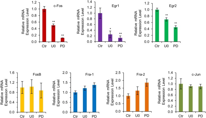
The above results suggest that both loss of Gfi1-mediated transcriptional repression and the augmented activation of Erk1/2 may contribute to the increased expression of c-Fos, Egr-1 and Egr-2 in the Lin− BM cells from Gfi1−/− mice. We therefore addressed whether inhibition of Erk1/2 signaling diminished the expression of c-Fos, Egr-1 and Egr-2 in Gfi1−/− BM cells. As shown in Fig. 6, treatment of Gfi1−/− Lin− BM cells with the specific Mek1/2 inhibitors U0126 or PD0325901 resulted in significantly reduced mRNA levels of c-Fos, Egr-1 and Egr-2, but had no effect on the expression of other Fos family members, including FosB, Fra-1 and Fra-2, and c-Jun.
Figure 6.
Mek1/2 inhibitors specifically inhibit the expression of Erk1/2 downstream targets c-Fos, Egr-1 and Egr-2. Lin− BM cells from Gfi1−/− mice were treated without (Ctr) or with Mek1/2 inhibitors U0126 (UO) or PD0325901 (PD) for 8 hours. The mRNA levels of indicated genes were assessed by qRT-PCR.
Mek1/2 inhibitors partially rescue neutrophil development of Lin− BM cells from Gfi1−/− mice
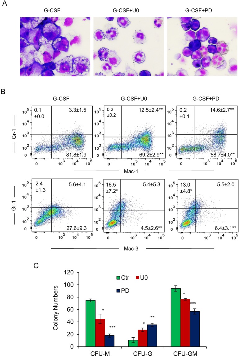
BM myeloid precursors from Gfi1−/− mice are unable to differentiate into mature neutrophils in vitro, but give rise to atypical monocytes/macrophages8,11. Consistent with the previous reports, Gfi1−/− Lin− BM cells developed into cells with an appearance reminiscent of monocytes/macrophages when cultured in G-CSF (Fig. 7). Because suppression of Erk1/2 signaling in Gfi1−/− Lin− BM cells reduced the expression of c-Fos, Egr-1 and Egr-2, we asked whether the Mek1/2 inhibitors rescued neutrophil development of Gfi1−/− BM cells. Interestingly, treatment of Gfi1−/− BM cells with U0126 or PD0325901 led to a significant shift towards neutrophil development, as evident from the neutrophil-like morphology (Fig. 7A) and reduced percentages of Gr-1-/Mac-1+ and Gr-1-/Mac-3+ monocytes with concomitant increase of Gr-1+/Mac-1+ cells and Gr-1+/Mac-3- neutrophils (Fig. 7B). Treatment with U0126 or PD0325901 also upregulated the expression of NE, LF and myeloperoxidase (MPO), but downregulated the expression of M-CSF and MMP12 (Suppl. Fig. 3). In methylcellulose colony formation assays, both U0126 and PD0325901 increased the numbers of CFU-G, but reduced the formation of CFU-M (Fig. 7C). Taken together, these data indicated that inhibition of Erk1/2 signaling resulted in partial restoration of neutrophil development in Gfi1−/− BM cells, presumably through suppressing the expression of c-Fos, Egr-1 and Egr-2.
Figure 7.
Mek1/2 inhibitors partially rescue neutrophil development in BM cells from Gfi1−/− mice. (A) Lin− cells from Gfi1−/− mice were cultured in G-CSF-containing medium without or with U0126 (U0) or PD0325901 (PD). Cell morphology was examined on day 3 (May Grünwald Giemsa staining). (B) Expression of Gr-1, Mac-1 and Mac-3 was analyzed by flow cytometry on day 3. Data are presented as percentage of positively stained cells (n = 3; mean ± SD). (C) BM cells from Gfi1−/− mice were cultured in methylcellulose medium containing IL-3, IL-6, SCF and G-CSF without or with U0 or PD. Colonies were counted 7 days later.
Discussion
Gfi1 supports the neutrophil development and antagonizes the alternative monocyte/macrophage fate. The molecular mechanism by which Gfi1 favors neutrophil over monocyte development is incompletely understood, but may involve Gfi1-mediated repression of Pu.1, Egr-2 and Csf1 as well as miR-21 and miR-196b17,24. It appears that Gfi1-mediated repression of Csf1 is important for its role in granulopoiesis as Csf1 ablation rescued granulopoiesis that was blocked by the DN Gfi1 N382S mutant in mouse BM cells11. In this paper, we have shown that Gfi1 promotes granulopoiesis independent of its effect on M-CSF signaling. We have further shown that Gfi1 binds to and represses c-Fos and Egr-1. These data indicate that c-Fos and Egr-1, along with the previously identified Egr-220, are Gfi1 target genes. c-Fos forms the AP-1 protein through heterodimerization with c-Jun. As c-Fos/AP1, Egr-1 and Egr-2 have been shown to promote monopoiesis25–29, it is likely that the effects of Gfi1 on neutrophil versus monocyte development are mediated in part through repression of c-Fos, Egr-1 and Egr-2.
We have also shown that Gfi1 ablation results in enhanced activation of Erk1/2 in response to G-CSF and M-CSF in mouse Lin− BM cells, indicating that Gfi1 negatively regulates Erk1/2 signaling. As c-Fos, Egr-1 and Egr-2 are the IEGs of the Erk1/2 signaling pathway22,23, this raises the possibility that Gfi1 may downregulate the expression of c-Fos, Egr-1 and Egr-2 in part through suppression of cytokine-induced activation of Erk1/2 signaling. Indeed, the augmented expression of c-Fos, Egr-1 and Egr-2 in the Lin− BM cells from Gfi1−/− cells mice was significantly attenuated upon suppression of Erk1/2 signaling using the Mek1/2 inhibitors. However, it remains to be determined how Gfi1 inhibits Erk1/2 signaling. As Gfi1 is a nuclear protein that functions as a transcriptional repressor, the effect of Gfi1 on Erk1/2 signaling is likely indirect. It is possible that Gfi1 may repress a positive regulator of the Erk1/2 pathway or indirectly increase the expression of a negative regulator of Erk1/2 signaling.
Mutations in GFI1 have been associated with SCN3,13,14. When expressed in mouse BM cells, the SCN-derived DN GFI1 mutant supported monopoiesis, but blocked neutrophil development in response to G-CSF11. It has been shown that Gfi1−/− myeloid precursors are intrinsically defective for neutrophil development and in vivo administration of G-CSF had no effect on neutropenia in Gfi1−/− mice7–9. In this aspect, it is noteworthy that the MEK1/2 inhibitors U0126 and PD0325901 partially rescued G-CSF-induced neutrophil development in Gfi1−/− BM cells, likely through downregulation of the expression of c-Fos, Egr-1 and Egr-2. It would be interesting to explore whether in vivo administration of Mek1/2 inhibitors alleviates neutropenia in Gfi1−/− mice; if it does, suppression of Erk1/2 signaling could represent a novel therapeutic approach in the treatment of SCN patients with GFI1 mutations.
Materials and Methods
Cell lines and cell culture
Murine myeloid 32D cells and multipotential FDCP-mix A4 cells expressing the different forms of G-CSFR have been described18,30,31. 32D and FDCP-mix cells were cultured as described18. Briefly, 32D cells were maintained in RPMI-1640 with 10% heat inactivated fetal bovine serum (HI-FBS), 10% WEHI-3B cell-conditioned media as a crude source of murine interleukin-3, and 1% penicillin/streptomycin (P/S). FDCP-mix A4 cells were maintained in IMDM medium supplemented with 15% horse serum and 10% WEHI-3B cell conditioned medium and P/S.
Construction of plasmids
Murine Egr-1 promoter fragment (from −1780 bp to + 21 bp) was generated by PCR from BAC plasmid (Clone# RP23-108C3, BACPAC Resources) and inserted into pGL3-basic plasmid. c-Fos promoter (from −1141 bp to + 19 bp) luciferase reporter construct was a generous gift from Dr. Wan-Wan Lin (National Taiwan University). The Dox-inducible Gfi1 expression construct pPMPrtTA-Gfi1-GFP has been described before19.
Mice, bone marrow cell isolation and colony assays
Gfi1 knockout mice8 were bred and housed in the animal facility at The University of Toledo. All experiments using mouse BM cells were approved by the Institutional Animal Care and Use Committee (IACUC) of The University of Toledo and were performed per the approved protocol. Bone marrow cells were isolated from 6- to 8-week-old C57BL/6 WT and Gfi1 mutant mice as previously described18. Lin− cells were purified using the mouse Lineage Cell Depletion kit (Miltenyi Biotec) and cultured in IMDM media with 10% FBS, 10 ng/ml IL-3, 20 ng/ml IL-6 and 25 ng/ml SCF (Peprotech). For colony forming assay, Gfi1 knockout mice were treated with 5-fluorouracil (50 mg/kg) intraperitoneally prior to isolation of BM cells 5 days later. Cells were cultured in IMDM media containing 10% FBS, 1% P/S, 25 ng/ml SCF, 10 ng/ml IL-3 and 20 ng/ml IL-6 for 1 hour, and then 104 cells were plated in Methylcellulose-based Media (R&D System) containing 10% FBS, IL-3, IL-6, SCF and G-CSF with or without indicated inhibitors. Colonies were counted on day 7.
Flow cytometry
Cells were first washed in PBS containing 2% horse serum and then blocked using Fc block (eBioscience) for 15 min. Subsequently, cells were incubated with isotype control FITC-conjugated anti-mouse IgG, antibodies against F4/80, Gr-1, Mac-1, Mac-3, G-CSFR or M-CSFR for 30 min prior to washing in PBS with 2% horse serum. Cells were analyzed by two-color flow cytometry on an LSR Fortessa (BD Biosciences) using FACSDiva and analyzed with FlowJo (Tree Star).
Western blot analysis
The experiments were performed as previously described18. Cells were lysed in SDS lysis buffer. Proteins were separated by SDS-PAGE and then transferred onto polyvinylidenedifluoride (PVDF) membranes. The membranes were incubated with the antibodies against phospho-Erk1/2, c-Fos, Egr-1, Egr-2, or β-actin (Cell Signaling), followed by detection of signals using enhanced chemiluminescence.
Transient transfection and luciferase reporter assay
Cells were transiently transfected by electroporation with the luciferase reporter constructs containing the c-Fos or Egr-1 promoter fragment. After recovering in complete culture medium for 16 hours, cells were washed and placed in RPMI-1640 medium containing 10% FBS and 10 ng/ml G-CSF for 8 hours. Cells were harvested and luciferase activities were measured using luciferase reporter kit and Molecular Devices Lmaxluminometer (Sunnyvale, CA).
Real-time reverse transcription polymerase chain reaction (qRT-PCR)
Total RNA was extracted using TRIzol reagent (Invitrogen) and reverse transcribed into cDNA using the GoScript™ Reverse Transcription System and Oligo(dT)15 primer (Promega, Madison, WI). The relative mRNA levels of the different genes were quantitated by qRT-PCR using the SsoFastTM EvaGreen Supermix® kit (Bio-Rad) following normalization to GAPDH mRNA expression.
Apoptosis assay
Apoptosis was examined using the Annexin V-PE apoptosis detection kit (BD Biosciences) as previously described18. Briefly, 0.3 × 10⁶ cells were collected and incubated with Annexin V-PE and 7 amino-actinomycin (7-AAD). Cells were analyzed by two-color flow cytometry as described above.
Chromatin immunoprecipitation assay (ChIP assay)
ChIP assays were performed essentially as described32. Briefly, 32D cells were fixed with 1% formaldehyde and then lysed in hypotonic buffer [5 mM Tris-HCl (pH 7.5), 85 mM KCl and 0.5% Nonidet P-40]. After centrifugation at 6000 rpm for 5 min, nuclei were lysed in ChIP lysis buffer [1% SDS, 10 mM EDTA, and 50 mMTris HCl (pH 7.5)] and sonicated to shear chromatin DNA to ~500-bp fragments. Nuclear lysates were precleared with protein A/G agarose beads and rabbit normal IgG for 1 h and subjected to immunoprecipitation using the anti Gfi1 or a species-matched irrelevant antibody. Precipitated DNA was examined by semi-quantitative PCR.
Statistics
Statistical analyses were performed using GraphPad Prism software (GraphPad Software, La Jolla, CA, USA). Data are presented as mean ± SD in the figures. A p value < 0.05 was considered significant and shown as * with P < 0.01 shown as ** and P < 0.001 as ***.
Supplementary information
Acknowledgements
We thank Dr. Wan-Wan Lin for providing the c-Fos promoter luciferase reporter construct, and Drs. Stuart H. Orkin and H. Leighton Grimes for providing Gfi1 knockout mice. This work was supported by grant R15HL135695 (F.D) from the National Heart, Lung, and Blood Institute, National Institutes of Health.
Author Contributions
Y.Z. and N.H. contributed equally to this work. Y.Z., N.H. and F.D. performed experiments and analyzed results. F.D. designed the study and wrote the paper. All authors reviewed and approved the manuscript.
Data Availability
No datasets were generated or analysed during the current study.
Competing Interests
The authors declare no competing interests.
Footnotes
Publisher’s note: Springer Nature remains neutral with regard to jurisdictional claims in published maps and institutional affiliations.
Yangyang Zhang and Nan Hu contributed equally.
Electronic supplementary material
Supplementary information accompanies this paper at 10.1038/s41598-018-37402-z.
References
- 1.Kazanjian A, Gross EA, Grimes HL. The growth factor independence-1 transcription factor: new functions and new insights. Crit Rev Oncol Hematol. 2006;59:85–97. doi: 10.1016/j.critrevonc.2006.02.002. [DOI] [PMC free article] [PubMed] [Google Scholar]
- 2.Moroy T, Khandanpour C. Growth factor independence 1 (Gfi1) as a regulator of lymphocyte development and activation. Seminars in immunology. 2011;23:368–378. doi: 10.1016/j.smim.2011.08.006. [DOI] [PubMed] [Google Scholar]
- 3.Moroy T, Vassen L, Wilkes B, Khandanpour C. From cytopenia to leukemia: the role of Gfi1 and Gfi1b in blood formation. Blood. 2015;126:2561–2569. doi: 10.1182/blood-2015-06-655043. [DOI] [PMC free article] [PubMed] [Google Scholar]
- 4.Zeng H, Yucel R, Kosan C, Klein-Hitpass L, Moroy T. Transcription factor Gfi1 regulates self-renewal and engraftment of hematopoietic stem cells. EMBO J. 2004;23:4116–4125. doi: 10.1038/sj.emboj.7600419. [DOI] [PMC free article] [PubMed] [Google Scholar]
- 5.Hock H, et al. Gfi-1 restricts proliferation and preserves functional integrity of haematopoietic stem cells. Natue. 2004;431:1002–1007. doi: 10.1038/nature02994. [DOI] [PubMed] [Google Scholar]
- 6.Khandanpour C, et al. Growth factor independence 1 protects hematopoietic stem cells against apoptosis but also prevents the development of a myeloproliferative-like disease. Stem Cells. 2011;29:376–385. doi: 10.1002/stem.575. [DOI] [PubMed] [Google Scholar]
- 7.Karsunky H, et al. Inflammatory reactions and severe neutropenia in mice lacking the transcriptional repressor Gfi1. Nat Genet. 2002;30:295–300. doi: 10.1038/ng831. [DOI] [PubMed] [Google Scholar]
- 8.Hock H, et al. Intrinsic requirement for zinc finger transcription factor Gfi-1 in neutrophil differentiation. Immunity. 2003;18:109–120. doi: 10.1016/S1074-7613(02)00501-0. [DOI] [PubMed] [Google Scholar]
- 9.Zhu J, Jankovic D, Grinberg A, Guo L, Paul WE. Gfi-1 plays an important role in IL-2-mediated Th2 cell expansion. Proc Natl Acad Sci USA. 2006;103:18214–18219. doi: 10.1073/pnas.0608981103. [DOI] [PMC free article] [PubMed] [Google Scholar]
- 10.Horman SR, et al. Gfi1 integrates progenitor versus granulocytic transcriptional programming. Blood. 2009;113:5466–5475. doi: 10.1182/blood-2008-09-179747. [DOI] [PMC free article] [PubMed] [Google Scholar]
- 11.Zarebski A, et al. Mutations in growth factor independent-1 associated with human neutropenia block murine granulopoiesis through colony stimulating factor-1. Immunity. 2008;28:370–380. doi: 10.1016/j.immuni.2007.12.020. [DOI] [PMC free article] [PubMed] [Google Scholar]
- 12.De La Luz Sierra M, et al. The transcription factor Gfi1 regulates G-CSF signaling and neutrophil development through the Ras activator RasGRP1. Blood. 2010;115:3970–3979. doi: 10.1182/blood-2009-10-246967. [DOI] [PMC free article] [PubMed] [Google Scholar]
- 13.Person RE, et al. Mutations in proto-oncogene GFI1 cause human neutropenia and target ELA2. Nat Genet. 2003;34:308–312. doi: 10.1038/ng1170. [DOI] [PMC free article] [PubMed] [Google Scholar]
- 14.Xia, J. et al. Prevalence of mutations in ELANE, GFI1, HAX1, SBDS, WAS and G6PC3 in patients with severe congenital neutropenia. Br J Haematol (2009). [DOI] [PMC free article] [PubMed]
- 15.Spooner CJ, Cheng JX, Pujadas E, Laslo P, Singh H. A recurrent network involving the transcription factors PU.1 and Gfi1 orchestrates innate and adaptive immune cell fates. Immunity. 2009;31:576–586. doi: 10.1016/j.immuni.2009.07.011. [DOI] [PMC free article] [PubMed] [Google Scholar]
- 16.Dahl R, Iyer SR, Owens KS, Cuylear DD, Simon MC. The transcriptional repressor GFI-1 antagonizes PU.1 activity through protein-protein interaction. J Biol Chem. 2007;282:6473–6483. doi: 10.1074/jbc.M607613200. [DOI] [PMC free article] [PubMed] [Google Scholar]
- 17.Velu CS, Baktula AM, Grimes HL. Gfi1 regulates miR-21 and miR-196b to control myelopoiesis. Blood. 2009;113:4720–4728. doi: 10.1182/blood-2008-11-190215. [DOI] [PMC free article] [PubMed] [Google Scholar]
- 18.Hu N, Qiu Y, Dong F. Role of Erk1/2 Signaling in the Regulation of Neutrophil Versus Monocyte Development in Response to G-CSF and M-CSF. J Biol Chem. 2015;290:24561–24573. doi: 10.1074/jbc.M115.668871. [DOI] [PMC free article] [PubMed] [Google Scholar]
- 19.Du P, Tang F, Qiu Y, Dong F. GFI1 is repressed by p53 and inhibits DNA damage-induced apoptosis. PLoS One. 2013;8:e73542. doi: 10.1371/journal.pone.0073542. [DOI] [PMC free article] [PubMed] [Google Scholar]
- 20.Laslo P, et al. Multilineage transcriptional priming and determination of alternate hematopoietic cell fates. Cell. 2006;126:755–766. doi: 10.1016/j.cell.2006.06.052. [DOI] [PubMed] [Google Scholar]
- 21.Khandanpour C, et al. Growth Factor Independence 1 Antagonizes a p53-Induced DNA Damage Response Pathway in Lymphoblastic Leukemia. Cancer Cell. 2013;23:200–214. doi: 10.1016/j.ccr.2013.01.011. [DOI] [PMC free article] [PubMed] [Google Scholar]
- 22.Buchwalter G, Gross C, Wasylyk B. Ets ternary complex transcription factors. Gene. 2004;324:1–14. doi: 10.1016/j.gene.2003.09.028. [DOI] [PubMed] [Google Scholar]
- 23.O’Donnell A, Odrowaz Z, Sharrocks AD. Immediate-early gene activation by the MAPK pathways: what do and don’t we know? Biochemical Society transactions. 2012;40:58–66. doi: 10.1042/BST20110636. [DOI] [PubMed] [Google Scholar]
- 24.Fraszczak J, Moroy T. The role of the transcriptional repressor growth factor independent 1 in the formation of myeloid cells. Curr Opin Hematol. 2017;24:32–37. doi: 10.1097/MOH.0000000000000295. [DOI] [PubMed] [Google Scholar]
- 25.Friedman AD. Transcriptional control of granulocyte and monocyte development. Oncogene. 2007;26:6816–6828. doi: 10.1038/sj.onc.1210764. [DOI] [PubMed] [Google Scholar]
- 26.Huber R, et al. Regulation of monocyte differentiation by specific signaling modules and associated transcription factor networks. Cell Mol Life Sci. 2014;71:63–92. doi: 10.1007/s00018-013-1322-4. [DOI] [PMC free article] [PubMed] [Google Scholar]
- 27.Lord KA, Abdollahi A, Hoffman-Liebermann B, Liebermann DA. Proto-oncogenes of the fos/jun family of transcription factors are positive regulators of myeloid differentiation. Mol Cell Biol. 1993;13:841–851. doi: 10.1128/MCB.13.2.841. [DOI] [PMC free article] [PubMed] [Google Scholar]
- 28.Cai DH, et al. C/EBP alpha:AP-1 leucine zipper heterodimers bind novel DNA elements, activate the PU.1 promoter and direct monocyte lineage commitment more potently than C/EBP alpha homodimers or AP-1. Oncogene. 2008;27:2772–2779. doi: 10.1038/sj.onc.1210940. [DOI] [PMC free article] [PubMed] [Google Scholar]
- 29.Hong S, Skaist AM, Wheelan SJ, Friedman AD. AP-1 protein induction during monopoiesis favors C/EBP: AP-1 heterodimers over C/EBP homodimerization and stimulates FosB transcription. J Leukoc Biol. 2011;90:643–651. doi: 10.1189/jlb.0111043. [DOI] [PMC free article] [PubMed] [Google Scholar]
- 30.Dong F, et al. Distinct cytoplasmic regions of the human granulocyte colony- stimulating factor receptor involved in induction of proliferation and maturation. Mol Cell Biol. 1993;13:7774–7781. doi: 10.1128/MCB.13.12.7774. [DOI] [PMC free article] [PubMed] [Google Scholar]
- 31.Zhuang D, Qiu Y, Haque SJ, Dong F. Tyrosine 729 of the G-CSF receptor controls the duration of receptor signaling: involvement of SOCS3 and SOCS1. J Leukoc Biol. 2005;78:1008–1015. doi: 10.1189/jlb.0105032. [DOI] [PubMed] [Google Scholar]
- 32.Basu S, Liu Q, Qiu Y, Dong F. Gfi-1 represses CDKN2B encoding p15INK4B through interaction with Miz-1. Proc Natl Acad Sci USA. 2009;106:1433–1438. doi: 10.1073/pnas.0804863106. [DOI] [PMC free article] [PubMed] [Google Scholar]
Associated Data
This section collects any data citations, data availability statements, or supplementary materials included in this article.
Supplementary Materials
Data Availability Statement
No datasets were generated or analysed during the current study.


