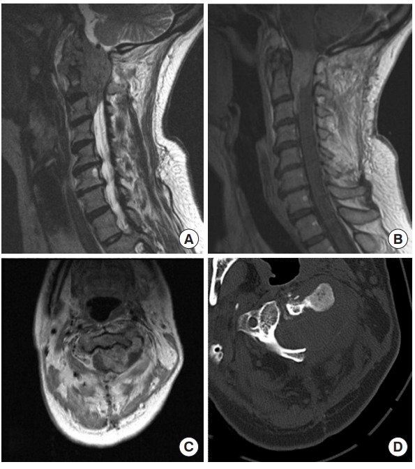
Fig. 1.

Preoperative images of case 1, a 46-year-old female who presented with cervical epidural sarcoma. (A) Sagittal T2, (B) sagittal gadolinium-enhanced T1, (C) axial gadolinium-enhanced T1 MRI, and (D) noncontrast axial computed tomography (CT) scan through the tumor at the level of C2. An enhancing epidural mass is seen extending from the foramen magnum down to the level of the C3 vertebra in the epidural space. The CT scan reveals significant bony erosion.
